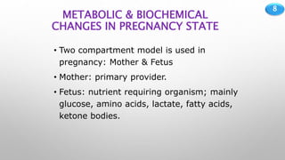
BIOCHEMICAL
CHANGES IN
PREGNANCY
DR. OFONMBUK UMOH
REGISTRAR, DEPARTMENT OF
CHEMICAL PATHOLOGY
2
OUTLINE
• Introduction
• Overview of anatomical &
Physiological changes in pregnancy
• Systemic biochemical changes
• Maternal and fetal health
assessment
• Complications of pregnancy
• Conclusion
• References
3
INTRODUTION
• Normal human pregnancy lasts approximately 40
weeks from the last menstrual period. During
pregnancy, a woman undergoes dramatic physiological
and hormonal changes in biological composition that
can be evaluated biochemically to monitor their
wellbeing, and for detection of any anomaly.
4
BRIEF OVERVIEW OF PREGNANCY
• A fertilized ovum becomes an embryo after
implantation in the uterus and development of a
placenta; then becomes a fetus at 10 weeks after most
primitive systemic structures have been formed.
• Pregnancy is divided into 3 trimesters of roughly 13
weeks each; and term pregnancy is adduced at 37 –
42 weeks of gestation.
5
ENDOMETRIAL CHANGES IN OVARIAN
CYCLE
6
7
8
METABOLIC & BIOCHEMICAL
CHANGES IN PREGNANCY STATE
• Two compartment model is used in
pregnancy: Mother & Fetus
• Mother: primary provider.
• Fetus: nutrient requiring organism; mainly
glucose, amino acids, lactate, fatty acids,
ketone bodies.
9
• 10-20 % increase in BMR by the end of 3rd
trimester
• Weight gain = 11 kgs, due to:
• Uterus
• Breast
• Increase blood volume
• Increased extravascular extracellular fluid
• Maternal reserves (increase in cellular water, fats
& proteins).
10
THE PLACENTA
• The placenta and umbilical cord forms the primary link
between mother and fetus
• …while keeping the maternal & fetal circulation
separate, providing nourishment to fetus, eliminating
fetal waste; and also produces hormones vital to
pregnancy.
• Chorionic gonadotropin
• Placental lactogen
• Placental steroids
• The
11
• The placenta is a highly active endocrine organ during
gestation, secreting a variety of hormones with
physiological effects.
• Efforts to study the action/function of these hormones in
driving physiological changes during pregnancy has been
attempted in two main ways.
• First, the expression and activity of the hormones have
been manipulated in vivo by either exogenously
administering or genetically manipulating the expression
of hormones and hormone receptors to study the
physiological activities.
12
• Secondly, hormone analogs have been manipulated
similarly in cultured cells and tissue explants to study
the cellular and molecular mechanisms by which they
modulate function.
• The effects of these hormones in non-pregnant study
groups is required as controls, to provide information on
the baseline and physiological changes that occur in the
absence and presence of these placental-derived
hormones respectively.
• These have not been very successful so far; and further
studies have been limited by ethical considerations.
13
Normal Placental Transport
• Not transported
• Most proteins
• Thyroid hormones
• Maternal IgM, IgA
• Maternal/Fetal RBCs
• Actively transported
• Glucose Calcium
• Many amino acids
• Limited passive
transport
• Unconjugated steroids
• Passive transport
• Oxygen
• Carbon dioxide
• Sodium & Chloride
• Urea Ethanol
• Molecules up to 5000 Da
having lipid solubility
• Receptor-mediated
endocytosis
• Insulin
• Maternal IgG
14
15
THE AMNIOTIC FLUID
• …a fluid-filled compartment, which the fetus lives
throughout intrauterine life.
• It increases gradually during pregnancy, to about 1400
ml at term. Its volume per time can be a guide to fetal
wellbeing:
• Oligohydramnios: low volume
• Polyhydramnios: high volume
16
Composition of amniotic fluid
17
Composition of amniotic fluid…contd
18
MATERNAL ADAPTATION
• Placental hormones interplay to mediate maternal
adaptations to pregnancy, parturition and lactation.
• Large quantities of estrogen, progesterone, placental
lactogen and corticosteroids produced during pregnancy
affects various metabolic, physiologic and endocrine
systems.
• There is:
• An increase in resistance to angiotensin
• A predominance of lipid metabolism over glucose use
• Increased synthesis by the liver, of fibrinogen, thyroid – and
19
General biochemical
picture…
• Electrolytes show little
change
• Urea nitrogen markedly
• Bicarbonate reduces as
much as 20% almost
throughout gestation.
• Creatine kinase markedly
incr
20
Renal changes
• Pregnancy increases the GFR to about 170
ml/min/1.73m2 by 20 weeks and therefore increases
the clearance of urea, creatinine and uric acid a
decrease in the concentration of these analytes during
pregnancy, until towards term when it normalizes.
• Glucosuria, up to 1000mg/d, due to incr GFR.
• …also, protein loss in urine can incr up to 300 mg/day.
21
Endocrine changes
• Progesterone prevent menses.
• 1,25 dihydroxyvitamin D is increased during pregnancy
which promotes increased intestinal absorption of
calcium and the transfer of calcium to the fetus.
• Estrogen stimulates an increase in plasma transport
proteins: thyroxine binding globulin, (TBG), cortisol
binding globulin (CBG) and sex hormone binding
globulin (SHBG).
22
• Estrogen also:
• Stimulates release of prolactin, up to 10 fold
• Suppress luteinizing and follicle stimulating hormone release
• The high concentration of TBG raises total T3 and T4,
but causes a slight decrease in fT4.
23
Hepatic changes
• Albumin synthesis decreases
• Alkaline phosphatase (ALP) activity almost triples, due
to ALP of placental origin.
• Hepatic clearance of cortisol decreases as estrogen
stimulates hepatic production of CBG. The diurnal
rhythm of cortisol is maintained.
24
Hematological changes
• Blood volume increases by up to 45%
• Plasma vol incr more rapidly than RBC mass
• Blood coagulation factors by up to 65% ESR,
increasing the risk of thromboembolism.
25
26
MATERNAL AND FETAL HEALTH ASSESSMENT
• Preconception evaluation should include: medical,
reproductive and family history; physical examination
and laboratory tests such as:
• Urinalysis, Hematocrit
• Blood group, rhesus factor & genotype
• Pap smear, HIV antibody
• Screening for TORCHES
• Gonococcus and chlamydia
• Hepatitis B surface antigen
• Depending on demographic risks:
• Genetic testing for cystic fibrosis, Thalassemias, IBEMs etc.
27
• In suspected pregnancy, following a missed menses,
some laboratory tests are conducted, which are useful
for managing a normal or abnormal pregnancy:
• Pregnancy test – hCG of about 25 IU/L (urine), or 10 IU/L
(serum).
• ...
• Other necessary obstetric maternal-fetal wellbeing
monitoring routine: USS, fetal HR, lie & movement, uterine
contractions etc.
28
Abnormal pregnancy biochemistry
• In event of certain risks in index pregnancy, screening
for:
• Neural tube defects: anencephaly, meningomyelocele.
Associated with folic acid deficiency. Elevated Alpha-
fetoprotein (AFP) in maternal serum – screening.
• Down’s syndrome: Trisomy 21.
• Phenotype – moderate to severe mental retardation, hypotonia,
congen. heart defect and flat facial profile.
• Triple test: serum hCG, AFP and unconjugated estriol, 15-18 weeks
GA. Inhibin A (Quadruple test). Amniocentesis, karyotyping.
• Respiratory distress syndr: aka hyaline membr disease.
About 10% of preterm births. Deficiency of pulmonary
surfactant  collapse of alveoli during expiration (fetal
29
• Gestational diabetes (GDM): defined as glucose
intolerance of variable degree, first recognition during
pregnancy.
• In pregnant mothers with metabolic syndrome; or previous
pregnancy history of GDM, macrosomia (≥ 4000 g) or stillbirth
• OGTT, 24-28 weeks gestation.
• Also, certain complications may arise in pregnancies,
such as:
• Ectopic pregnancy: serial quantitative hCG measurement with
slow rate of increase. Normally, hCG should double every 2-3
days.
30
• Preeclampsia: HTN, proteinuria, edema
• HELLP syndrome: RUQ pain, thrombocytopenia, DIC,
very high LDH, ALT & AST, usually 5-10x URL
• Hemolytic disease of the newborn: when maternal
antibodies reacts with antigen on fetal erythrocytes.
• Synonyms: Rhesus D isoimmunization, erythroblastosis fetalis.
• Can cause hydrops fetalis, hemolytic disease of the newborn,
severe NNJ and kernicterus.
• Serial monitoring of amniotic fluid bilirubin, fetal exchange
blood transfusion.
• Administration of anti-RhD immunoglobulin RhoGAM to
sensitized mothers.
31
• Liver disease of pregnancy:
• fatty liver of pregnancy: micro-vesicular fat in hepatocytes.
Rapid onset of abdominal pain, malaise, nausea/vomiting,
mild elevation of liver enzymes, bilirubin >6mg/dl, life-
threatening hypoglycemia, hyperuricemia, renal failure.
• cholestasis of pregnancy: diffuse pruritus, pale stool, dark
urine. Serum bilirubin >5mg/dl, ALP 2-4x URL. Elevated
prothrombin time.
32
CONCLUSION
• Pregnancy is an awe-inspiring process of
growth and development with diverse changes
in physiology and biochemical features.
Awareness of these changes and pregnancy-
unique disorders is critical in the proper
management of pregnancy.
33
REFERENCES
• Tietz Textbook of Clinical Chemistry and Molecular
Diagnostics; fifth Ed., by Burtis et al.
• Martin A. Crook; Clinical Chemistry and Metabolic
Medicine, 8th Edition.
• Clinical Chemistry; Principles, Techniques &
Correlations, 7th Ed., by Bishop et al.
• Oxford Handbook of Clinical Pathology; 17th Edition,
Oxford University Press, London.
34
THANK YOU
FOR
LISTENING

Biochemical changes in pregnancy

  • 1.
    BIOCHEMICAL CHANGES IN PREGNANCY DR. OFONMBUKUMOH REGISTRAR, DEPARTMENT OF CHEMICAL PATHOLOGY
  • 2.
    2 OUTLINE • Introduction • Overviewof anatomical & Physiological changes in pregnancy • Systemic biochemical changes • Maternal and fetal health assessment • Complications of pregnancy • Conclusion • References
  • 3.
    3 INTRODUTION • Normal humanpregnancy lasts approximately 40 weeks from the last menstrual period. During pregnancy, a woman undergoes dramatic physiological and hormonal changes in biological composition that can be evaluated biochemically to monitor their wellbeing, and for detection of any anomaly.
  • 4.
    4 BRIEF OVERVIEW OFPREGNANCY • A fertilized ovum becomes an embryo after implantation in the uterus and development of a placenta; then becomes a fetus at 10 weeks after most primitive systemic structures have been formed. • Pregnancy is divided into 3 trimesters of roughly 13 weeks each; and term pregnancy is adduced at 37 – 42 weeks of gestation.
  • 5.
  • 6.
  • 7.
  • 8.
    8 METABOLIC & BIOCHEMICAL CHANGESIN PREGNANCY STATE • Two compartment model is used in pregnancy: Mother & Fetus • Mother: primary provider. • Fetus: nutrient requiring organism; mainly glucose, amino acids, lactate, fatty acids, ketone bodies.
  • 9.
    9 • 10-20 %increase in BMR by the end of 3rd trimester • Weight gain = 11 kgs, due to: • Uterus • Breast • Increase blood volume • Increased extravascular extracellular fluid • Maternal reserves (increase in cellular water, fats & proteins).
  • 10.
    10 THE PLACENTA • Theplacenta and umbilical cord forms the primary link between mother and fetus • …while keeping the maternal & fetal circulation separate, providing nourishment to fetus, eliminating fetal waste; and also produces hormones vital to pregnancy. • Chorionic gonadotropin • Placental lactogen • Placental steroids • The
  • 11.
    11 • The placentais a highly active endocrine organ during gestation, secreting a variety of hormones with physiological effects. • Efforts to study the action/function of these hormones in driving physiological changes during pregnancy has been attempted in two main ways. • First, the expression and activity of the hormones have been manipulated in vivo by either exogenously administering or genetically manipulating the expression of hormones and hormone receptors to study the physiological activities.
  • 12.
    12 • Secondly, hormoneanalogs have been manipulated similarly in cultured cells and tissue explants to study the cellular and molecular mechanisms by which they modulate function. • The effects of these hormones in non-pregnant study groups is required as controls, to provide information on the baseline and physiological changes that occur in the absence and presence of these placental-derived hormones respectively. • These have not been very successful so far; and further studies have been limited by ethical considerations.
  • 13.
    13 Normal Placental Transport •Not transported • Most proteins • Thyroid hormones • Maternal IgM, IgA • Maternal/Fetal RBCs • Actively transported • Glucose Calcium • Many amino acids • Limited passive transport • Unconjugated steroids • Passive transport • Oxygen • Carbon dioxide • Sodium & Chloride • Urea Ethanol • Molecules up to 5000 Da having lipid solubility • Receptor-mediated endocytosis • Insulin • Maternal IgG
  • 14.
  • 15.
    15 THE AMNIOTIC FLUID •…a fluid-filled compartment, which the fetus lives throughout intrauterine life. • It increases gradually during pregnancy, to about 1400 ml at term. Its volume per time can be a guide to fetal wellbeing: • Oligohydramnios: low volume • Polyhydramnios: high volume
  • 16.
  • 17.
  • 18.
    18 MATERNAL ADAPTATION • Placentalhormones interplay to mediate maternal adaptations to pregnancy, parturition and lactation. • Large quantities of estrogen, progesterone, placental lactogen and corticosteroids produced during pregnancy affects various metabolic, physiologic and endocrine systems. • There is: • An increase in resistance to angiotensin • A predominance of lipid metabolism over glucose use • Increased synthesis by the liver, of fibrinogen, thyroid – and
  • 19.
    19 General biochemical picture… • Electrolytesshow little change • Urea nitrogen markedly • Bicarbonate reduces as much as 20% almost throughout gestation. • Creatine kinase markedly incr
  • 20.
    20 Renal changes • Pregnancyincreases the GFR to about 170 ml/min/1.73m2 by 20 weeks and therefore increases the clearance of urea, creatinine and uric acid a decrease in the concentration of these analytes during pregnancy, until towards term when it normalizes. • Glucosuria, up to 1000mg/d, due to incr GFR. • …also, protein loss in urine can incr up to 300 mg/day.
  • 21.
    21 Endocrine changes • Progesteroneprevent menses. • 1,25 dihydroxyvitamin D is increased during pregnancy which promotes increased intestinal absorption of calcium and the transfer of calcium to the fetus. • Estrogen stimulates an increase in plasma transport proteins: thyroxine binding globulin, (TBG), cortisol binding globulin (CBG) and sex hormone binding globulin (SHBG).
  • 22.
    22 • Estrogen also: •Stimulates release of prolactin, up to 10 fold • Suppress luteinizing and follicle stimulating hormone release • The high concentration of TBG raises total T3 and T4, but causes a slight decrease in fT4.
  • 23.
    23 Hepatic changes • Albuminsynthesis decreases • Alkaline phosphatase (ALP) activity almost triples, due to ALP of placental origin. • Hepatic clearance of cortisol decreases as estrogen stimulates hepatic production of CBG. The diurnal rhythm of cortisol is maintained.
  • 24.
    24 Hematological changes • Bloodvolume increases by up to 45% • Plasma vol incr more rapidly than RBC mass • Blood coagulation factors by up to 65% ESR, increasing the risk of thromboembolism.
  • 25.
  • 26.
    26 MATERNAL AND FETALHEALTH ASSESSMENT • Preconception evaluation should include: medical, reproductive and family history; physical examination and laboratory tests such as: • Urinalysis, Hematocrit • Blood group, rhesus factor & genotype • Pap smear, HIV antibody • Screening for TORCHES • Gonococcus and chlamydia • Hepatitis B surface antigen • Depending on demographic risks: • Genetic testing for cystic fibrosis, Thalassemias, IBEMs etc.
  • 27.
    27 • In suspectedpregnancy, following a missed menses, some laboratory tests are conducted, which are useful for managing a normal or abnormal pregnancy: • Pregnancy test – hCG of about 25 IU/L (urine), or 10 IU/L (serum). • ... • Other necessary obstetric maternal-fetal wellbeing monitoring routine: USS, fetal HR, lie & movement, uterine contractions etc.
  • 28.
    28 Abnormal pregnancy biochemistry •In event of certain risks in index pregnancy, screening for: • Neural tube defects: anencephaly, meningomyelocele. Associated with folic acid deficiency. Elevated Alpha- fetoprotein (AFP) in maternal serum – screening. • Down’s syndrome: Trisomy 21. • Phenotype – moderate to severe mental retardation, hypotonia, congen. heart defect and flat facial profile. • Triple test: serum hCG, AFP and unconjugated estriol, 15-18 weeks GA. Inhibin A (Quadruple test). Amniocentesis, karyotyping. • Respiratory distress syndr: aka hyaline membr disease. About 10% of preterm births. Deficiency of pulmonary surfactant  collapse of alveoli during expiration (fetal
  • 29.
    29 • Gestational diabetes(GDM): defined as glucose intolerance of variable degree, first recognition during pregnancy. • In pregnant mothers with metabolic syndrome; or previous pregnancy history of GDM, macrosomia (≥ 4000 g) or stillbirth • OGTT, 24-28 weeks gestation. • Also, certain complications may arise in pregnancies, such as: • Ectopic pregnancy: serial quantitative hCG measurement with slow rate of increase. Normally, hCG should double every 2-3 days.
  • 30.
    30 • Preeclampsia: HTN,proteinuria, edema • HELLP syndrome: RUQ pain, thrombocytopenia, DIC, very high LDH, ALT & AST, usually 5-10x URL • Hemolytic disease of the newborn: when maternal antibodies reacts with antigen on fetal erythrocytes. • Synonyms: Rhesus D isoimmunization, erythroblastosis fetalis. • Can cause hydrops fetalis, hemolytic disease of the newborn, severe NNJ and kernicterus. • Serial monitoring of amniotic fluid bilirubin, fetal exchange blood transfusion. • Administration of anti-RhD immunoglobulin RhoGAM to sensitized mothers.
  • 31.
    31 • Liver diseaseof pregnancy: • fatty liver of pregnancy: micro-vesicular fat in hepatocytes. Rapid onset of abdominal pain, malaise, nausea/vomiting, mild elevation of liver enzymes, bilirubin >6mg/dl, life- threatening hypoglycemia, hyperuricemia, renal failure. • cholestasis of pregnancy: diffuse pruritus, pale stool, dark urine. Serum bilirubin >5mg/dl, ALP 2-4x URL. Elevated prothrombin time.
  • 32.
    32 CONCLUSION • Pregnancy isan awe-inspiring process of growth and development with diverse changes in physiology and biochemical features. Awareness of these changes and pregnancy- unique disorders is critical in the proper management of pregnancy.
  • 33.
    33 REFERENCES • Tietz Textbookof Clinical Chemistry and Molecular Diagnostics; fifth Ed., by Burtis et al. • Martin A. Crook; Clinical Chemistry and Metabolic Medicine, 8th Edition. • Clinical Chemistry; Principles, Techniques & Correlations, 7th Ed., by Bishop et al. • Oxford Handbook of Clinical Pathology; 17th Edition, Oxford University Press, London.
  • 34.

Editor's Notes

  • #11 HCG – stim ovary to pdce progest > prevent menstruation …which has lactogenic, metabolic/aldosterone stim. effects on mother; as well as somatotropic & erythropoietic effect on fetus, …estrogen & progest: which ensures uterine growth & adeq bld ss
  • #14 The placenta is an effective barrier to the movement of large proteins & hydrophobic subst bound to pl. pr
  • #16 -It maintains a constant temperature, cushions against injuries and provides a medium where the fetus moves. Fetus swallows it & urinates into it. -Oligo…bilat renal agenesis, atresia or IUGR -Polyhy…maternal DM, multiple preg, Rh-isoimmuniz, esophag. atresia
  • #19 Normal = uneventful preg.
  • #20 There is incr GFR due to hyper-dynamic state
  • #21 …which incr gluc to the kidneys and lowers the renal gluc threshold.
  • #23 …although preg is an euthyr state, post partum thyr dysfxn is common.
  • #26 …hyper-reninaemic hyperaldosteronism.
  • #28 …other pre-conception screenings
  • #29 -Downs…there are a barrage of other aneuploidies to be screened for, in at-risk groups. -normally, surfactants coat the alveolar epith & reduce surface tension during expiration. …assessment of FLM
  • #30 -failure of neural tube fusion at the brain/spinal cord -Ectopic preg: implantatn of fertilized ovum at location other than the uterus. Tubal rupture common. Lower abd pain, vag bleeding & adnexal mass -Severe nausea, vomiting, malnutrition.
  • #31 -usu in late 2t or 3t of preg. Can cause convulsion (eclampsia) -life threatening complicaon to Pre-eclampsia
  • #32 -usu 3t of pregn. Hyperuricemia from severe hepatic tissue destruction -typcal features of cholestasis/obstructive jaundice… …..due to Vit K malabsorption.