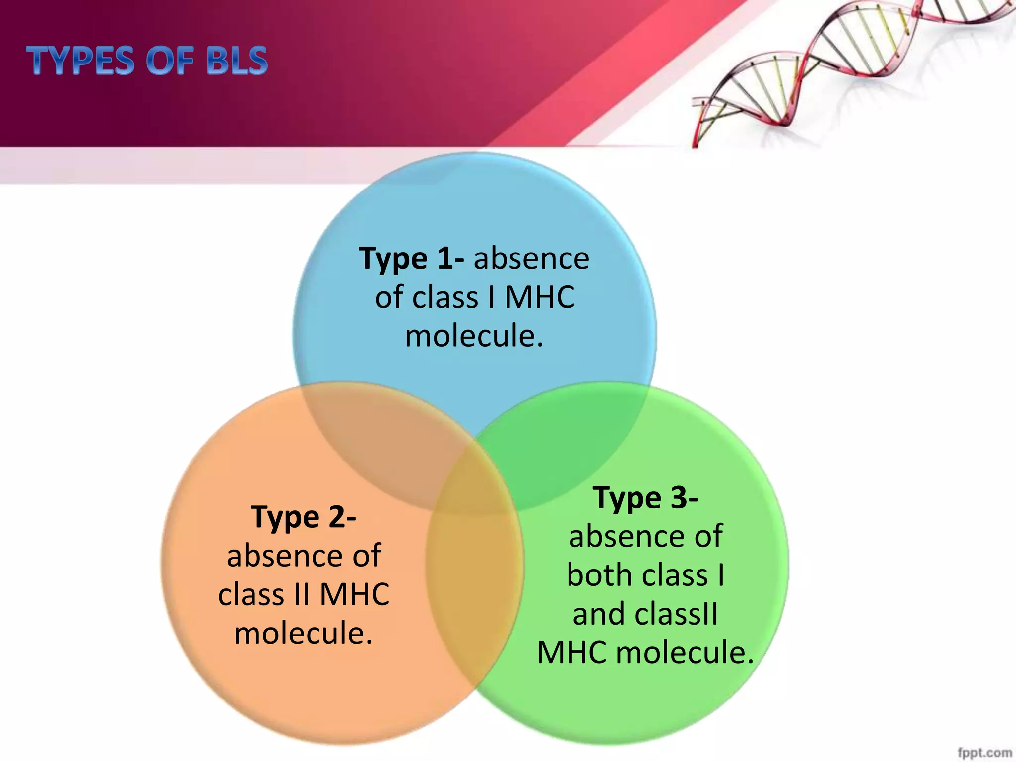
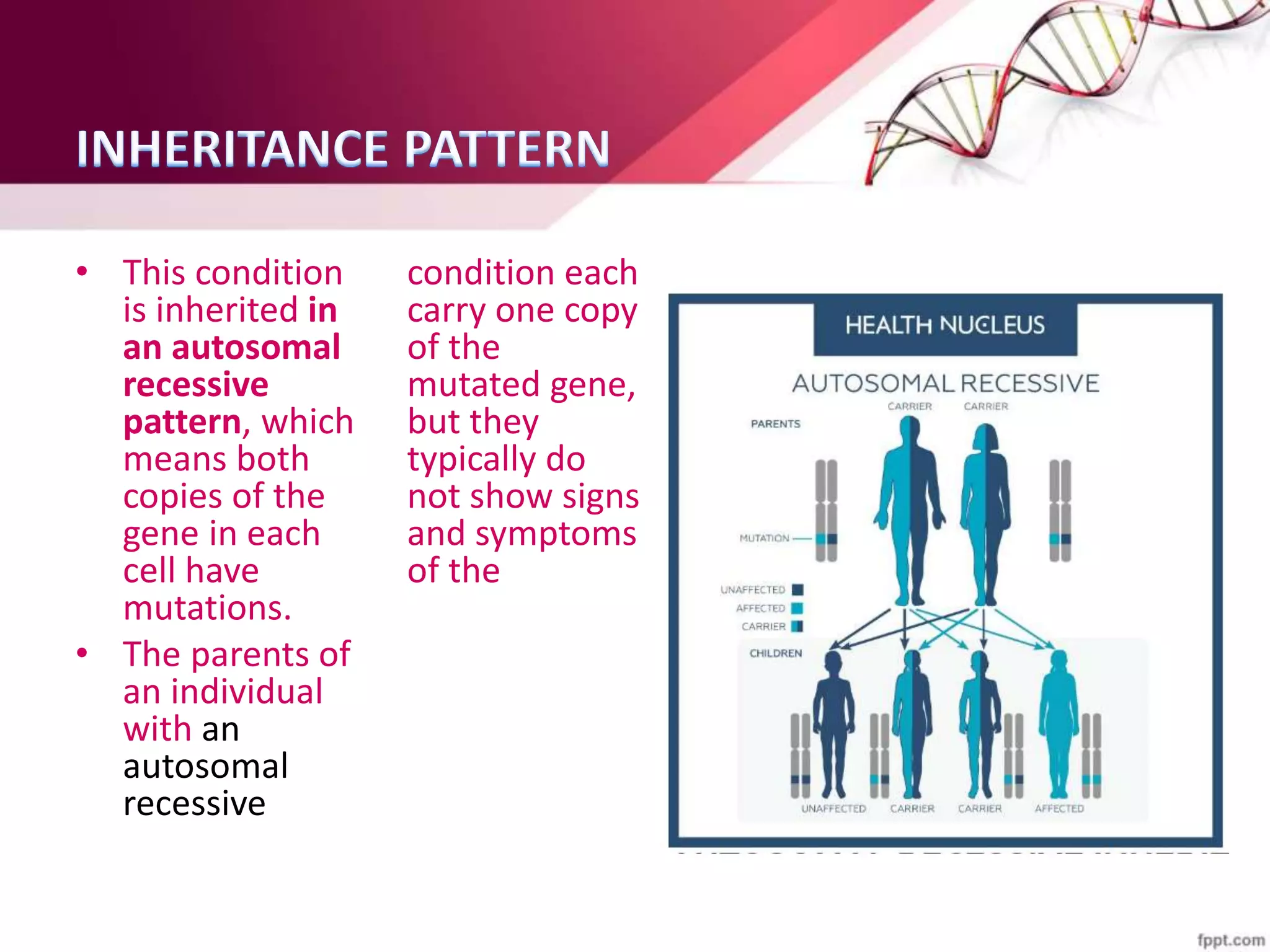
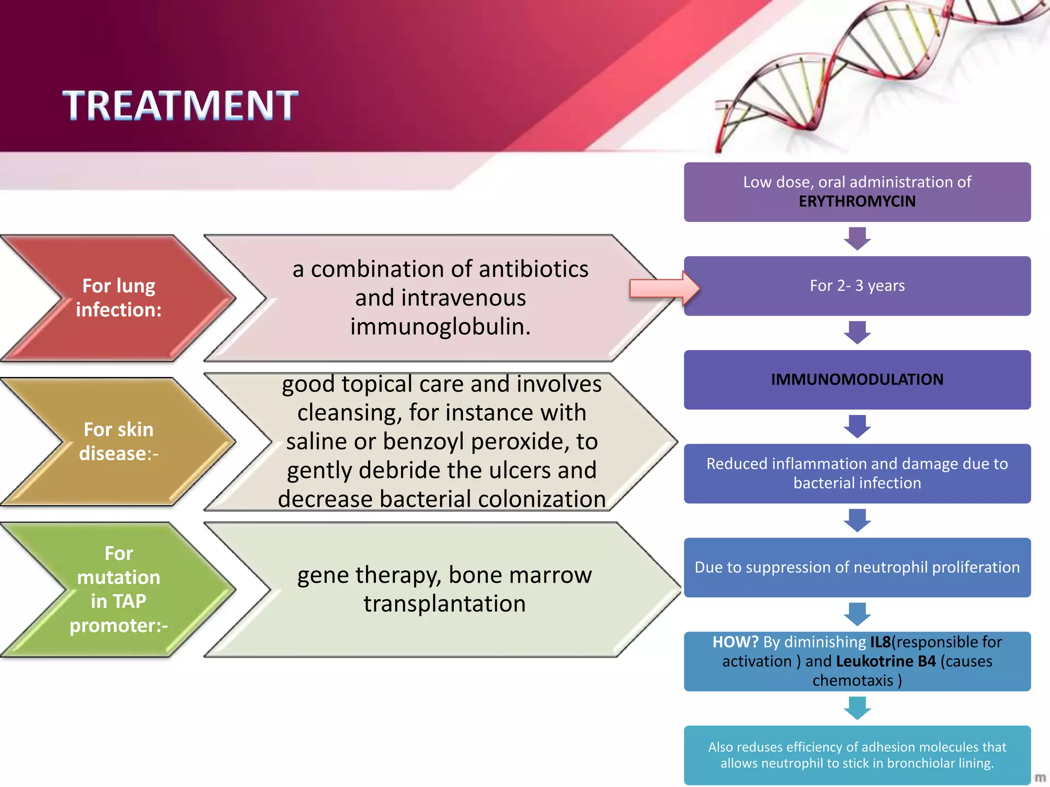
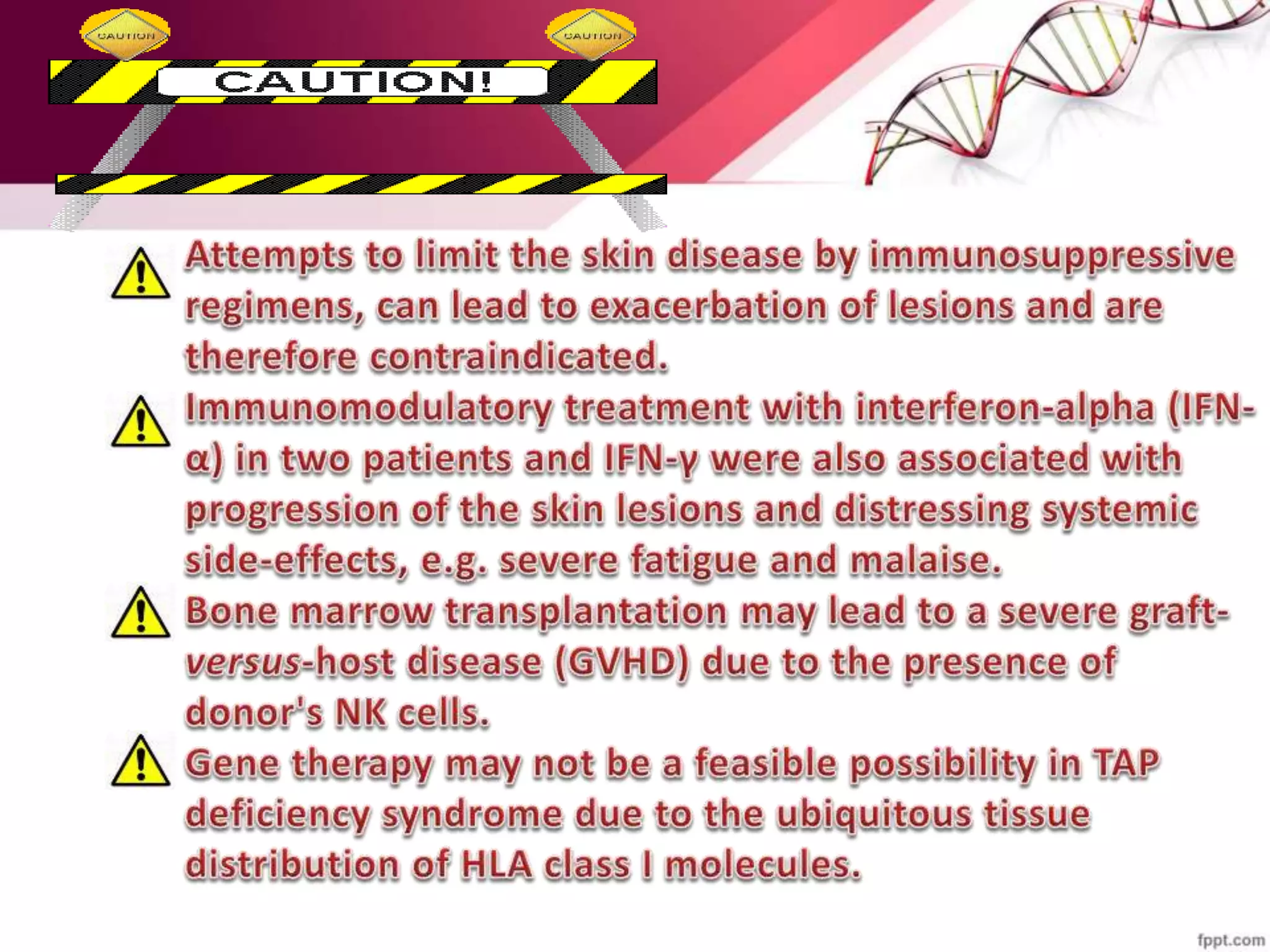
• A rare recessive genetic immuno-deficiency
disorder.
• There is either partial or complete absence of MHC
or HLA on the surface of lymphocytes. i.e lyphocytes
are BARE
• This leads to fragile immune system, much
resembling to SCID.
Type 1- absence
of class I MHC
molecule.
Type 3-
absence of
both class I
and classII
MHC molecule.
Type 2-
absence of
class II MHC
molecule.
Type 2- BLS
• Possibly due to a defect in gene
activation and/or a defect in
accessibility of the promoter
protein; mutations in any of
four genes can cause the BLS:
• Class-II trans-activator (CIITA)
located at 16p13.13;
• Regulatory factor of the X
box 5 (RFX5)located at 1q21.3
• RFX-associated protein
(RFXAP)located at 19p13.11;
• RFX ankyrin repeats
(RFXANK)located at 13q13.3.
• More Common(At least 100
cases have been reported in the
medical literature)
Type 1- BLS
• The genetic basis for BLS-I is
due to defects in the MHC
genes themselves.
• associated with TAP2, TAP1, or
TAPBP deficiencies.
• Rare(About 30 affected
individuals have been described
in the medical literature.)
• Characterized by the low expression of MHC I proteins in
the cells of the patients.
• Cellular abnormalities:
Increased number and heightened
activity of NK cells
Increased number of  T cells
Decreased number of CD8+ T cells
NK cell recognition of target
cells
NK cells recognize and kill infected and tumor cells by their
absence of MHC class I
(a) Normal cells present a ligand for the activating (killing)
receptor on NK cells AND a ligand for the inhibitory
receptor (class I MHC serves as this second ligand)
(b) When viruses infect cells, MHC class I expression is
reduced to evade CTLs - This makes them a prime target for
elimination by NK cells
INCREASED  T-cell and DECEASED
 T-cells
• There is lineage commitment at
DN2 stage.
• After birth the development of 
T-cells is 3 X more favored over 
T-cells.
• DP  T-cells to becomes SP T-cells
when they interact with MHC I or
MHC II.
 T-cells
interacts with
phosphorylated
metabolites
produced by
microbes.
Since in this case
the MHC I are
absent on the
lymphocytes so
there is decrease
in number of CD8+
 T-cells on the
other hand
increase in  T-
cells.
• The disorder is due to defect in the TAP1 or TAP 2 gene.
• The TAP1 and TAP 2 together complex to form Transporter
associated with antigen processing complex(TAP complex).
Absence of
susceptibility to severe
viral infection.
When viruses infect
cells, MHC class I
expression is reduced
So, there are not
enough CTLs to
combat the virus
entered into the body.
Bacterial/viral infection
in the upper respiratory
tract-chronic purulent
rhinitis
Inflammation in the
nasal passage.
Symptoms: stuffy
nose, nasal discharge,
feeling of mucus in
back of throat(post
nasal drip).
Dilation of the
bronchial tubes,
i.e. Bronchiectasis
Actually Bronchiectasis is
thought to be caused by
the
mutation/recombination
in Class I HLA region.
The region near HLA-B, at
Chromosome-6p21.3
This region also have
genes for TAP protein,
involved in BLS I
Necrotizing skin
lesions
Due to absence of MHC on
the lymphocytes, KIRs are
not bound to their ligands.
This activates NK cells and
their activity heightens,
therefore causing to attack
own cells and causing
necrosis like effects
• This condition
is inherited in
an autosomal
recessive
pattern, which
means both
copies of the
gene in each
cell have
mutations.
• The parents of
an individual
with an
autosomal
recessive
condition each
carry one copy
of the
mutated gene,
but they
typically do
not show signs
and symptoms
of the
condition.
For lung
infection:
a combination of antibiotics
and intravenous
immunoglobulin.
For skin
disease:-
good topical care and involves
cleansing, for instance with
saline or benzoyl peroxide, to
gently debride the ulcers and
decrease bacterial colonization
For
mutation
in TAP
promoter:-
gene therapy, bone marrow
transplantation
Low dose, oral administration of
ERYTHROMYCIN
For 2- 3 years
IMMUNOMODULATION
Reduced inflammation and damage due to
bacterial infection
Due to suppression of neutrophil proliferation
HOW? By diminishing IL8(responsible for
activation ) and Leukotrine B4 (causes
chemotaxis )
Also reduses efficiency of adhesion molecules that
allows neutrophil to stick in bronchiolar lining.
Bare Lymphocyte Symdrome

Bare Lymphocyte Symdrome

  • 2.
    • A rarerecessive genetic immuno-deficiency disorder. • There is either partial or complete absence of MHC or HLA on the surface of lymphocytes. i.e lyphocytes are BARE • This leads to fragile immune system, much resembling to SCID.
  • 3.
    Type 1- absence ofclass I MHC molecule. Type 3- absence of both class I and classII MHC molecule. Type 2- absence of class II MHC molecule.
  • 4.
    Type 2- BLS •Possibly due to a defect in gene activation and/or a defect in accessibility of the promoter protein; mutations in any of four genes can cause the BLS: • Class-II trans-activator (CIITA) located at 16p13.13; • Regulatory factor of the X box 5 (RFX5)located at 1q21.3 • RFX-associated protein (RFXAP)located at 19p13.11; • RFX ankyrin repeats (RFXANK)located at 13q13.3. • More Common(At least 100 cases have been reported in the medical literature) Type 1- BLS • The genetic basis for BLS-I is due to defects in the MHC genes themselves. • associated with TAP2, TAP1, or TAPBP deficiencies. • Rare(About 30 affected individuals have been described in the medical literature.)
  • 5.
    • Characterized bythe low expression of MHC I proteins in the cells of the patients. • Cellular abnormalities: Increased number and heightened activity of NK cells Increased number of  T cells Decreased number of CD8+ T cells
  • 6.
    NK cell recognitionof target cells NK cells recognize and kill infected and tumor cells by their absence of MHC class I (a) Normal cells present a ligand for the activating (killing) receptor on NK cells AND a ligand for the inhibitory receptor (class I MHC serves as this second ligand) (b) When viruses infect cells, MHC class I expression is reduced to evade CTLs - This makes them a prime target for elimination by NK cells
  • 7.
    INCREASED  T-celland DECEASED  T-cells • There is lineage commitment at DN2 stage. • After birth the development of  T-cells is 3 X more favored over  T-cells. • DP  T-cells to becomes SP T-cells when they interact with MHC I or MHC II.  T-cells interacts with phosphorylated metabolites produced by microbes. Since in this case the MHC I are absent on the lymphocytes so there is decrease in number of CD8+  T-cells on the other hand increase in  T- cells.
  • 8.
    • The disorderis due to defect in the TAP1 or TAP 2 gene. • The TAP1 and TAP 2 together complex to form Transporter associated with antigen processing complex(TAP complex).
  • 9.
    Absence of susceptibility tosevere viral infection. When viruses infect cells, MHC class I expression is reduced So, there are not enough CTLs to combat the virus entered into the body. Bacterial/viral infection in the upper respiratory tract-chronic purulent rhinitis Inflammation in the nasal passage. Symptoms: stuffy nose, nasal discharge, feeling of mucus in back of throat(post nasal drip).
  • 10.
    Dilation of the bronchialtubes, i.e. Bronchiectasis Actually Bronchiectasis is thought to be caused by the mutation/recombination in Class I HLA region. The region near HLA-B, at Chromosome-6p21.3 This region also have genes for TAP protein, involved in BLS I Necrotizing skin lesions Due to absence of MHC on the lymphocytes, KIRs are not bound to their ligands. This activates NK cells and their activity heightens, therefore causing to attack own cells and causing necrosis like effects
  • 11.
    • This condition isinherited in an autosomal recessive pattern, which means both copies of the gene in each cell have mutations. • The parents of an individual with an autosomal recessive condition each carry one copy of the mutated gene, but they typically do not show signs and symptoms of the condition.
  • 12.
    For lung infection: a combinationof antibiotics and intravenous immunoglobulin. For skin disease:- good topical care and involves cleansing, for instance with saline or benzoyl peroxide, to gently debride the ulcers and decrease bacterial colonization For mutation in TAP promoter:- gene therapy, bone marrow transplantation Low dose, oral administration of ERYTHROMYCIN For 2- 3 years IMMUNOMODULATION Reduced inflammation and damage due to bacterial infection Due to suppression of neutrophil proliferation HOW? By diminishing IL8(responsible for activation ) and Leukotrine B4 (causes chemotaxis ) Also reduses efficiency of adhesion molecules that allows neutrophil to stick in bronchiolar lining.