Bar charts quickstudy nervous system

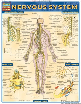
This document provides a detailed overview of the human nervous system including the cerebellum, spinal cord, plexuses, nerves and dermatomes. The first part features labeled diagrams of the cervical and lumbosacral plexuses, showing the formation and branches of nerves in these regions. The second section depicts spinal nerve roots and the overall structure of a nerve. The third section outlines the peripheral nervous system, displaying spinal and autonomic ganglia. The final section is a chart of cutaneous innervation, identifying dermatome regions and distributions of peripheral nerves across the body surface. In summary, the document comprehensively maps the central and peripheral components of the human nervous system through multiple detailed illustrations and diagrams.

Recommended
More Related Content
Viewers also liked
Viewers also liked (14)
Similar to Bar charts quickstudy nervous system
Similar to Bar charts quickstudy nervous system (20)
More from Abdallah Yakoub
More from Abdallah Yakoub (20)
Bar charts quickstudy nervous system
- 1. WORLD’S #1 ACADEMIC OUTLINE BarCharts, Inc.® CERVICOBRACHIAL PLEXUS LUMBOSACRAL PLEXUS Cerebellum 1st cervical vertebrae (transverse process)** Trace of the mandible Supraclavicular n. 1st cervical n. 7th cervicle vertebrae (pedicle & transverse process)** 1st thoracic vertebrae Upper trunk (pedicle)** Middle trunk Trace of the scapula Cervical Inferior trunk plexus Lateral cord Posterior cord 8th cervical n. Medial cord 1st thoracic n. Humerus Spinal cord Musculocutaneous n. Trace of the scapula Cervical plexus C1-C4 Cervical n.n. C1-C8 Brachial plexus C5-T1 C1 C2 C3 C4 C5 C6 C7 C8 T1 T2 T3 T4 Thoracic T5 n.n. T1-T12 T6 Brain 1st lumbar vertebrae (pedicle)** 5th lumbar vertebrae (pedicle)** Iliohypogastric n. Ilioinguinal n. Genitofemoral n. Lateral femoral cutaneous n. Trace of the pelvis Intercostal n.n. Femoral n. Superior gluteal n. Inferior gluteal n. Brachial plexus Posterior femoral cutaneous n. Musculocutaneous n. T10 Ulnar n. Iliohypogastric n. Inferior rectal n. Dorsal n. of penis (clitoris) Deep branch Ilioinguinal n. m. = muscle n. = nerve n.n. = nerves ** = cut Pudendal n. Median n. Ulnar n. Posterior brachial cutaneous n. Femur Muscular branches Subcostal n. Median n. Coccygeal n. Sciatic n. Radial n. Cauda equina Radial n. Cutaneous n. of forearm T9 Sacral plexus L5-S4 Sacral n.n. S1-S5 L5 S1 S2 S3 S4 S5 Axillary n. Conus medullaris Axillary n. T8 T11 Thoracic n.n. T12 T1-T12 L1 Lumbar plexus L2 T12-L4 L3 Lumbar n.n. L4 L1-L5 Sacrum, is made up of 5 fused vertebrae (pedicles)** Trace of the spinal column T7 12th thoracic vertebrae (pedicle)** Perineal n. Superficial branch Muscular branches Lumbar plexus Trace of the pelvis Dorsal branch Sacral plexus Superior & inferior gluteal n.n. Palmar branch Dorsal digital n.n. Sciatic n. Femoral n. Palmar digital n.n. of the median n. Dorsal digital n.n. SPINAL CORD Dorsal root (sensory) Palmar digital n.n. of the ulnar n. Posterior femoral cutaneous n. NERVE STRUCTURE Femoral n. Nucleolus Nucleus Muscular branches Axon hillock Pudendal n. White matter (sensory) Ventral root (motor) Synapse Spinous process Common peroneal (fibular) n. White matter (motor) Myelin sheath Dendrites Tibial n. Pia mater Sensory ganglion Axon (n. fiber) Saphenous n. Gray matter Muscular branches Motor n. fibers Nissl substance Schwann cell nucleus Arachnoid matter Superior articular process Cell body Deep peroneal n. N. fibers Pedicle Transverse process Striated m. Saphenous n. Dura mater Gray & white rami communicantes Sympathetic ganglion Medial dorsal cutaneous n. Axonal terminal Telodendria Common dorsal digital n.n. Common plantar digital n.n. Schwann cell Endoneurium Perineurium Fascicle Myofibrils Lateral plantar n. Inferior articular process & facet Impulse direction Neurilemma (sheath of Schwann) Node of Ranvier (axon) Blood vessels Sympathetic trunk Medial plantar n. Intervertebral disc Proper plantar digital n.n. Vertebral body (centrum) NERVOUS SYSTEM Epineurium to get more free charts visit us http://scientific4you.blogspot.com
- 2. CUTANEOUS INNERVATION: DERMATOMES & PERIPHERAL NERVE DISTRIBUTIONS C2 V1 V2 V3 C3 C4 Greater occipital n. Lesser occipital n. Ophthalmic n. Maxillary n. Trigeminal n. Mandibular n. Lesser occipital n. Transverse cervical n. C3 Supraclavicular n.n. Axillary n. Lower lateral brachial cutaneous n. Medial antebrachial cutaneous n.n. T5 T6 T7 T8 T9 T10 Lateral antebrachial cutaneous n.n. Iliohypogastric n. Genitofemoral & ilioinguinal n. T11 T12 L1 S2 L2 Ulnar palmar n. Radial n. superbrachial branch Median n.: palmar branch Digital branches S3 Genitofemoral n. L3 Palmar digital branch Dorsal n. of penis Lateral femoral cutaneous n. Scrotal branch of perineal n. L4 C6 C7 C8 Laterial cutaneous rami of thoracic spinal n.n. Axillary n. Intercostobrachial & medial brachial cutaneous n.n. Posterior brachial cutaneous n. Inferior lateral cutaneous n. Lateral antebrachial cutaneous n. Posterior antebrachial cutaneous n. Medial antebrachial cutaneous n. Radial n. superficial & dorsal digital branches S4 S5 Co1 Ulnar n. Median n. L1 Anterior femoral cutaneous n. Obturator n. L2 Common peroneal n. Infrapatellar branch of the saphenous n. L5 Posterior cutaneous rami of thoracic spinal n.n. C4 C5 C6 C7 C8 T1 T2 T3 T4 T5 T6 T7 T8 T9 T10 T11 T12 L1 L2 L3 L4 L5 S1 S2 S3 Medial brachial cutaneous & intercostobrachial n.n. T4 C8 Posterior branches of occipital n. C2 T3 C7 Great auricular n. Intercostal n.n. 1. Anterior cutaneous rami 2. Lateral cutaneous rami T2 C6 Greater occipital n. Transverse cervical n. Supraclavicular n.n. T1 C5 T1 C8 = Ophthalmic n. = Maxillary n. = Mandibular n. = Cervical = Thoracic = Lumbar = Sacrum = Coccyx Great auricular n. C5 C6 V1 V2 V3 C T L S Co L3 Ilioinguinal n. Superior cluneal n.n. Inferior & medial cluneal n.n. Lateral femoral cutaneous n.n. Annococcygeal n. Posterior scrotal (labial) n. Posterior femoral cutaneous n. Lateral sural cutaneous n. Obturator n. Medial crural cutaneous branches of the saphenous n. L4 S1 L5 Infrapatellar branch of the saphenous n. S2 Superficial peroneal (fibular) n. Common peroneal n. Lateral sural cutaneous n. S1 Sural n. L4 Deep peroneal (fibular) n. Medial crural cutaneous branches of the saphenous n. Sural n. Superficial peroneal n. Tibial n. L5 Lateral plantar n. Medial plantar n. U.S.$3.95 / CAN.$5.95 ISBN-13: 978-142320741-2 ISBN-10: 142320741-6 Customer Hotline: 1-800-230-9522 CREDITS Images ® Vincent Perez perezstudio.com free downloads & hundreds of titles at quickstudy.com NOTE TO STUDENT Use this comprehensive study guide in the classroom, in the gym, at home or anywhere you need complete nervous system information. This guide is not designed to take the place of classroom attendance. All rights reserved. No part of this publication may be reproduced or transmitted in any form, or by any means, electronic or mechanical, including photocopy, recording, or any information storage & retrieval system, without written permission from the publisher. ©1999, 2003, 2005 BarCharts Inc. 0608 to get more free charts visit us http://scientific4you.blogspot.com