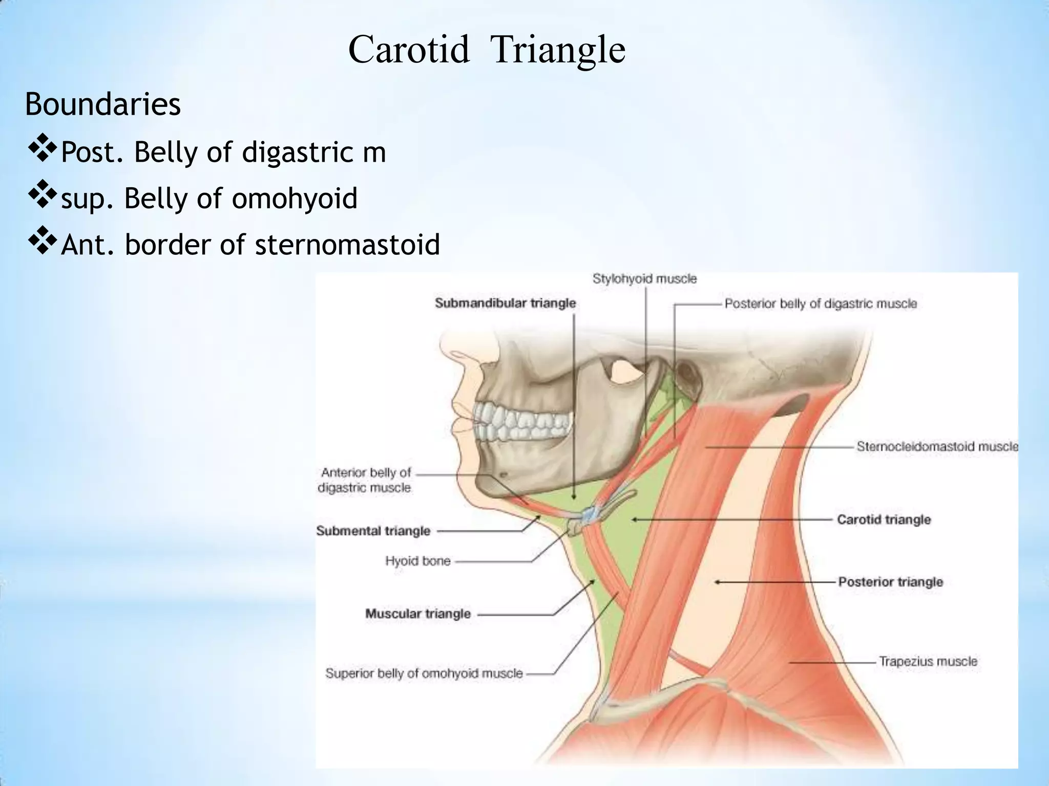
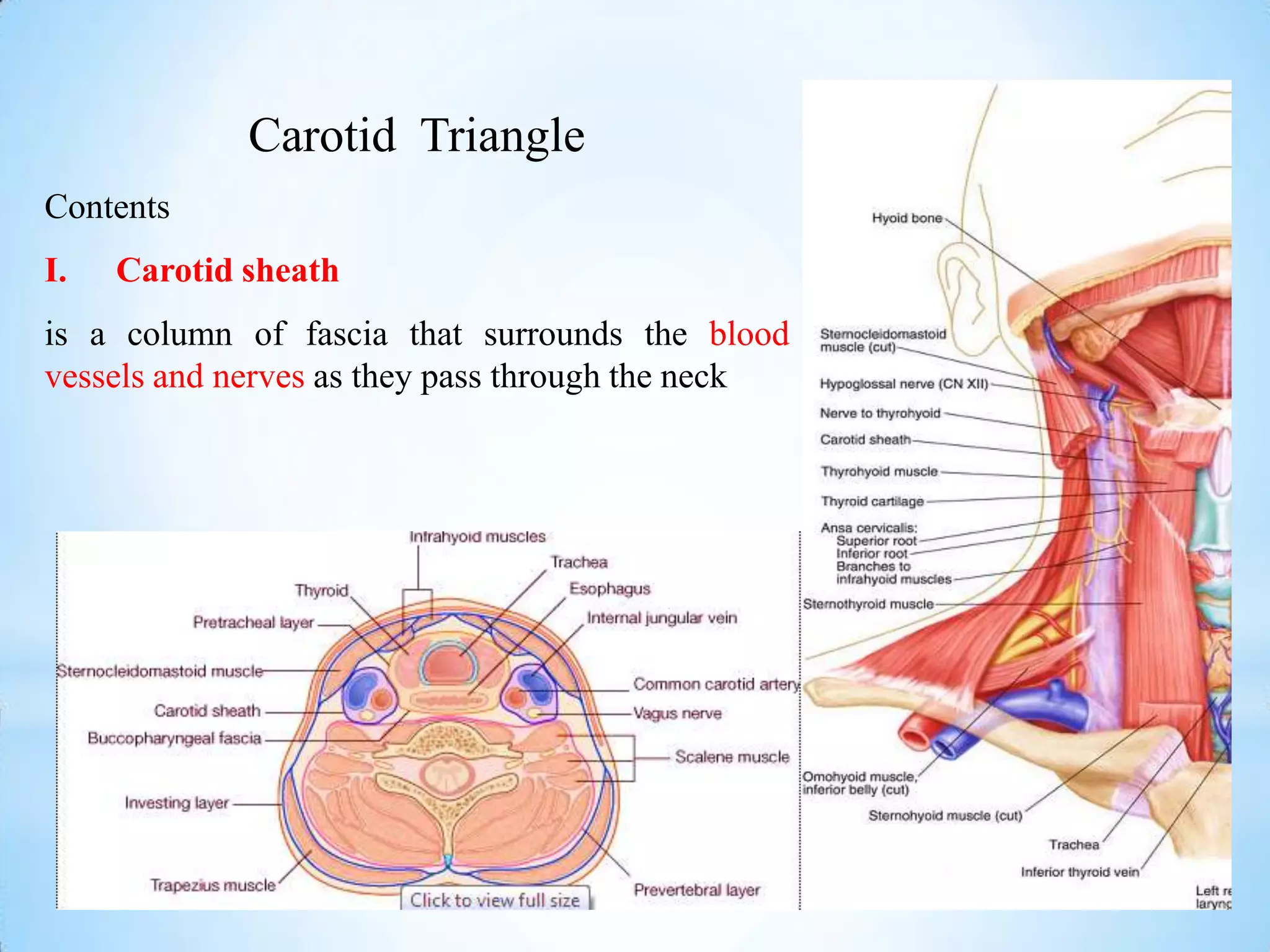
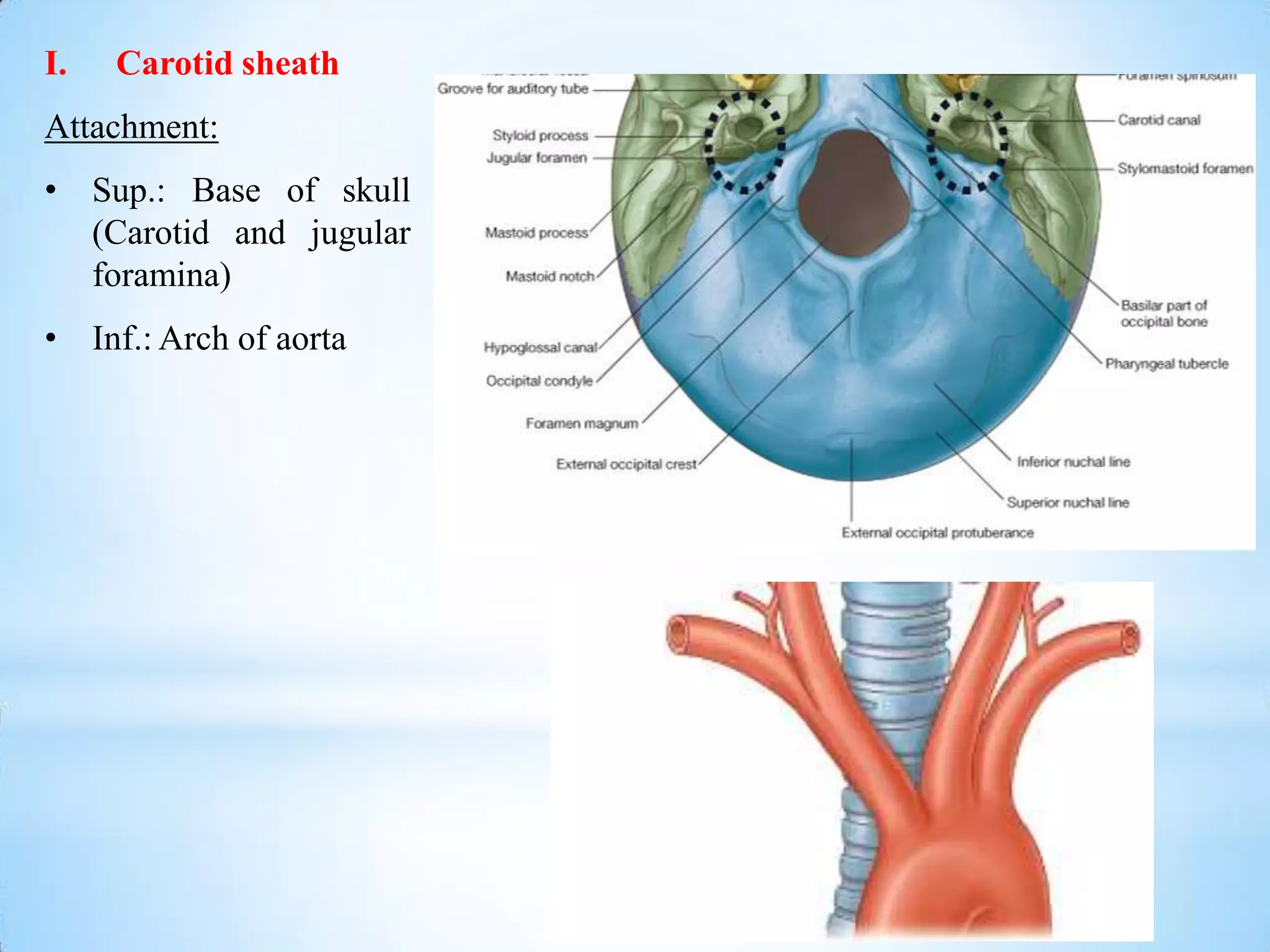
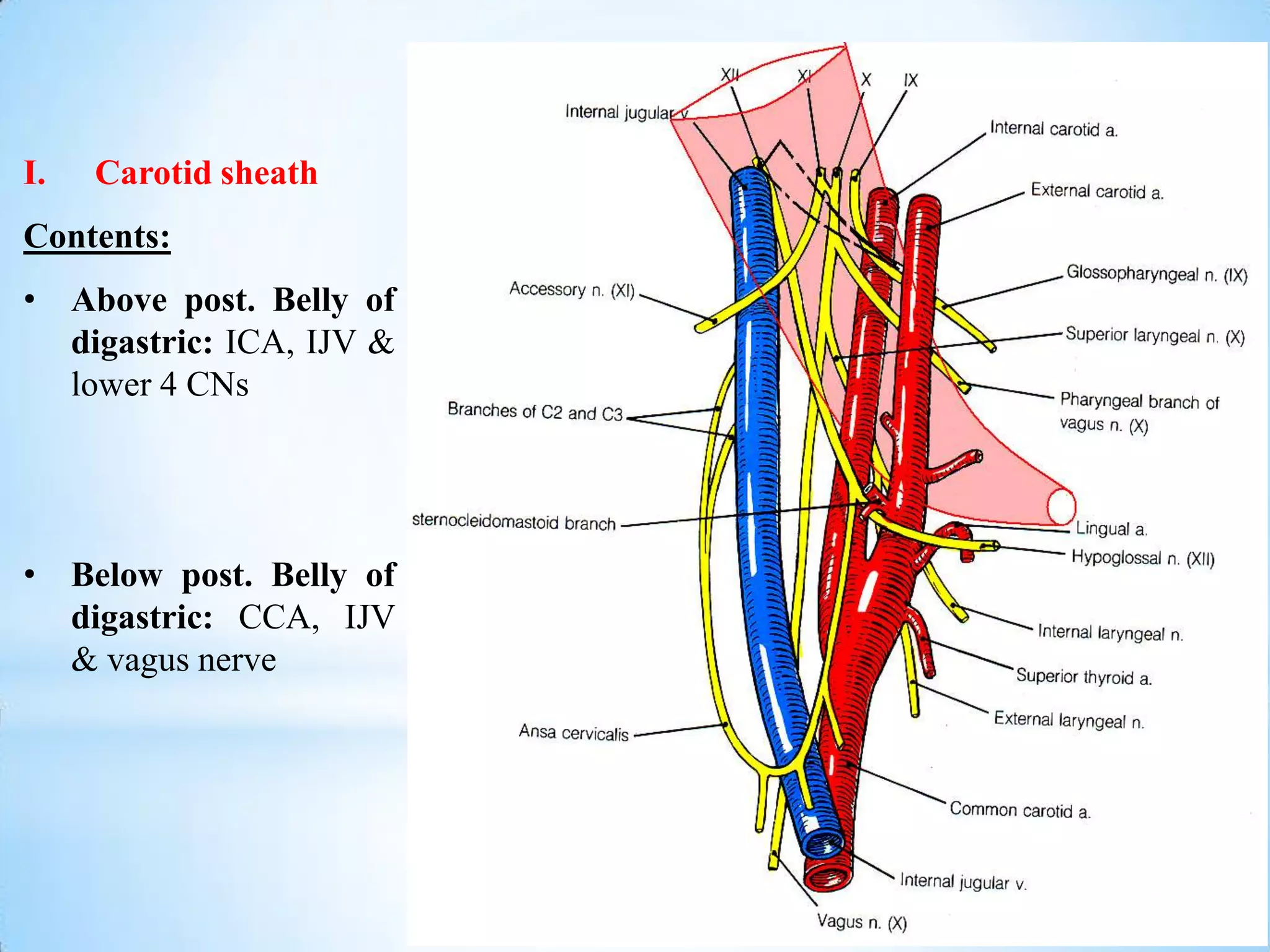
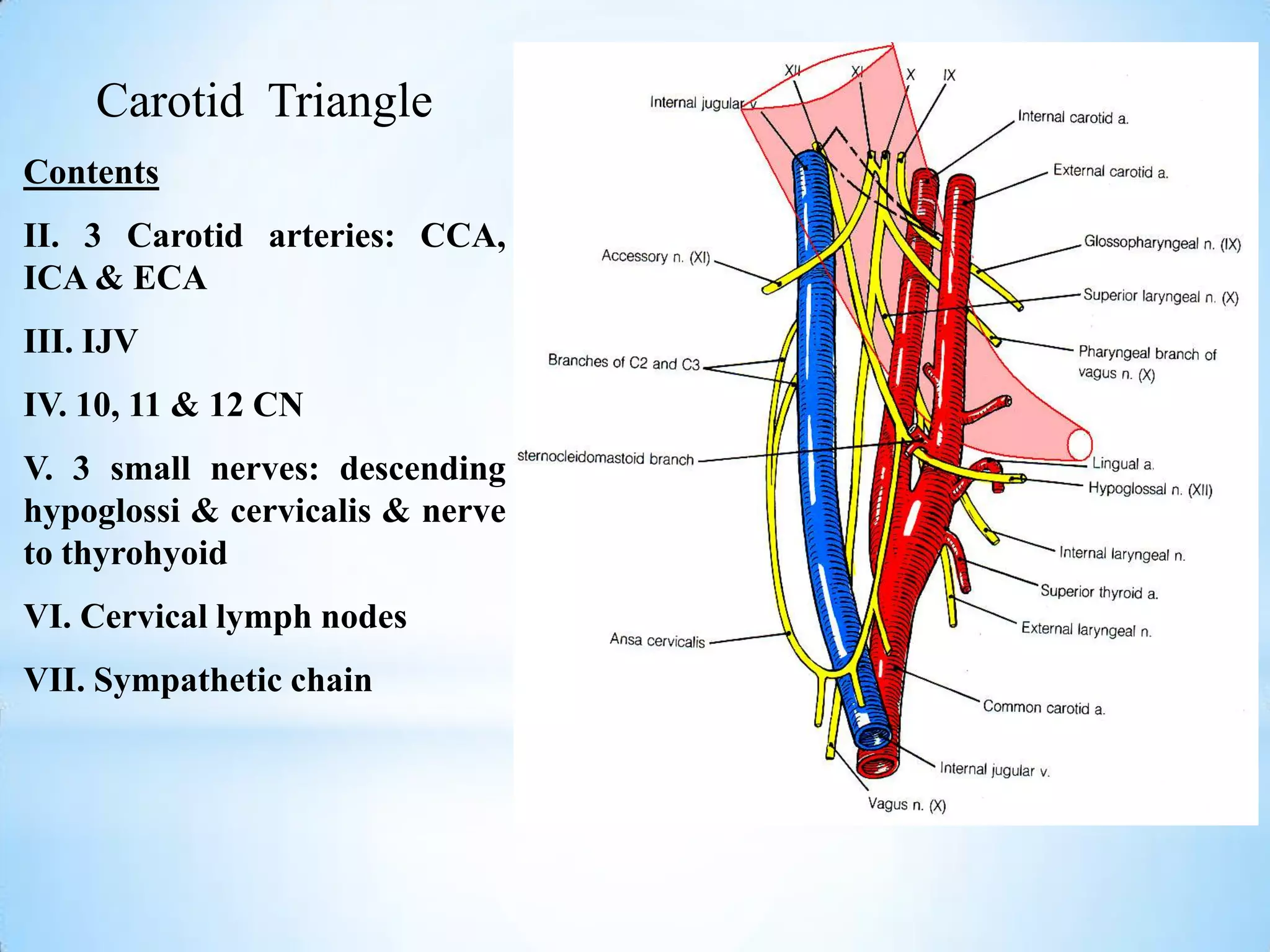
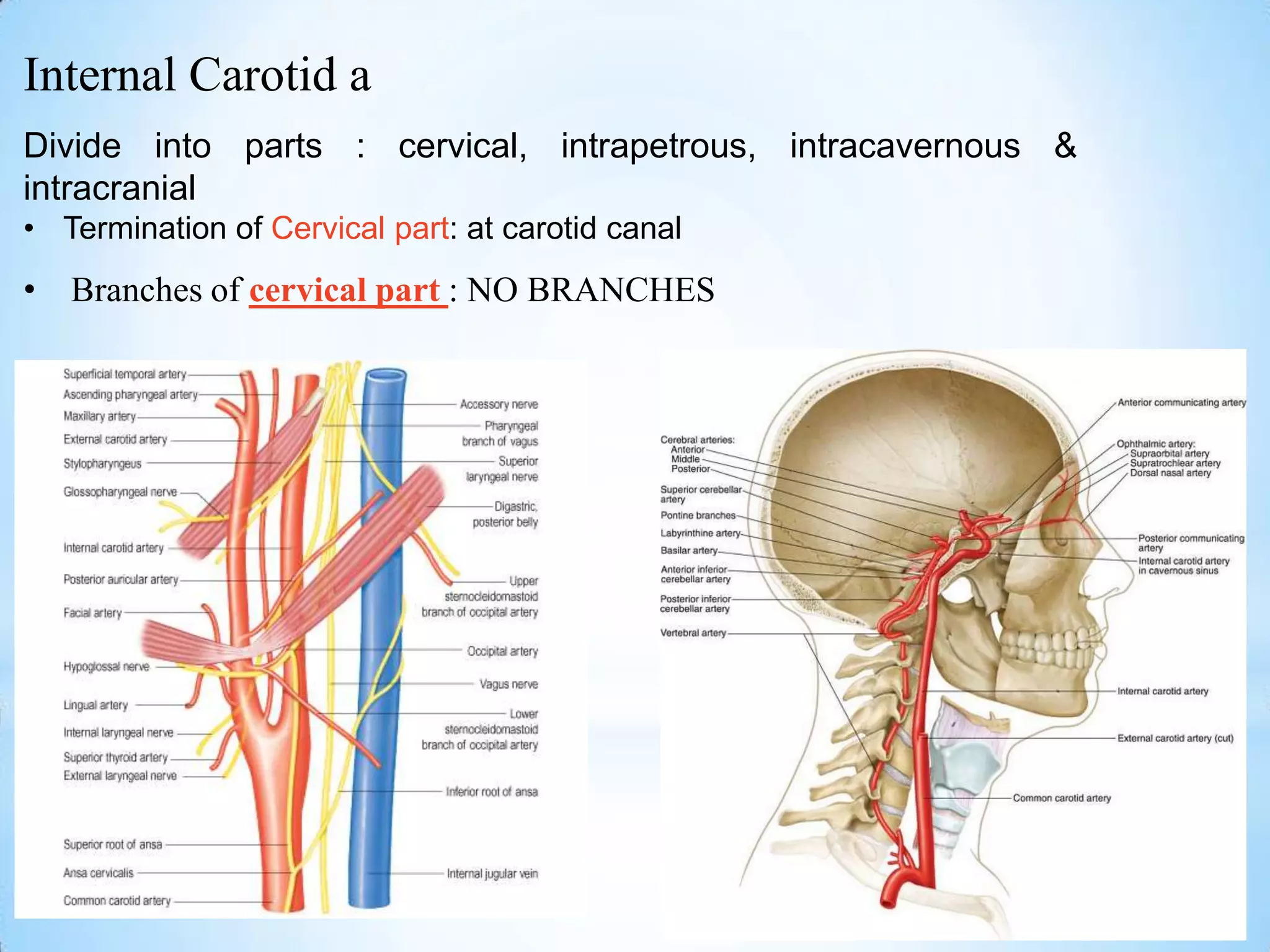
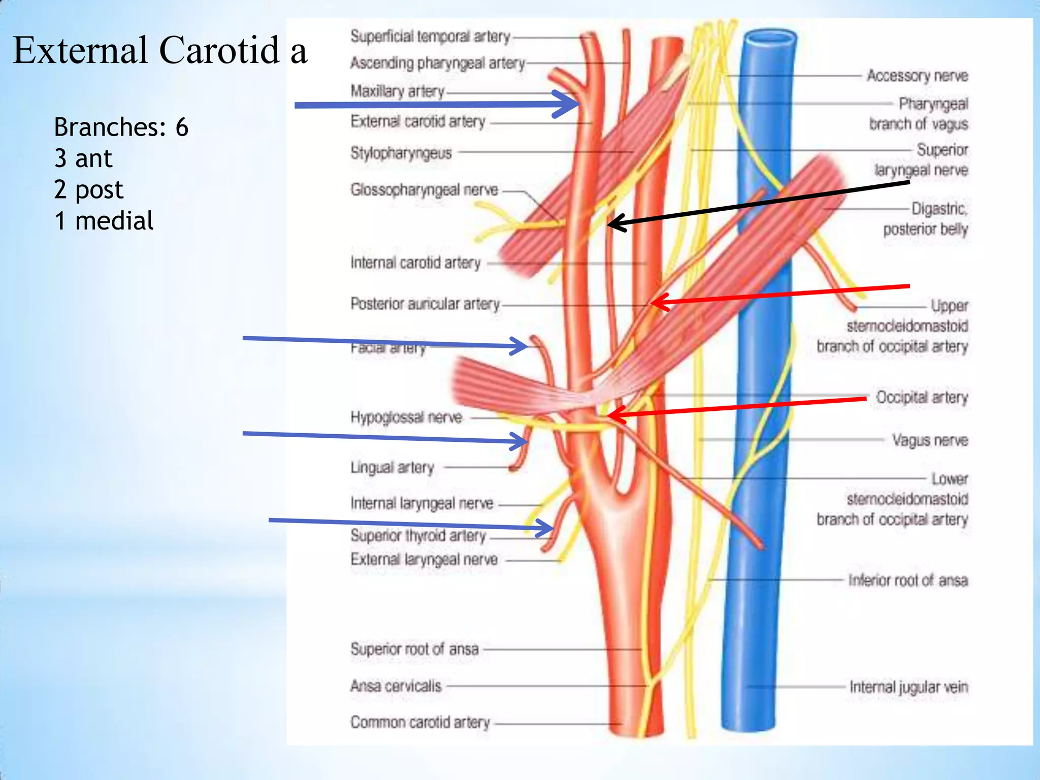
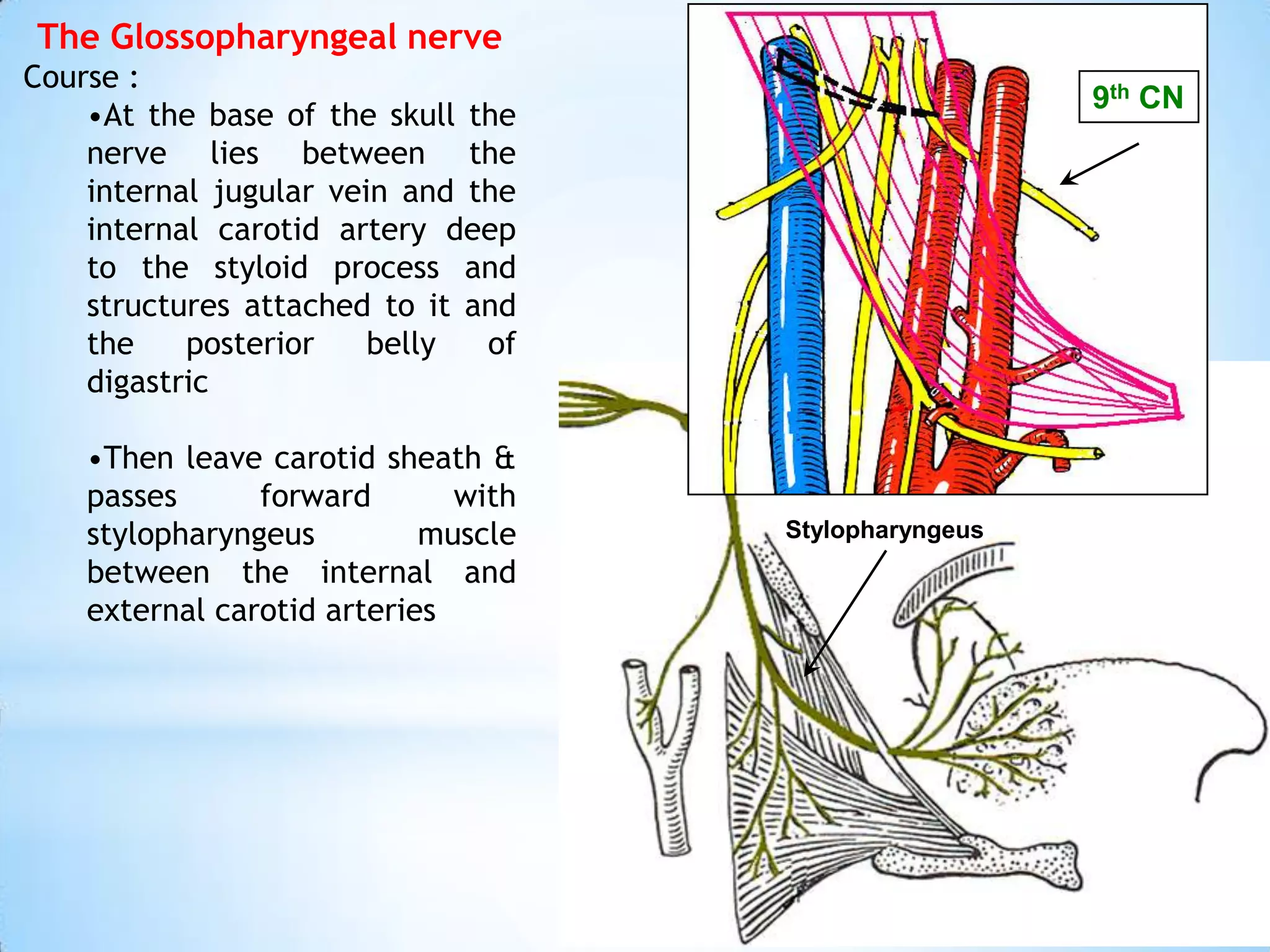
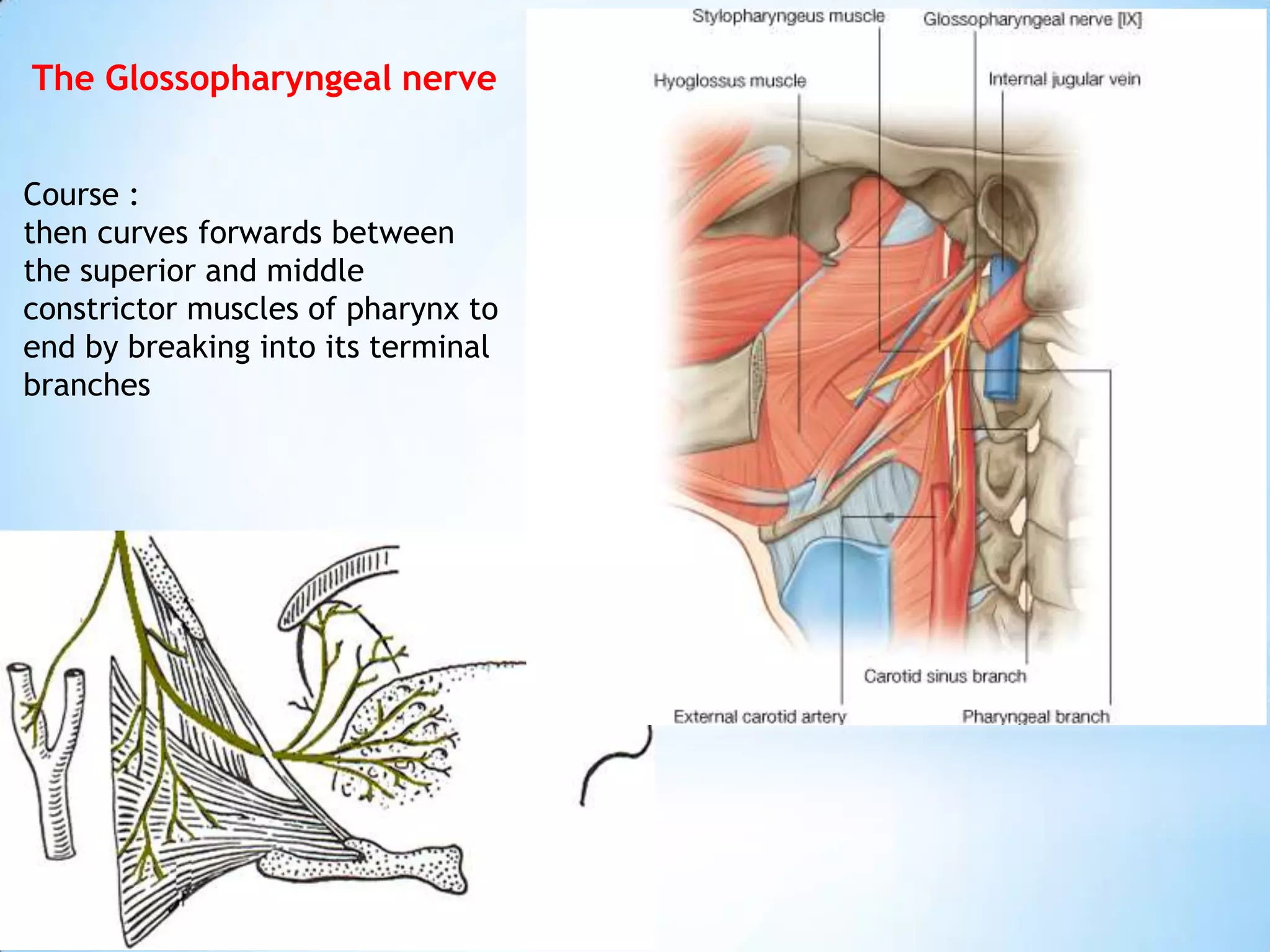
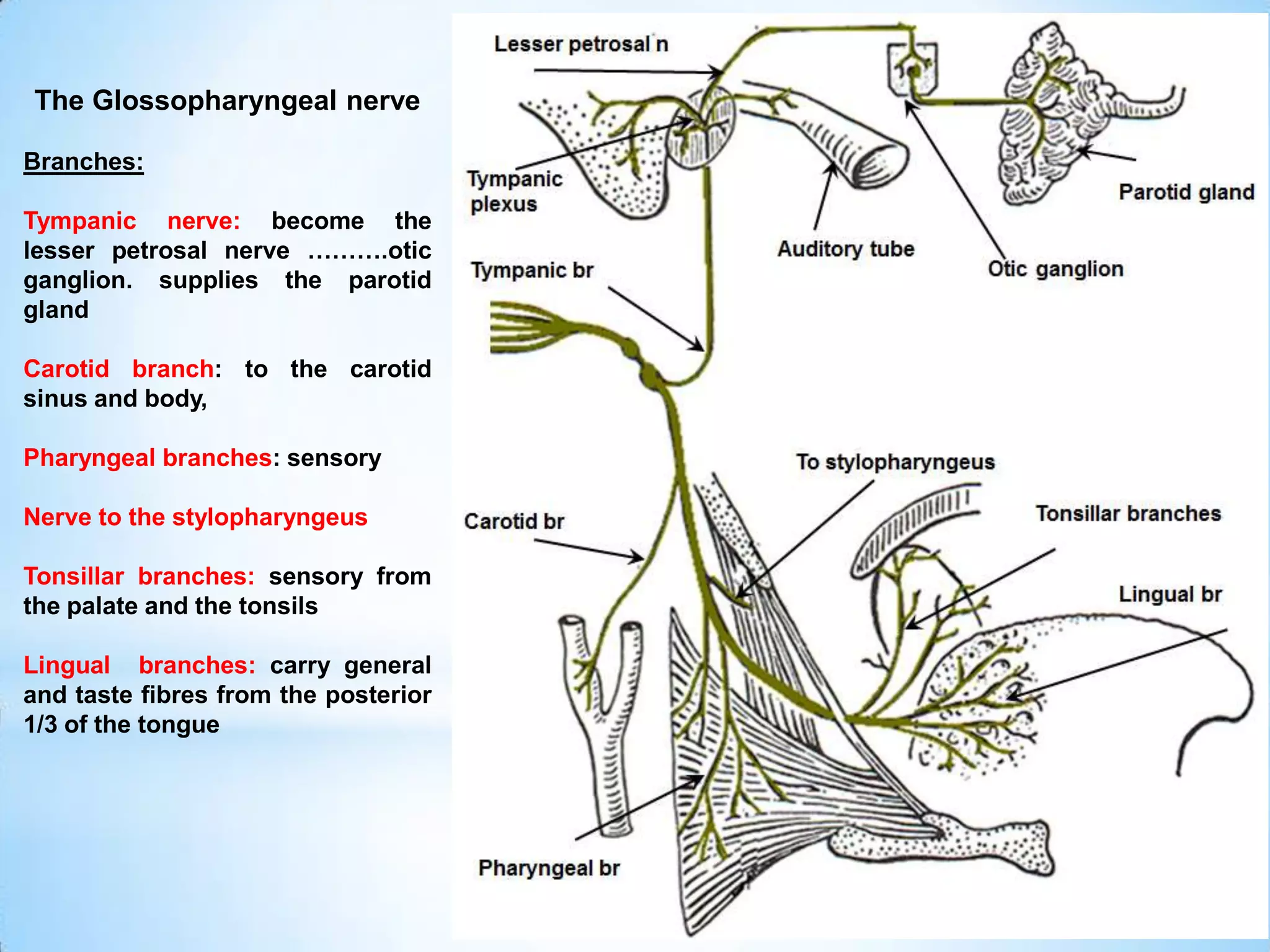
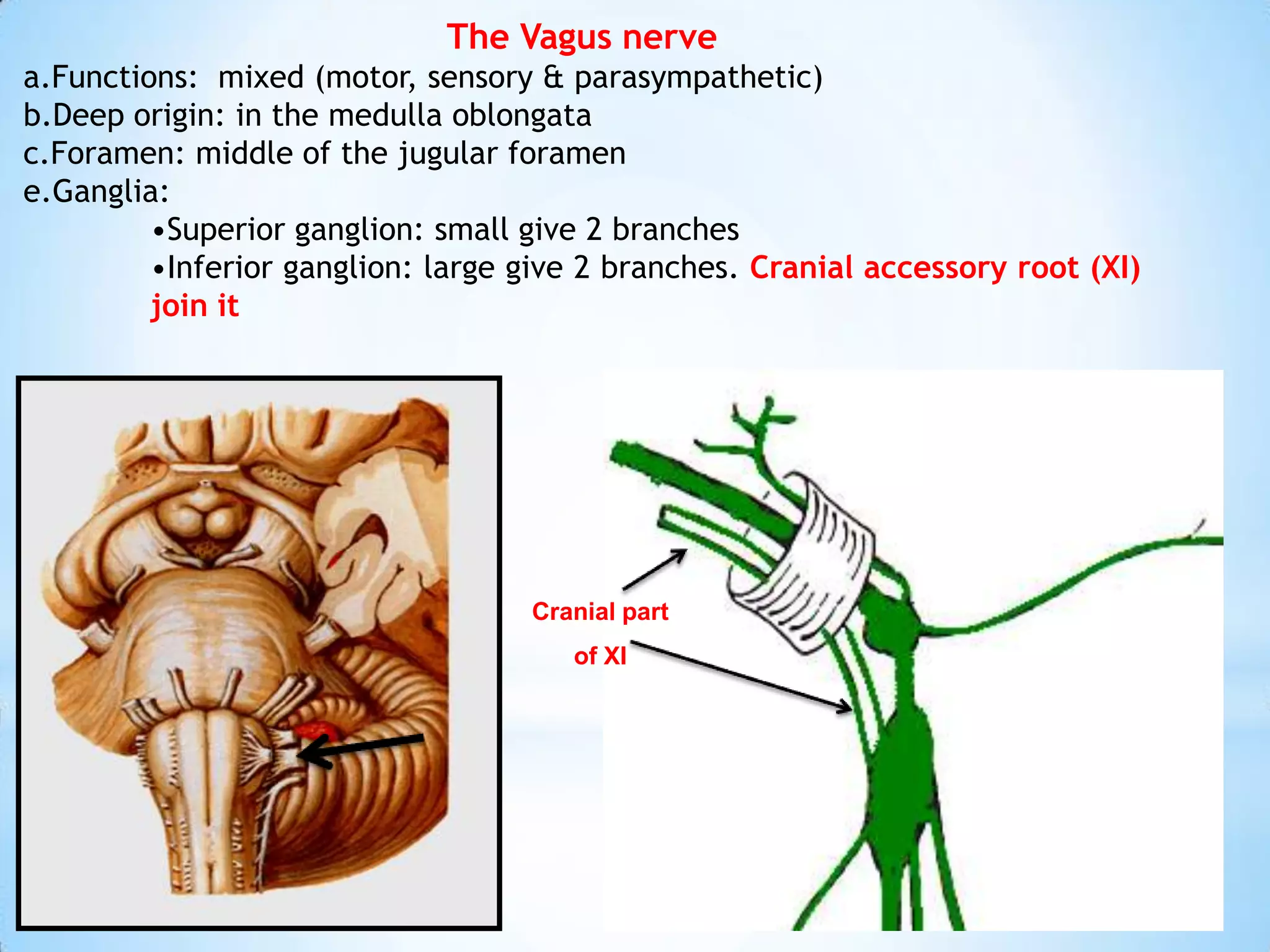
Carotid Triangle
Boundaries
Post. Belly of digastric m
sup. Belly of omohyoid
Ant. border of sternomastoid
Carotid Triangle
Contents
I.   Carotid sheath
is a column of fascia that surrounds the blood
vessels and nerves as they pass through the neck
I.   Carotid sheath
Attachment:
• Sup.: Base of skull
  (Carotid and jugular
  foramina)
• Inf.: Arch of aorta
I.   Carotid sheath
Contents:
• Above post. Belly of
  digastric: ICA, IJV &
  lower 4 CNs


• Below post. Belly of
  digastric: CCA, IJV
  & vagus nerve
Carotid Triangle
Contents
II. 3 Carotid arteries: CCA,
ICA & ECA
III. IJV
IV. 10, 11 & 12 CN
V. 3 small nerves: descending
hypoglossi & cervicalis & nerve
to thyrohyoid
VI. Cervical lymph nodes
VII. Sympathetic chain
Common Carotid a
Origin:
• Rt CCA: from innominate a
• Lt CCA: from arch of aorta
Common Carotid a
Termination: at C3-4 or at upper border of thyroid cartilage by dividing into 2
terminal branches ICA & ECA
Branches: ICA and ECA
Internal Carotid a
Origin: at C3-4 or at upper border of thyroid cartilage from CCA
Termination: intracranially by dividing into anterior and middle cerebral a
Internal Carotid a
Divide into parts : cervical, intrapetrous, intracavernous &
intracranial
• Termination of Cervical part: at carotid canal
• Branches of cervical part : NO BRANCHES
External Carotid a
Origin: at C3-4 or at upper border of thyroid
cartilage from CCA
Termination:     in parotid g. by dividing into
superficial temporal a & maxillary a
External Carotid a
  Branches: 6
  3 ant
  2 post
  1 medial
Internal jugular
           vein
Beginning: sigmoid venous
sinus
Termination: join subclavian
V to form innominate V
 Has 2 dilatations (sup. &
  inf. Bulbs)
Tributaries:
1.    Inf. Petrosal sinus
2.    Pharyngeal V
3.    Lingual V
4.    Common facial V
5.    Sup. Thyroidal V
6.    Middle thyroidal V
The Glossopharyngeal nerve
a.Functions: mixed (motor, sensory & parasympathetic)
b.Deep origin: in the medulla oblongata
c.Foramen: middle of the jugular foramen
e.Ganglia:
    •Superior ganglion: small and has no branches
    •Inferior ganglion: large give tympanic branch
The Glossopharyngeal nerve
Course :
                                                       9th CN
    •At the base of the skull the
    nerve lies between the
    internal jugular vein and the
    internal carotid artery deep
    to the styloid process and
    structures attached to it and
    the    posterior    belly  of
    digastric

    •Then leave carotid sheath &
    passes      forward       with
    stylopharyngeus         muscle   Stylopharyngeus
    between the internal and
    external carotid arteries
The Glossopharyngeal nerve


Course :
then curves forwards between
the superior and middle
constrictor muscles of pharynx to
end by breaking into its terminal
branches
The Glossopharyngeal nerve

Branches:

Tympanic nerve: become the
lesser petrosal nerve ……….otic
ganglion. supplies the parotid
gland

Carotid branch: to the carotid
sinus and body,

Pharyngeal branches: sensory

Nerve to the stylopharyngeus

Tonsillar branches: sensory from
the palate and the tonsils

Lingual branches: carry general
and taste fibres from the posterior
1/3 of the tongue
The Vagus nerve
a.Functions: mixed (motor, sensory & parasympathetic)
b.Deep origin: in the medulla oblongata
c.Foramen: middle of the jugular foramen
e.Ganglia:
         •Superior ganglion: small give 2 branches
         •Inferior ganglion: large give 2 branches. Cranial accessory root (XI)
         join it


                                   Cranial part
                                      of XI
Vagus nerve                 10th CN
Course :
 At the base of the skull the nerve
lies between the internal jugular
vein and the internal carotid
artery deep to the styloid process
and structures attached to it and
the posterior belly of digastric
Vagus nerve
Course :

The nerve descends vertically
within     the    carotid    sheath
between the common carotid
artery and the internal jugular
vein until it reaches the root of
the neck, it crosses in front of the
first part of the subclavian artery
to the thorax
Vagus nerve

Branches:

I. From sup. ganglion
Meningeal n
Auricular n

II. From inf. ganglion
Pharyngeal n (motor)
Superior laryngeal n (mixed)

III. Cardiac branch (parasym.)

IV. Recurrent laryngeal n (mixed)

3 carotid triangle___9th_10th_cn

  • 2.
    Carotid Triangle Boundaries Post. Bellyof digastric m sup. Belly of omohyoid Ant. border of sternomastoid
  • 3.
    Carotid Triangle Contents I. Carotid sheath is a column of fascia that surrounds the blood vessels and nerves as they pass through the neck
  • 4.
    I. Carotid sheath Attachment: • Sup.: Base of skull (Carotid and jugular foramina) • Inf.: Arch of aorta
  • 5.
    I. Carotid sheath Contents: • Above post. Belly of digastric: ICA, IJV & lower 4 CNs • Below post. Belly of digastric: CCA, IJV & vagus nerve
  • 6.
    Carotid Triangle Contents II. 3Carotid arteries: CCA, ICA & ECA III. IJV IV. 10, 11 & 12 CN V. 3 small nerves: descending hypoglossi & cervicalis & nerve to thyrohyoid VI. Cervical lymph nodes VII. Sympathetic chain
  • 7.
    Common Carotid a Origin: •Rt CCA: from innominate a • Lt CCA: from arch of aorta
  • 8.
    Common Carotid a Termination:at C3-4 or at upper border of thyroid cartilage by dividing into 2 terminal branches ICA & ECA Branches: ICA and ECA
  • 9.
    Internal Carotid a Origin:at C3-4 or at upper border of thyroid cartilage from CCA Termination: intracranially by dividing into anterior and middle cerebral a
  • 10.
    Internal Carotid a Divideinto parts : cervical, intrapetrous, intracavernous & intracranial • Termination of Cervical part: at carotid canal • Branches of cervical part : NO BRANCHES
  • 11.
    External Carotid a Origin:at C3-4 or at upper border of thyroid cartilage from CCA Termination: in parotid g. by dividing into superficial temporal a & maxillary a
  • 13.
    External Carotid a Branches: 6 3 ant 2 post 1 medial
  • 14.
    Internal jugular vein Beginning: sigmoid venous sinus Termination: join subclavian V to form innominate V  Has 2 dilatations (sup. & inf. Bulbs) Tributaries: 1. Inf. Petrosal sinus 2. Pharyngeal V 3. Lingual V 4. Common facial V 5. Sup. Thyroidal V 6. Middle thyroidal V
  • 16.
    The Glossopharyngeal nerve a.Functions:mixed (motor, sensory & parasympathetic) b.Deep origin: in the medulla oblongata c.Foramen: middle of the jugular foramen e.Ganglia: •Superior ganglion: small and has no branches •Inferior ganglion: large give tympanic branch
  • 17.
    The Glossopharyngeal nerve Course: 9th CN •At the base of the skull the nerve lies between the internal jugular vein and the internal carotid artery deep to the styloid process and structures attached to it and the posterior belly of digastric •Then leave carotid sheath & passes forward with stylopharyngeus muscle Stylopharyngeus between the internal and external carotid arteries
  • 18.
    The Glossopharyngeal nerve Course: then curves forwards between the superior and middle constrictor muscles of pharynx to end by breaking into its terminal branches
  • 19.
    The Glossopharyngeal nerve Branches: Tympanicnerve: become the lesser petrosal nerve ……….otic ganglion. supplies the parotid gland Carotid branch: to the carotid sinus and body, Pharyngeal branches: sensory Nerve to the stylopharyngeus Tonsillar branches: sensory from the palate and the tonsils Lingual branches: carry general and taste fibres from the posterior 1/3 of the tongue
  • 20.
    The Vagus nerve a.Functions:mixed (motor, sensory & parasympathetic) b.Deep origin: in the medulla oblongata c.Foramen: middle of the jugular foramen e.Ganglia: •Superior ganglion: small give 2 branches •Inferior ganglion: large give 2 branches. Cranial accessory root (XI) join it Cranial part of XI
  • 21.
    Vagus nerve 10th CN Course : At the base of the skull the nerve lies between the internal jugular vein and the internal carotid artery deep to the styloid process and structures attached to it and the posterior belly of digastric
  • 22.
    Vagus nerve Course : Thenerve descends vertically within the carotid sheath between the common carotid artery and the internal jugular vein until it reaches the root of the neck, it crosses in front of the first part of the subclavian artery to the thorax
  • 23.
    Vagus nerve Branches: I. Fromsup. ganglion Meningeal n Auricular n II. From inf. ganglion Pharyngeal n (motor) Superior laryngeal n (mixed) III. Cardiac branch (parasym.) IV. Recurrent laryngeal n (mixed)