Download to read offline
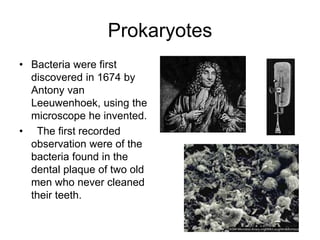
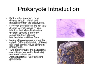
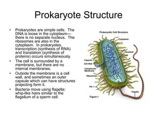
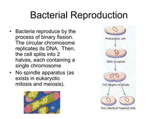
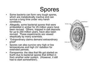


Prokaryotes include bacteria and archaea. Bacteria were first observed in 1674 and include diverse species found in many habitats. Prokaryotes are single-celled without internal membranes and reproduce through binary fission. Major groups include eubacteria like E. coli and archaea found in extreme environments like hot springs or salt lakes. Bacteria play important roles in ecosystems through processes like nitrogen fixation and photosynthesis.


