![Copyright © Lippincott Williams & Wilkins. Unauthorized reproduction of this article is prohibited.
patient cooperation with intact electrophysiologic moni-
toring. Obviously, the above-mentioned conditions often
contradict each other, making ‘anesthetic’ management
for awake neurosurgical procedures one of the most
challenging in anesthesia practice.
The kind of neurologic monitoring required for func-
tional neurosurgery depends on the procedure and the
area of interest in the brain. An assessment of cognitive
function, speech, vision, and motor function is required in
surgery for resection of brain tumors or epileptic focus
located close to the eloquent cortex (Broca’s and
Wernicke’s speech areas in the dominant frontal and
temporal lobe, motor strip, and visual cortex). During
implantation of a deep brain stimulator (DBS) for a patient
with movement disorder, an assessment of changes in
tremor or spasticity/rigidity in a fully awake patient is
mandatory. Intraoperative electrophysiologic testing for
tumor and epilepsy surgery includes electroencephalo-
graphy (EEG) and electrocorticography (ECocG) for
cortical brain mapping. The microelectrode recording
(MER) of impulses of subcortical areas, including sub-
thalamic nucleus, is used during DBS implantation.
Patient positioning for functional neurosurgery makes
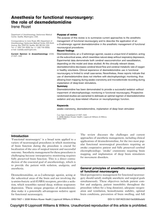
access to the patient’s airway difficult, further complicat-
ing anesthesia management (Fig. 1).
Intraoperative ‘anesthetic’ management of awake craniot-
omy has changed during the last two decades. Anesthesia
medications with rapid onset of action and short half-lives
such as propofol andremifentanil replaced previously used
neuroleptanalgesia with fentanyl and droperidol. Multiple
anesthestic techniques are used for awake craniotomies:
monitored anesthesia care, conscious sedation, the
‘asleep–awake’ and ‘asleep–awake–asleep’ techniques
[1
]. The ‘asleep–awake–asleep’ technique is currently
considered as the most popular one [1
]. The first ‘asleep’
stage is provided with heavy sedation or even general
anesthesia in the beginning of the surgery during a
patient’s positioning, fixation of the head with pins, cra-
niotomy or burr holes, and final exposure of the brain. Skin
infiltration with local anesthetic at pin insertion sites and
the skin incision, or performance of a scalp nerve block,
are essential for pain relief and reducing opiates-related
complications [2]. A variety of different medications
and combinations of medications has been reported to
be successfully used: propofol, remifentanil, fentanyl,
midzolam [1
,3,4]. For airway management, oral or naso-
pharyngeal airways, oral and nasal endotracheal tubes,
laryngeal mask (LMA) as well as unprotected airway have
all been used with different degrees of success [1
]. For the
next awake stage, the patient must be brought to full
consciousness. In this awake phase, mapping of the brain
with electrocorticography during cognitive testing is per-
formed to delineate the area to be resected. The transition
from the ‘asleep’ to the ‘awake’ phase is the most challen-
ging task in the anesthetic management because of poten-
tial complications during the awakening and manipulation
of the airway, while the brain is exposed. Coughing,
Valsalva maneuver, vomiting and movement during extu-
bation of the airway may lead to disastrous complications
such as bleeding, brain swelling, venous air embolism, and
potentially death. Consequently, many anesthesiologists
538 Neuroanaesthesia
Figure 1 Positioning of patient for functional neurosurgery
Positioning
Head fixation
Pins
Mayfield
frame
Head position
Neutral
Flexion
Extension
Lateral flexion
Pins
Stereotactic
frame
Hoarshoe
gelpad
Supine SittingLateral
Body positioning
OR Table
90˚ 180˚
OR, operating room.](data:image/gif;base64,R0lGODlhAQABAIAAAAAAAP///yH5BAEAAAAALAAAAAABAAEAAAIBRAA7)
Recommended
Recommended
More Related Content
What's hot
What's hot (20)
Viewers also liked
Viewers also liked (14)
Similar to Dexmedetomidina Neuroanestesia
Similar to Dexmedetomidina Neuroanestesia (20)
More from guestc3bf72
More from guestc3bf72 (20)
Recently uploaded
Recently uploaded (20)
Dexmedetomidina Neuroanestesia
- 1. Copyright © Lippincott Williams & Wilkins. Unauthorized reproduction of this article is prohibited. Anesthesia for functional neurosurgery: the role of dexmedetomidine Irene Rozet Introduction ‘Functional neurosurgery’ is a broad term applied to a variety of neurosurgical procedures in which monitoring of brain function during the procedure is crucial for localization of the area of surgical interest and successful outcome. Anesthetic management for these procedures is challenging because intraoperative monitoring requires fully preserved brain function. This is a direct contra- diction of the essential goal of anesthesiology, which is to provide the patient with sedation, analgesia, and anesthesia. Dexmedetomidine, an a-2-adrenergic agonist, acting at the subcortical areas of the brain and not involving the g-amino-butyric acid (GABA) receptors, provides a seda- tion, which resembles natural sleep, without respiratory depression. These unique properties of dexmedetomi- dine make it a potentially advantageous sedative agent for functional neurosurgery. The review discusses the challenges and current approaches of anesthetic management, including clinical applications of dexmedetomidine, for the two most pop- ular functional neurosurgical procedures requiring an awake cooperative patient and fully preserved cerebral electrophysiology: ‘awake’ craniotomy requiring brain mapping, and implantation of deep brain stimulators for movement disorders. General principles of anesthetic management of functional neurosurgery Ideal perioperative management for functional neurosur- gery should satisfy multiple anesthetic and surgical goals simultaneously. These objectives include patient com- fort and analgesia, patient immobility throughout the procedure (often for a long duration), adequate oxygen- ation and ventilation, hemodynamic stability, optimal brain conditions, prevention of brain swelling, and full Department of Anesthesiology, Harborview Medical Center, Seattle, Washington, USA Correspondence to Irene Rozet, MD, Department of Anesthesiology, Harborview Medical Center, 325 Ninth Avenue, Box 359724, Seattle, WA 98104, USA Tel: +1 206 744 3059; fax: +1 206 744 8090; e-mail: irozet@u.washington.edu Current Opinion in Anaesthesiology 2008, 21:537–543 Purpose of review The purpose of this review is to summarize current approaches to the anesthetic management of functional neurosurgery and to describe the application of an a-2-adrenergic agonist dexmedetomidine in the anesthetic management of functional neurosurgical procedures. Recent findings Dexmedetomidine, an a-2-adrenergic agonist, causes a unique kind of sedation, acting on the subcortical areas, which resembles natural sleep without respiratory depression. Experimental data demonstrate both cerebral vasoconstriction and vasodilatation, depending on the model and dose studied. At the clinically relevant doses, dexmedetomidine decreases cerebral blood flow and cerebral metabolic rate of oxygen in healthy volunteers. Clinical experience of dexmedetomidine use in functional neurosurgery is limited to small case-series. Nevertheless, these reports indicate that use of dexmedetomidine does not interfere with electrophysiologic monitoring, thus allowing brain mapping during awake craniotomy and microelectrode recording during implantation of deep-brain stimulators. Summary Dexmedetomidine has been demonstrated to provide a successful sedation without impairment of electrophysiologic monitoring in functional neurosurgery. Prospective randomized studies are warranted to delineate an optimal regimen of dexmedetomidine sedation and any dose-related influence on neurophysiologic function. Keywords awake craniotomy, dexmedetomidine, implantation of deep brain stimulator Curr Opin Anaesthesiol 21:537–543 ß 2008 Wolters Kluwer Health | Lippincott Williams & Wilkins 0952-7907 0952-7907 ß 2008 Wolters Kluwer Health | Lippincott Williams & Wilkins DOI:10.1097/ACO.0b013e32830edafd
- 2. Copyright © Lippincott Williams & Wilkins. Unauthorized reproduction of this article is prohibited. patient cooperation with intact electrophysiologic moni- toring. Obviously, the above-mentioned conditions often contradict each other, making ‘anesthetic’ management for awake neurosurgical procedures one of the most challenging in anesthesia practice. The kind of neurologic monitoring required for func- tional neurosurgery depends on the procedure and the area of interest in the brain. An assessment of cognitive function, speech, vision, and motor function is required in surgery for resection of brain tumors or epileptic focus located close to the eloquent cortex (Broca’s and Wernicke’s speech areas in the dominant frontal and temporal lobe, motor strip, and visual cortex). During implantation of a deep brain stimulator (DBS) for a patient with movement disorder, an assessment of changes in tremor or spasticity/rigidity in a fully awake patient is mandatory. Intraoperative electrophysiologic testing for tumor and epilepsy surgery includes electroencephalo- graphy (EEG) and electrocorticography (ECocG) for cortical brain mapping. The microelectrode recording (MER) of impulses of subcortical areas, including sub- thalamic nucleus, is used during DBS implantation. Patient positioning for functional neurosurgery makes access to the patient’s airway difficult, further complicat- ing anesthesia management (Fig. 1). Intraoperative ‘anesthetic’ management of awake craniot- omy has changed during the last two decades. Anesthesia medications with rapid onset of action and short half-lives such as propofol andremifentanil replaced previously used neuroleptanalgesia with fentanyl and droperidol. Multiple anesthestic techniques are used for awake craniotomies: monitored anesthesia care, conscious sedation, the ‘asleep–awake’ and ‘asleep–awake–asleep’ techniques [1 ]. The ‘asleep–awake–asleep’ technique is currently considered as the most popular one [1 ]. The first ‘asleep’ stage is provided with heavy sedation or even general anesthesia in the beginning of the surgery during a patient’s positioning, fixation of the head with pins, cra- niotomy or burr holes, and final exposure of the brain. Skin infiltration with local anesthetic at pin insertion sites and the skin incision, or performance of a scalp nerve block, are essential for pain relief and reducing opiates-related complications [2]. A variety of different medications and combinations of medications has been reported to be successfully used: propofol, remifentanil, fentanyl, midzolam [1 ,3,4]. For airway management, oral or naso- pharyngeal airways, oral and nasal endotracheal tubes, laryngeal mask (LMA) as well as unprotected airway have all been used with different degrees of success [1 ]. For the next awake stage, the patient must be brought to full consciousness. In this awake phase, mapping of the brain with electrocorticography during cognitive testing is per- formed to delineate the area to be resected. The transition from the ‘asleep’ to the ‘awake’ phase is the most challen- ging task in the anesthetic management because of poten- tial complications during the awakening and manipulation of the airway, while the brain is exposed. Coughing, Valsalva maneuver, vomiting and movement during extu- bation of the airway may lead to disastrous complications such as bleeding, brain swelling, venous air embolism, and potentially death. Consequently, many anesthesiologists 538 Neuroanaesthesia Figure 1 Positioning of patient for functional neurosurgery Positioning Head fixation Pins Mayfield frame Head position Neutral Flexion Extension Lateral flexion Pins Stereotactic frame Hoarshoe gelpad Supine SittingLateral Body positioning OR Table 90˚ 180˚ OR, operating room.
- 3. Copyright © Lippincott Williams Wilkins. Unauthorized reproduction of this article is prohibited. try to avoid airway instrumentation. When mapping is completed, either sedation or analgesia or both may be provided again for the ‘asleep’ phase. Airway instrumenta- tion when the patient’s head is in pins and covered by the surgical drapes may be difficult, especially if emergency intubation of the trachea is required. In implantations of DBS, the first ‘asleep’ phase usually requires less time than the first ‘asleep’ phase in awake craniotomy. DBS implantation does not require wide exposure of the brain. Only burr holes are needed, which can be performed under local infiltration of the scalp. Although the use of various sedative techniques, includ- ing propofol infusion, fentanyl, remifentanil, and inhala- tional anesthetics have been reported [5,6 ], many surgeons prefer to avoid any sedation because of the extreme sensitivity of subcortical areas to the GABA- ergic medications, which may completely abolish MER recording and tremor. When localization of the DBS electrode is finalized, the patient’s head is disengaged from the frame, and the patient may then be anesthe- tized for the implantation of the generator into the chest wall. Potential risks involved in anesthetic management for awake neurosurgical procedures are summarized in Table 1. Unfortunately, the actual incidence of compli- cations and therefore the optimal anesthetic management are largely unknown due to the lack of prospective randomized studies. Currently available data are incon- clusive because the studies differ in: anesthetic manage- ment, criteria applied for definitions of complications, and study design. The largest retrospective chart review of awake craniotomy by Skucas and Artru [3] reported that airway problems occurred in 2% out of 332 cases of asleep–awake–asleep technique using only propofol infusion in patients with unsecured airway undergoing epilepsy surgery. Manninen et al. [4] prospectively randomized 50 patients undergoing tumor surgery with brain mapping to receive sedation with propofol and remifentanil infusion, or propofol and fentanyl, and observed an incidence of respiratory complications as high as 18% in this cohort, without any difference between the two groups. Respiratory complications were defined as any of following: a decrease in respiratory rate, oxygen desaturation, or airway obstruction. Because the majority of surgeons request the avoidance of any sedation, the most significant challenge in DBS implan- tations is the maintainance of hemodynamic stability and the prevention/treatment of intraoperative hypertension, a known risk factor for developing intracerebral hemor- rhage. In the prospective study involving 128 patients receiving DBS implantations for Parkinson’s disease with intermittent infusion of propofol, about 60% of patients developed intraoperative hypertension [7]. Intracerebral hemorrhage is considered to be the most severe compli- cation, with an incidence of 3.3–6% reported in the prospective studies [8–10]. The incidence of respiratory complications in DBS implantations is unknown. Benzodiazepines, barbiturates, and opioids may deliver patient comfort and hemodynamic stability, but they may also depress electyrophysiologic activity of the brain, interfere with mapping, and cause respiratory depression and airway compromise. To achieve a balance between uneventful awakening and avoidance of respiratory com- plications, the pharmacokinetic-guided titration of propo- folandremifentanilusingthetargetcontrolinfusiondevice has been advocated in patients with Parkinson’s disease [11]andforawakecraniotomies[12].Thesedevicesarenot yet approved for use in the United States. In addition to obesity [3] and age [6 ], which were suggested by the retrospective chart reviews as risk factors, other patient-related risk factors are less clear. Previously, obstructive sleep apnea was considered as a potential risk factor for perioperative respiratory complications [1 ]. However, there are no data to support this. There is also a paucity of data to predict who would be an ideal candi- date for awake functional neurosurgery. According to the Anesthesia for functional neurosurgery Rozet 539 Table 1 Potential risks and complications expected in awake functional neurosurgery Undersedation Oversedation General General Pain Drowsiness Discomfort Restlessness Restlessness Impaired cognition, lack of cooperation, inability to perform mapping Anxiety Inability to stay still Respiratory Voluntary movements Airway obstruction Respiratory depression Hypoventilation Respiratory Hypercarbia Inability to clear secretions (PD) Oxygen desaturation Coughing Apnea Dyspnea–hyperventilation Need for airway manipulations and for providing an emergency airway Coughing and Valsalva during transition from ‘asleep’ to ‘awake’ stage Neurological Seizures Neurological Brain swelling Brain swelling Bleeding Seizures Bleeding Hemodynamic Arterial hypertension Hemodynamic Tachycardia, arrhythmia Arterial hypotension Gastrointestinal Gastrointestinal Nausea, vomiting Nausea, vomiting Aspiration of gastric contents Involuntary movements Shivering Sedative-induced dyskinesias Arterial hypotension PD, Parkinson’s disease.
- 4. Copyright © Lippincott Williams Wilkins. Unauthorized reproduction of this article is prohibited. current data, in most instances, only the lack of patient cooperation will preclude awake neurosurgery. However, factors including abnormal anatomy of the airway, pre- sence of gastroesophageal reflux and obesity should be considered as potential risk factors. Dexmedetomidine: a unique sedative agent Dexmedetomidine is a selective agonist of a2-adrenergic receptors (a2-ARs) with high affinity to a2-ARs (a2:a1 effectratio of1620:1),whichiseighttimeshigher thanwith clonidine. Dexmedetomidine has a rapid onset of action. It undergoes biotransformation in the liver, and the kidneys excrete about 95% of its metabolites. Its distribution half- life is 6 min, and a clearance half-life is 2 h. Presynaptic and postsynaptic a2-ARs are distributed over vital organs (heart, pancreas, kidneys), blood vessels and the central and the peripheral nervous system. Stimulation of postsynaptic a2-ARs leads to hyperpolarization of neuronal membrane, whereas stimulation of presynaptic a2-ARs reduces the release of norepinephrine. In the spinal cord, a2-ARs are predominantly postsynaptic, and located in the dorsal horn. Activation of spinal a2-ARs inhibits nociception, which most likely explains the analgetic properties of dexmedetomidine. Both presyn- aptic and postsynaptic a2-ARs are widely distributed in the brain, particularly in the pons and medulla. The major site of noradrenergic innervation in the brain with the highest concentration of presynaptic a2-ARs is the locus ceruleus, which is responsible for arousal, sleep, anxiety, and withdrawal symptoms from drug addiction. As a result, the central effect of dexmedetomidine, which is mani- fested by anxiolysis and sedation, is noncortical and sub- cortical in origin. It does not involve the GABA system and consequently does not cause cognitive impairment or disinhibition, differentiating dexmedetomidine from all GABA-mimetic sedatives and anesthetics. The mechanisms of anesthetic-induced effects on uncon- sciousness and amnesia are not clear. Recently, both cortex [13] and thalamus [14] have been suggested to be primarily responsible for the anesthetic-induced unconsciousness. The unusual subcortical form of dex- medetomidine-induced sedation is characterized by an easy and quick arousal, resembling natural sleep. The neuroprotective properties of dexmedetomidine have been demonstrated in various animal models of cerebral ischemia [15–17]. There are recent experi- mental data suggesting that in addition to a2-ARs, the neuroprotective effect of dexmedetomidine may include other pathways in the brain, independent of a2-ARs, and most probably involve I1-imidazoline receptors in the brainstem and hippocampus [18]. Further in-vivo studies are required to support this suggestion and to elucidate mechanisms of dexmedetomidine-induced neuroprotection. Postsynaptic a2-ARs are widely presented in smooth muscles of conductance and resistance vessels. Both vaso- dilatating [19–21] and vasoconstricting [21–24] effects of dexmedetomidine on cerebral and spinal arteries and venules have been reported in animal studies. These contradictory results may be explained by the differences between models, animal species, and the dose of dexme- detomidine, as well as experimental conditions and back- ground anesthesia, all of which can potentially modify vascular reactivity to the drug. Current data suggest that dexmedetomidine-induced cerebral vasoconstriction has a direct nonendothelium-dependent mechanism, whereas dexmedetomidine-induced vasodilatation may be endo- thelium dependent and involve nitric oxide pathways. However, the exact mechanisms of dexmedetomidine’s effect on the cerebral vasculature have not been comple- tely elucidated yet. Regardless of the mechanisms involved, current human studies on healthy volunteers clearly demonstrate that dexmedetomidine decreases cerebral blood flow (CBF) [25,26 ]. Using a PET-scan in awake healthy volunteers, Prielipp et al. [25] demonstrated a decrease in global CBF by about 30% with intravenous infusion of dexmedetomi- dine of only 0.2mg/kg/hour for 30min. Prielipp et al. [25] suggested cerebral vasoconstriction as an underlying mechanism of decreased CBF, which is in agreement with the previous in-vitro data and one in-vivo study in dogs [27]; however, a recent study by Drummond et al. [26 ] demonstrated a simultaneous decrease of CBF and CMRO2 with dexmedetomidine in healthy volunteers. In this study [26 ] CBF was estimated by measuring blood flow velocity in the middle cerebral arteries (Vmca) using Transcranial Doppler Ultrasonography (TCD) and CMRO2 was calculated by measuring oxygen saturation in the jugular bulb. In healthy volunteers, dexmedetomidine also preserves cerebral autoregulation but slightly decreases carbon dioxide reactivity (A.M. Lam, personal communication). Although both anticonvulsant and proconvulsant effects of dexmedetomidine have been shown in animal studies [28–31], there are no data supporting proconvulsant effects of dexmedetomidine in humans; however, a-2 agonists may produce epileptiform activity in some patients with epilepsy. The underlying mechanism of this phenomenon was suggested to resemble the epilepti- form activity during sleep deprivation, as a-2 agonists modulate pathways of natural sleep [32]. The effect of dexmedetomidine on ventilation is minimal. Initially, administration of dexmedetomidine increases arterial carbon dioxide (PaCO2), but it also leads to 540 Neuroanaesthesia
- 5. Copyright © Lippincott Williams Wilkins. Unauthorized reproduction of this article is prohibited. an increase in respiratory rate, a process described as ‘hypercapnic arousal phenomenon’. Hypercapnic arousal phenomenon is a specific respiratory response to dexme- detomidine and is associated with partial awakening and hyperventilation in response to an increase in PaCO2, without significant respiratory depression even at the deep level of sedation [33]. This phenomenon makes dexme- detomidine a unique sedative agent lacking respiratory depressive properties of central origin, and, in contrast, it causes a natural sleep-like sedation without respiratory depression and with an easy arousal. Bradycardia and decrease in blood pressure (BP) are the most common hemodynamic effects of dexmedetomi- dine. However, a biphasic effect on BP, contingent on the dose and the rate of infusion, may also be observed, with a decrease in BP at low doses but an increase in BP at high doses. Clinical application of dexmedetomidine to functional neurosurgery The first successful use of dexmedetomidine in functional neurosurgery was published in 2001 by Bekker et al. [34], who reported sedation with dexmedetomidine in a patient requiring language mapping for tumor resection. It was followed by the discouraging report of the inability to perform neurocognitive testing with dexmedetomidine by Bustillo et al. [35]. This most probably occurred because of concomitant use of fentanyl and midazolam. A number of recent case series clearly demonstrate that dexmedeto- midine may be successfully used in functional neurosur- gery [6 ,36,37 ,38,39 ]. Souter et al. [37 ] were the first to report the use of dexmedetomidine not just for sedation at the first ‘asleep’ phase in awake craniotomy but during language mapping and electrocorticography (ECoG) recording. It was suc- cessfully used in six patients with seizure disorders, with three of them receiving dexmedetomidine only. All three patients received continuous infusion of dexmedetomi- dine at 0.3–0.7 mg/kg/hour, which was titrated to main- tain sedation, guided by modified OAA/S score. One patient developed a subclinical seizure detected by EEG while being sedated with dexmedetomidine, thus allowing the investigators to conclude that dexmedeto- midine does not supress epileptiform activity and can be used in patients with seizure disorders requiring brain mapping [37 ]. To prove this, Talke et al. [39 ] prospec- tively observed EEGs in five patients with intractable seizures who received 0.5 mg/kg/hour of dexmedetomi- dine, followed by a bolus of 0.5 mg/kg, and found that there was no decrease in epileptiform activity. Moreover, in some foci an increase in epileptiform activity was observed. Oda et al. [40 ] evaluated the influence of dexmedetomidine on the ECoG recording in 11 patients with temporal lobe epilepsy, anesthetized with 2.5% of sevoflurane and hyperventilated to PaCO2 of 30 mmHg, and demonstrated that the median frequency of ECoG recording did not change at a plasma level of dexmede- tomidine of 0.5 ng/ml, but decreased at 1.5 ng/ml. How- ever, there was no change in spike activity. It is possible that concomitant use of sevoflurane and hyperventilation could potentially affect anticonvulsant or proconvulsant activity of dexmedetomidine. To verify the dose-depen- dent anticonvulsant versus proconvulsant effect of dex- medetomidine, and its effect on the seizure activity when other anesthetics are used concomitantly, prospective randomized trials are required. As mentioned above, most neurosurgeons are reluctant to use any kind of sedation for DBS implantations when MERs are utilized for thepositioningof theDBS electrode because of the profound suppressive effect of GABA-ergic medications on the basal ganglia. At first, dexmedetomi- dine had been reported to provide adequate sedation without impairment of MER recordings in a series of 11 patients undergoing DBS implantations for Parkinson’s disease [38], when dexmedetomidine was titrated to main- tain sedation and guided by the modified OAA/S score. When this cohort of patients was compared with the historical control cases, dexmedetomidine provided better hemodynamic stability and patient satisfaction [38]. Recently, Elias et al. [41 ] demonstrated the depression of MER recording with moderate/heavy sedation with dexmedetomidine, defined as sleepy and unarousable state, with a bispectral index less than 80. On the contrary, in lightly sedated patients with a bispectral index higher than 80, no depression of MER recording has been observed. This was associated with the dexmedetomidine infusion of 0.1–0.4mg/kg/hour [41 ]. Although dexmede- tomidine has theoretical advantages over GABA-ergic medications such as lack of respiratory depression, hemo- dynamic stability, and possible ability to suppress GABA- ergic drugs-induced dyskinesias [42], the optimal sedative dose of dexmedetomidine, the incidence of complications as well as benefits of dexmedetomidine use in DBS implantationsremainunknown andshould beinvestigated prospectively. Conclusion Current data on the anesthetic techniques for functional neurosurgery is provided by the retrospective chart reviews and small prospective case series. With conven- tional sedation for the awake craniotomy and implanation of DBS, the patient is potentially at risk for respiratory depression, discomfort and arterial hypertension. Dexmedetomidine is a unique sedative agent, which does not cause respiratory depression. It has been shown to be safe in both awake craniotomy and DBS implan- Anesthesia for functional neurosurgery Rozet 541
- 6. Copyright © Lippincott Williams Wilkins. Unauthorized reproduction of this article is prohibited. tations in small retrospective case series. The optimal dose regimen of dexmedetomidine for functional neuro- surgery is unknown. Prospective randomized trials are necessary to elucidate: an optimal anesthetic technique for awake craniotomy and DBS implantation; incidence of complications, including respiratory complications and hemodynamic stability; safety and usefulness of combinations of differ- ent anesthetics; dose–response of dexmedetomidine on the electrocorticography and MER recording. Acknowledgement The author is grateful to Professor AM Lam for providing his data on the influence of dexmedetomidine on the cerebral autoregulation and carbon dioxide reactivity in healthy volunteers, and to Professor CM Bernards for providing his data and input on the influence of sedation and opioids on the respiratory depression in patients with sleep apnea syndrome. References and recommended reading Papers of particular interest, published within the annual period of review, have been highlighted as: of special interest of outstanding interest Additional references related to this topic can also be found in the Current World Literature section in this issue (p. 684). 1 Erickson KM, Cole DJ. Anesthetic considerations for awake craniotomy for epilepsy. Anesthesiol Clin 2007; 25:535–555. This is the most recent meticulous review on the anesthetic techniques for awake craniotomies. 2 Ayoub C, Girard F, Boudreault D, et al. A comparison between scalp nerve block and morphine for transitional analgesia after remifentanil-based anesthesia in neurosurgery. Anesth Analg 2006; 103:1237–1240. 3 Skucas AP, Artru AA. Anesthetic complications of awake craniotomies for epilepsy surgery. Anesth Analg 2006; 102:882–887. 4 Manninen PH, Balki M, Lukitto K, Bernstein M. Patient satisfaction with awake craniotomy for tumor surgery: a comparison of remifentanil and fentanyl in conjunction with propofol. Anesth Analg 2006; 102:237–242. 5 Venkatraghavan L, Manninen P, Mak P, et al. Anesthesia for functional neuro- surgery: review of complications. J Neurosurg Anesthesiol 2006; 18:64–67. 6 Khatib R, Ebrahim Z, Rezai A, et al. Perioperative events during deep brain stimulation: the experience at cleveland clinic. J Neurosurg Anesthesiol 2008; 20:36–40. Large retrospective chart review of complications of DBS implantations in 250 patients, showing age as an independent risk factor. 7 Santos P, Valero R, Arguis MJ, et al. Preoperative adverse events during stereotactic microelectrode-guided deep brain surgery in Parkinson’s dis- ease. Rev Esp Anestesiol Reanim 2004; 51:523–530. 8 Deep-Brain Stimulation for Parkinson’s Disease Study Group. Deep-brain stimulation of the subthalamic nucleus or the pars interna of the globus pallidus in Parkinson’s disease. N Engl J Med 2001; 345:956–963. 9 Tir M, Devos D, Blond S, et al. Exhaustive, one-year follow-up of subthalamic nucleus deep brain stimulation in a large, single-center cohort of parkinsonian patients. Neurosurgery 2007; 61:297–304. 10 Binder DK, Rau GM, Starr PA. Risk factors for hemorrhage during micro- electrode-guided deep brain stimulator implantation for movement disorders. Neurosurgery 2005; 56:722–732. 11 Fabregas N, Rapado J, Gambus PL, et al. Modeling of the sedative and airway obstruction effects of propofol in patients with Parkinson disease undergoing stereotactic surgery. Anesthesiology 2002; 97:1378–1386. 12 Lobo F, Beiras A. Propofol and remifentanil effect-site concentrations estimated by pharmacokinetic simulation and bispectral index monitoring during craniot- omy with intraoperative awakening for brain tumor resection. J Neurosurg Anesthesiol 2007; 19:183–189. 13 Velly LJ, Rey MF, Bruder NJ, et al. Differential dynamic of action on cortical and subcortical structures of anesthetic agents during induction of anesthesia. Anesthesiology 2007; 107:202–212. 14 Alkire MT, McReynolds JR, Hahn EL, Trivedi AN. Thalamic microinjection of nicotine reverses sevoflurane-induced loss of righting reflex in the rat. Anesthesiology 2007; 107:264–272. 15 Hoffman WE, Kochs E, Werner C, et al. Dexmedetomidine improves neu- rologic outcome from incomplete ischemia in the rat. Reversal by the alpha 2-adrenergic antagonist atipamezole. Anesthesiology 1991; 75:328– 332. 16 Kuhmonen J, Pokorny J, Miettinen R, et al. Neuroprotective effects of dexme- detomidine in the gerbil hippocampus after transient global ischemia. Anesthesiology 1997; 87:371–377. 17 Kuhmonen J, Haapalinna A, Sivenius J. Effects of dexmedetomidine after transient and permanent occlusion of the middle cerebral artery in the rat. J Neural Transm 2001; 108:261–271. 18 Dahmani S, Paris A, Jannier V, et al. Dexmedetomidine increases hippocampal phosphorylated extracellular signal-regulated protein kinase 1 and 2 content by an alpha 2-adrenoceptor-independent mechanism: evidence for the involvement of imidazoline I1 receptors. Anesthesiology 2008; 108:457– 466. 19 Bryan RM Jr, Steenberg ML, Eichler MY, et al. Permissive role of NO in alpha 2-adrenoceptor-mediated dilations in rat cerebral arteries. Am J Physiol 1995; 269:1171–1174. 20 Bryan RM Jr, Eichler MY, Swafford MW, et al. Stimulation of alpha 2 adreno- ceptors dilates the rat middle cerebral artery. Anesthesiology 1996; 85:82–90. 21 Iida H, Ohata H, Iida M, et al. Direct effects of alpha1- and alpha2-adrenergic agonists on spinal and cerebral pial vessels in dogs. Anesthesiology 1999; 91:479–485. 22 Ganjoo P, Farber NE, Hudetz A, et al. In vivo effects of dexmedetomidine on laser-Doppler flow and pial arteriolar diameter. Anesthesiology 1998; 88:429–439. 23 Ishiyama T, Dohi S, Iida H, et al. Mechanisms of dexmedetomidine-induced cerebrovascular effects in canine in vivo experiments. Anesth Analg 1995; 81:1208–1215. 24 Iida H, Iida M, Ohata H, et al. Hypothermia attenuates the vasodilator effects of dexmedetomidine on pial vessels in rabbits in vivo. Anesth Analg 2004; 98:477–482. 25 Prielipp RC, Wall MH, Tobin JR, et al. Dexmedetomidine-induced sedation in volunteers decreases regional and global cerebral blood flow. Anesth Analg 2002; 95:1052–1059. 26 Drummond JC, Dao AV, Roth DM, et al. Effect of dexmedetomidine on cerebral blood flow velocity, cerebral metabolic rate, and carbon dioxide response in normal humans. Anesthesiology 2008; 108:225–232. Healthy volunteers study demonstrated a concomitant decrease of cerebral metabolic rate of oxygen and cerebral blood flow with dexmedetomidine. 27 Zornow MH, Fleischer JE, Scheller MS, et al. Dexmedetomidine, an alpha 2-adrenergic agonist, decreases cerebral blood flow in the isoflurane- anesthetized dog. Anesth Analg 1990; 70:624–630. 28 Mirski MA, Rossell LA, McPherson RW, Traystman RJ. Dexmedetomidine decreases seizure threshold in a rat model of experimental generalized epilepsy. Anesthesiology 1994; 81:1422–1428. 29 Whittington RA, Virag L, Vulliemoz Y, et al. Dexmedetomidine increases the cocaine seizure threshold in rats. Anesthesiology 2002; 97:693–700. 30 Tanaka K, Oda Y, Funao T, et al. Dexmedetomidine decreases the convulsive potency of bupivacaine and levobupivacaine in rats: involvement of alpha2- adrenoceptor for controlling convulsions. Anesth Analg 2005; 100:687– 696. 31 Halonen T, Kotti T, Tuunanen J, et al. Alpha 2-adrenoceptor agonist, dexme- detomidine, protects against kainic acid-induced convulsions and neuronal damage. Brain Res 1995; 693:217–224. 32 Nelson LE, Lu J, Guo T, et al. The alpha2-adrenoceptor agonist dexmede- tomidine converges on an endogenous sleep-promoting pathway to exert its sedative effects. Anesthesiology 2003; 98:428–436. 33 Hsu YW, Cortinez LI, Robertson KM, et al. Dexmedetomidine pharmacody- namics: part I: crossover comparison of the respiratory effects of dexmedeto- midine and remifentanil in healthy volunteers. Anesthesiology 2004; 101: 1066–1076. 34 Bekker AY, Kaufman B, Samir H, Doyle W. The use of dexmedetomidine infusion for awake craniotomy. Anesth Analg 2001; 92:1251–1253. 35 Bustillo MA, Lazar RM, Finck AD, et al. Dexmedetomidine may impair cognitive testing during endovascular embolization of cerebral arteriovenous malforma- tions: a retrospective case report series. J Neurosurg Anesthesiol 2002; 14:209–212. 36 Ard JL Jr, Bekker AY, Doyle WK. Dexmedetomidine in awake craniotomy: a technical note. Surg Neurol 2005; 63:114–116. 542 Neuroanaesthesia
- 7. Copyright © Lippincott Williams Wilkins. Unauthorized reproduction of this article is prohibited. 37 Souter MJ, Rozet I, Ojemann JG, et al. Dexmedetomidine sedation during awake craniotomy for seizure resection: effects on electrocorticography. J Neurosurg Anesthesiol 2007; 19:38–44. Dexmedetomidine was successfully used during brain mapping. 38 Rozet I, Muangman S, Vavilala MS, et al. Clinical experience with dexmede- tomidine for implantation of deep brain stimulators in Parkinson’s disease. Anesth Analg 2006; 103:1224–1228. 39 Talke P, Stapelfeldt C, Garcia P. Dexmedetomidine does not reduce epilepti- form discharges in adults with epilepsy. J Neurosurg Anesthesiol 2007; 19:195–199. Prospective study in five patients, showing some proconvulsant effect of dexme- detomidine. 40 Oda Y, Toriyama S, Tanaka K, et al. The effect of dexmedetomidine on electrocorticography in patients with temporal lobe epilepsy under sevoflur- ane anesthesia. Anesth Analg 2007; 105:1272–1277. Prospective study showing depressive effect of dexmedetomidine on ECoG under sevoflurane anesthesia. 41 Elias WJ, Durieux ME, Huss D, Frysinger RC. Dexmedetomidine and arousal affect subthalamic neurons. Mov Disord 2008. Retospective study showing that the suppressing effect of dexmedetomidine on the microelectrode recording in DBS implantations is associated with heavy sedation. 42 Deogaonkar A, Deogaonkar M, Lee JY, et al. Propofol-induced dyskinesias controlled with dexmedetomidine during deep brain stimulation surgery. Anesthesiology 2006; 104:1337–1339. Anesthesia for functional neurosurgery Rozet 543