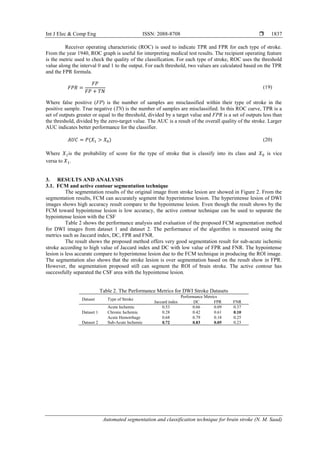
This document presents an automated technique for the segmentation and classification of brain stroke lesions using diffusion-weighted imaging (DWI) to aid in diagnosis. The method combines fuzzy c-means and active contour techniques for segmentation, along with a bagged tree classifier for lesion classification, achieving an overall classification accuracy of 90.8%. Performance metrics, including the Jaccard index and dice coefficient, indicate the proposed algorithm's effectiveness in identifying various types of strokes.
![International Journal of Electrical and Computer Engineering (IJECE)
Vol. 9, No. 3, June 2019, pp. 1832~1841
ISSN: 2088-8708, DOI: 10.11591/ijece.v9i3.pp1832-1841 1832
Journal homepage: http://iaescore.com/journals/index.php/IJECE
Automated segmentation and classification technique
for brain stroke
N. S. M. Noor1
, N. M. Saad2
, A. R. Abdullah3
, N. M. Ali4
1,2
Fakulti Kejuruteraan Elektronik dan Kejuruteraan Komputer, Universiti Teknikal Malaysia Melaka, Malaysia
3,4
Fakulti Kejuruteraan Elektrik, Universiti Teknikal Malaysia Melaka, Malaysia
Article Info ABSTRACT
Article history:
Received Sep 2, 2018
Revised Dec 16, 2018
Accepted Jan 26, 2019
Difussion-Weighted Imaging (DWI) plays an important role in the diagnosis
of brain stroke by providing detailed information regarding the soft tissue
contrast in the brain organ. Conventionally, the differential diagnosis of brain
stroke lesions is performed manually by professional neuroradiologists
during a highly subjective and time- consuming process. This study proposes
a segmentation and classification technique to detect brain stroke lesions
based on diffusion-weighted imaging (DWI). The type of stroke lesions
consists of acute ischemic, sub-acute ischemic, chronic ischemic and acute
hemorrhage. For segmentation, fuzzy c-Means (FCM) and active contour is
proposed to segment the lesion’s region. FCM is implemented with active
contour to separate the cerebral spinal fluid (CSF) with the hypointense
lesion. Pre-processing is applied to the DWI for image normalization,
background removal and image enhancement. The algorithm performance
has been evaluated using Jaccard Index, Dice Coefficient (DC) and both false
positive rate (FPR) and false negative rate (FNR). The average results for the
Jaccard index, DC, FPR and FNR are 0.55, 0.68, 0.23 and 0.23, respectively.
First statistical order method is applied to the segmentation result to obtain
the features for the classifier input. For classification technique, bagged tree
classifier is proposed to classify the type of stroke. The accuracy results for
the classification is 90.8%. Based on the results, the proposed technique has
potential to segment and classify brain stroke lesion from DWI images.
Keywords:
Brain imaging
Classification
Diffusion-weighted imaging
(DWI)
Segmentation
Stroke
Copyright © 2019 Institute of Advanced Engineering and Science.
All rights reserved.
Corresponding Author:
N. M. Saad,
Fakulti Kejuruteraan Elektronik dan Kejuruteraan Komputer,
Universiti Teknikal Malaysia Melaka, Melaka, Malaysia.
Email: norhashimah@utem.edu.my
1. INTRODUCTION
Nowadays, medical imaging tool plays an important role in viewing the internal tissue of human
brain. In brain stroke diagnosis, medical imaging such as MRI offers fast imaging tool with high resolution
and nonionizing radiation [1]. The MRI imaging tool is able to detect 85% stroke survivor from ischemic
stroke and 15-20% stroke survivor from hemorrhagic stroke [2]. DWI is one of the MRI modality that shows
high sensitivity (88-100%) and specificity (95-100%) in detecting early ischemic stroke [3].
To fully utilize medical imaging, new algorithms and methods are urged in the medical imaging
field for utilizing very large amounts of images wisely [4]. However, due to the large sample of images, the
diagnosis process is time consuming and tiredness [5]. A computer aided diagnosis is needed for
neuroradiologist to interpret the images more easily. Many researchers have studies machine learning
technique in brain imaging [6]. Machine learning technique is believed to perform tasks and solve problems
related to poor quality brain imaging samples by providing accurate representation and prior knowledge
modeling. Thus, it plays an important role in brain imaging where it has become one of the most promising
research areas in computer aided detection and diagnosis.](https://image.slidesharecdn.com/4317267-200724040925/85/Automated-segmentation-and-classification-technique-for-brain-stroke-1-320.jpg)
![Int J Elec & Comp Eng ISSN: 2088-8708
Automated segmentation and classification technique for brain stroke (N. M. Saad)
1833
Zhang Y. et al. [7] proposed kernel support vector machine to detect Alzheimer’s disease (AD) and
classify the disease between normal elder control (NC). Ramani R.G. et al. [8] proposed Naive Bayes,
Support Vector Machine and random tree to analyze the machine learning technique in classifying normal
and abnormal brain image. Mudali D. et al. [9] proposed decision tree method to interpret the diagnosis of
neurodegenerative brain diseases. Meet Oza et al. [10] proposed random forest classifier to classify brain
tumor into benign or malign cases. Fratello M. et al. [11] proposed random forest to classify two type of
disease from individual lateral sclerosis (ALS) patients, Parkinson’s disease (PD) patients and healthy
control (HC) subjects.
In this paper, segmentation and classification technique for brain stroke using DWI image is
proposed. The purpose of this study is to develop an automatic stroke segmentation and classification using
DWI images. The proposed segmentation analysis is based on the fuzzy c-Means (FCM) and active contour
technique. Active contour is integrated in the analysis framework to separate the cerebral spinal fluid (CSF)
with the hypointense lesion and increase the segmentation accuracy. The performance of the segmentation
techniques is evaluated based on Jaccardindex, Dice Coefficient (DC), false positive rate (FPR) and false
negative rate (FNR). After the segmentation result is obtain, the result image is extracted using first order
statistical method. These features are used for the bagged tree classifier input. The performance of the
classifier technique is evaluated based on the accuracy (ACC).
This paper contains five section. Section 2 discussed the flow process of the proposed methodology
in detail. Section 3 proposed the brain stroke analysis technique used to segment and classify the brain stroke
lesion. Section 4 discussed the result from the segmentation result and Section 5 conclude
the work presented.
2. RESEARCH METHOD
2.1. Proposed analysis framework
The flow process of this study starts with image preprocessing where image normalization, image
enhancement and background removal are applied. After that, FCM is applied for image segmentation stage
in order to extract the ROI of the stroke lesion. Then, the active contour is used to separate the CSF with the
hypointense stroke lesion region. Hyperintense is referred to bright lesion such as acute ischemic stroke,
acute hemorrhage stroke and sub-acute ischemic stroke lesion where hypointense is referred to dark lesion
such as chronic ischemic stroke lesion. The segmentation result is extracted using first order statistical
method for the input classifier. After the features are obtained, bagged tree classifier is used to classify the
type of strokes lesion. Last but not least, the performance of the brain image is evaluated based on the
Jaccard index, DC, FPR, FNR, ACC.
2.2. Imaging parameter
This paper focused on two brain stroke datasets from DWI images. Dataset 1 contains 61 samples
from the General Hospital of Kuala Lumpur. Dataset 2 contains 69 samples from the online database of
Ischemic Stroke Lesion Segmentation (ISLES). The dataset only focused on four types of brain stroke
lesion which are acute ischemic stroke, chronic ischemic stroke, acute hemorrhage stroke and sub-acute
ischemic stroke. The acute ischemic stroke, chronic ischemic stroke and acute hemorrhage stroke images
were gained from the General Hospital of Kuala Lumpur (HKL). The sub-acute ischemic stroke images
were gained from the public online Ischemic Stroke Lesion Segmentation (ISLES) challenge 2015 [12].
Table 1. The Brain Stroke Datasets
Stroke Datasets
Dataset 1 (clinical) Dataset 2 (ISLES) [12]
Acute Ischemic Chronic Ischemic Acute Hemorrhage Sub-acute Ischemic
Number of Samples 20 33 8 69
2.3. Image pre-processing
The pre-processing stage is the stage where the images need to undergo pre-processing stages to
acquire better segmentation [13]-[15]. Three algorithms were applied to the DWI image which is image
normalization, background removal and image enhancement [16]. These images are converted into the
desired form in which the intensity is adjusted and the noise is removed.
2.4. Fuzzy c-means
FCM is a clustering method where it divides a group of pixels into homogenous cluster and assigns
the pixels according to their category. It allows pixels’ points to be assigned to multiple clusters and each](https://image.slidesharecdn.com/4317267-200724040925/85/Automated-segmentation-and-classification-technique-for-brain-stroke-2-320.jpg)
![ ISSN: 2088-8708
Int J Elec & Comp Eng, Vol. 9, No. 3, June 2019 : 1832 - 1841
1834
pixel point has a degree of membership in a cluster to which it belongs. In this segmentation technique, the
pixel point is select base on the center of three cluster which are lower, middle and higher cluster. Each data
point of the cluster should equal to one. It is based on minimization of the following objective function:
𝐽 𝑚 = ∑ ∑ 𝑢𝑖𝑗
𝑚
‖𝑥𝑖 − 𝑐𝑗‖
2
𝐶
𝑗=1
𝑁
𝑖=1
, 1 ≤ 𝑚 < 0 (1)
where m is any real number greater than 1, iju is the degree of membership of ix in the cluster j, ix is the i
th of d-dimensional measured data, jc is the d-dimension center of the cluster, and is any norm
expressing the similarity between any measured data and the center. Fuzzy partitioning is carried out through
an iterative optimization of the objective function shown above, with the update of membership iju and the
cluster centers jc by:
𝑢𝑖𝑗 =
1
∑ (
‖𝑥𝑖 − 𝑐𝑗‖
‖𝑥𝑖 − 𝑐𝑗‖
)
2
𝑚−1
𝑐
𝑘=1
; 𝑐𝑗 =
∑ 𝑢𝑖𝑗
𝑚
. 𝑥𝑖
𝑁
𝑖=1
∑ 𝑢𝑖𝑗
𝑚𝑁
𝑖=1 (2)
This iteration will stop when,
𝑚𝑎𝑥𝑖𝑗{|𝑢𝑖𝑗
(𝑘+1)
| − 𝑢𝑖𝑗
(𝑘)
} < 𝜀 (3)
where is a termination criterion between 0 and 1, whereas k are the iteration steps.
2.5. Active contour
In DWI chronic stroke lesion image, the cerebrospinal fluid (CSF) share the similar intensity level
with the stroke lesion. Due to this matter, FCM method has failed to segment the lesion since the algorithm in
the cluster cannot be differentiated. To improve this performance, the CSF area is removed by using the
active contour method.
Active contour is a method that create boundaries in an image. It uses computer generated curves to
detect and locate object. This method is often using in medical images to find the boundaries of an organ in
the images. The image is classified into two part which are object region and background region. In this
method the region is represented as the inside and outside regions of the zero-level set.
Level set framework take two signs positive and negative to divide image domain. The image
domain is separated into two disjoint region Ω1 and Ω2. Local intensity clustering property means that the
intensities in the neighborhood 0 𝑦 can be divided into N clusters, with center 𝐶𝑖, 𝑖 = 1,2, … , 𝑁. It can be
written as:
𝐹𝑦 = ∑ ∫ |𝐼(𝑥) − 𝑐𝑖|2
𝑚𝑖(𝑥)
0 𝑦
𝑁
𝑖=1
𝑑𝑥 (4)
Where 𝐶𝑖 the cluster in the center of the i-th cluster is, 𝑚𝑖 is the membership function of the region, i.e.
𝑚𝑖(𝑥) = 1 for 𝑥 ∈ Ω𝑖 and𝑚𝑖(𝑥) = 0 for 𝑥 ∉ Ω𝑖. Or the corresponding equation can be witten as:
𝐸 𝑎𝑐𝑡𝑖𝑣𝑒 𝑐𝑜𝑛𝑡𝑜𝑢𝑟=∫ [𝐸 𝑖𝑛𝑡(𝑣(𝑠))+𝐸 𝑒𝑥𝑡(𝑣(𝑠))] 𝑑𝑠
𝑠 𝑚
0
(5)
Where the position of active contour is describe parametrically by v(s) = (x(s),y(s)), 𝐸𝑖𝑛𝑡 represents internal
potential energy of the contour, 𝐸𝑒𝑥𝑡 is the energy that models external constraints impose onto the contour
shape. G(y-x) is the Gaussian kernel applied as window function showed.
𝐺(𝑑) = { 𝑒𝑎
1
−
|𝑑|2
2𝜎2
𝑓𝑜𝑟 |𝑑| ≤ 𝜌
0 𝑜𝑡ℎ𝑒𝑟𝑤𝑖𝑠𝑒
(6)](https://image.slidesharecdn.com/4317267-200724040925/85/Automated-segmentation-and-classification-technique-for-brain-stroke-3-320.jpg)
![Int J Elec & Comp Eng ISSN: 2088-8708
Automated segmentation and classification technique for brain stroke (N. M. Saad)
1835
Where 𝑎is a constant, d is a distance between x and y points. 𝜎 is a standard deviation or scale parameter of
Gaussian function, and 𝜌 is a radius of neighboring pixels. The radius of the neighborhood should be choose
appropriately based on the degree of intensity in-homogeneity.
The next step is contour construction to define initial shape around the object that will serve as an
initialization set up. Last but not least, the greedy method is applied to simplify the implementation of the
minimization of energy without having to perform an optimization algorithm method like gradient descent
[17]-[21]. The function of this method is by finding for each point of the contour the closest local energy
minimizing neighbor will converge to the overall global minimum of the contour. The method using 2
equations which are:
𝐸𝑖𝑛𝑡 =
(𝛼(𝑠)|𝑉𝑠(𝑆)|2
+ 𝛽(𝑠)|𝑉𝑠(𝑆)|2)
2
(7)
Where 𝛼(𝑠)represent adjusts the elasticity of the active contour and 𝛽(𝑠)is adjusts the stiffness of the
active contour.
𝐸𝑒𝑥𝑡 = −𝛿 ∫ ‖∇(𝐺 𝑛 ∗ 𝐼)‖2
𝐵
𝐴
(𝛾(𝑠))𝑑𝑠 (8)
Where 𝛿 is a real weighting value which for obvious reason would be positive, 𝐺𝑛 is a Gaussian weighted
kernel of dimension n, I represent the input image, ∇is the spatial gradient function and 𝛾(𝑠) is the
contour function.
2.6. First order statistical method
First order statistical order method is applied at the ROI to obtain several features. Since this ROI
depends on the signal of intensity, the ROI is classified into two parts of the image which are hyperintense
and hypointense lesion. Mean, median and mode are used to separate the image between hyperintense and
hypointense lesion. For hyperintense lesion, standard deviation is assigned while in hypointense lesion mean
of region boundary is assigned. The standard deviation and mean of boundary are used to differentiate each
characteristic of stroke lesion.
The calculation for the first order method is shows as below:
𝜇 =
1
𝑁
∑ 𝑃(𝑖)
𝑁−1
𝑖=1
(9)
Median =
1
2
(𝑛 + 1)𝑡ℎ
𝑣𝑎𝑙𝑢𝑒
(10)
𝜎 = √
1
𝑁
∑(𝑃(𝑖) − 𝜇)2
𝑁−1
𝑖=0
(11)
Pearson mode skewness =
|𝑚𝑜𝑑𝑒 − 𝜇|
𝜎
(12)
Compactness =
(𝐿𝑒𝑛𝑔𝑡ℎ (𝑏𝑜𝑢𝑛𝑑𝑎𝑟𝑦))2
𝐴𝑟𝑒𝑎
(13)
Where P(i) is the probability intensity level of the ROI, N be the total number of gray levels in the
entire ROI.
2.7. Bagged tree classifier
Bagged tree classifier is a method that perform a collection of data by analyze the whole data than
individually. This classifier builds and trains a variety of decision trees and produces classes from the mode
of individual tree class. It applies general techniques by resampling data from the previous learning. Assume
that For b= 1,..,B; where B is the bagging classifier (B times) that repeat the train set of 𝐷 = 𝑑1, … , 𝑑 𝑛 with](https://image.slidesharecdn.com/4317267-200724040925/85/Automated-segmentation-and-classification-technique-for-brain-stroke-4-320.jpg)
![ ISSN: 2088-8708
Int J Elec & Comp Eng, Vol. 9, No. 3, June 2019 : 1832 - 1841
1836
the response 𝑅 = 𝑟1, … , 𝑟𝑛.Each individual tree class will observe the data from the previous data of tree class
and make their own decision. The decision that make is then is selected and being compare with the other
tree classes by average the data. For unseen data (x’
), the data is select by using the mod of data in
the tree class.
𝑓̂ =
1
𝐵
∑ 𝑓𝑏(𝑥′)
𝐵
𝑏=1
(14)
In this experiment, the bagged tree classifiers train 129 data for the number of branch node splits per
tree. Figure 1 shows the bagged tree diagram.
Figure 1. Bagged tree classifier diagram
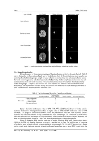
2.8. Performance evaluation
The performance evaluationis calculated from the segmentation results with the neuroradiologist
manual reference. Jaccard index, Dice coefficient (DC), false positive rate (FPR) and false negative rate
(FNR) are used as the performance metrics [16]. From this calculation, the DWI segmentation result can be
fully segment.
The performance verification for classification take part in the statistical calculation and shown in
confusion matrix attributes using MATLAB APPS. Number of observations, true positive rate (TPR), false
negative rate (FNR), positive predictive values (PPV) and false discovery rate (FDR) is used to identify the
performance of the classifier. Below is the statistical calculation for performance verification.
𝑇𝑃𝑅 =
𝑇𝑃
𝑃
(15)
𝑇𝑁𝑅 = 1 − 𝑇𝑃𝑅
(16)
Where true positive (TP) is the number of samples are correctly classified within their type of stroke in the
positive sample (P). Positive predictive value (PPV) as shown in Equation (17) is calculated as the
probability that correctly classify the type of stroke. The false positive (FP) is misclassified of stroke.
PPV =
𝑇𝑃
𝑇𝑃 + 𝐹𝑃
(17)
Lastly the performance of the classifier will be verified using Acc to as shown in Equation (18).
𝐴 𝑐𝑐 =
𝑇𝑃 + 𝑇𝑁
𝑃 + 𝑁
(18)](https://image.slidesharecdn.com/4317267-200724040925/85/Automated-segmentation-and-classification-technique-for-brain-stroke-5-320.jpg)
![ ISSN: 2088-8708
Int J Elec & Comp Eng, Vol. 9, No. 3, June 2019 : 1832 - 1841
1840
ACKNOWLEDGEMENTS
The authors would like to thank to the Universiti Teknikal Malaysia Melaka (UTeM), Rehabilitation
Engineering & Assistive Technology (REAT) research group under Center for Robotics & Industrial
Automation (CeRIA), Advanced Digital Signal Processing (ADSP) Lab and Ministry of Higher Education
(MOHE), Malaysia for sponsoring this work under project FRGS/1/TK04/FKE-CeRIA/F00334 and the use
of the existing facilities to complete this project.
REFERENCES
[1] Gomes J, Wachsman AM,“Handbook of Clinical Nutrition and Stroke,”New York: Humana Press, Totowa, NJ,pp.
15-31,2013.
[2] Kanchana R, Menaka R.,“Computer Reinforced Analysis for Ischemic Stroke Recognition: A Review”,Indian
Journal of Science and Technology,vol.8,no. 35, pp. 1-9,December 2015.
[3] Potente M, Gerhardt H, Carmeliet P.,“Basic and Therapeutic Aspects of Angiogenesis”,Cell, vol.146, no. 6, pp.
873-877, Sep 2011.
[4] Wernick MN, Yang Y, Brankov JG, Yourganov G, Strother SC.,“Machine Learning in Medical Imaging”,IEEE
Signal Processing Magazine, vol. 27 no.4, pp.25-38, Jul 2010.
[5] Khademi A, Venetsanopoulos A, Moody AR, “Robust White Matter Lesion Segmentation in FLAIR MRI”,IEEE
Transactions on Biomedical Engineering., vol.59, no. 3, pp. 860-71, Mar 2012.
[6] Suzuki K., “Pixel-based Machine Learning in Medical Imaging” Journal of Biomedical Imaging, vol 1, Jan 2012.
[7] Zhang Y, Dong Z, Phillips P, Wang S, Ji G, Yang J, Yuan TF,“Detection of Subjects and Brain Regions Related to
Alzheimer's Disease Using 3D MRI Scans Based on Eigenbrain and Machine Learning”,Frontiers in
Computational Neuroscience, vol. 2, Jun 2015.
[8] Ramani RG, Sivaselvi K.,“Classification of Pathological Magnetic Resonance Images of Brain Using Data Mining
Techniques”,InRecent Trends and Challenges in Computational Models (ICRTCCM), 2017 Second International
Conference, 2017,pp. 77-82, IEEE.
[9] Mudali D, Teune LK, Renken RJ, Leenders KL, Roerdink JB.,“Classification of Parkinsonian Syndromes from
FDG-PET Brain Data Using Decision Trees with SSM/PCA Features”,Computational and Mathematical Methods
in Medicine, 2015.
[10] Oza M, Kapdi R, Student B.,“Brain Tumor Disease Identification Using Random Forest Classifiers”, Brain, vol. 7
no. 1, Sep 2015.
[11] Fratello M, Caiazzo G, Trojsi F, Russo A, Tedeschi G, Tagliaferri R, Esposito F.,“Multi-View Ensemble
Classification of Brain Connectivity Images for Neurodegeneration Type Discrimination”, Neuroinformatics, vol.
15, no. 2, pp.199-213, April 2017.
[12] Maier O, Menze BH, von der Gablentz J, Häni L, Heinrich MP, Liebrand M, Winzeck S, Basit A, Bentley P, Chen
L, Christiaens D,“ISLES 2015-A Public Evaluation Benchmark for Ischemic Stroke Lesion Segmentation from
Multispectral MRI”, Medical Image Analysis, vol. 1, no. 35, pp. 250-690, Jan 2017.
[13] Liu J, Li M, Wang J, Wu F, Liu T, Pan Y,“A Survey of MRI-based Brain Tumor Segmentation Methods”,Tsinghua
Science and Technology, vol. 19, no. 6, pp.578-95, Dec 2014.
[14] Baraiya N, Modi H., “Comparative study of Different Methods for Brain Tumor Extraction from MRI Images
Using Image Processing”, Indian Journal of Science and Technology,Vol. 9, no. 4, Jan 2016.
[15] Rahman NA, Saad NM, Abdullah AR, Wahab FA., “The Internet of Things Beverages Bottle Shape Defect
Detection using Naïve Bayes Classifier,” International Journal of Human and Technology Interaction (IJHaTI),
vol. 2, no. 1, pp. 71-6, Apr 2018.
[16] Saad N.M, Noor NS, Abdullah AR, Muda S, Muda AF, Musa H., “Segmentation and Classification Analysis
Techniques for Stroke based on Diffusion-Weighted Images”, IAENG International Journal of Computer Science,
vol. 44, no. 3, Aug 2017.
[17] Abdullah AR, Abidullah NA, Shamsudin NH, Ahmad NH, Jopri MH.,“Power Quality Signals Classification
System Using Time-Frequency Distribution”, InApplied Mechanics and Materials, Trans Tech Publications, vol.
494, pp. 1889-1894, 2014.
[18] Abdullah AR, Abidullah NA, Shamsudin NH, Ahmad NH, Jopri MH.,“Performance Verification of Power Quality
Signals Classification System”, InApplied Mechanics and Materials, Trans Tech Publications, vol. 752,
pp. 1158-1163, 2015.
[19] Jopri MH, Abdullah AR, Kassim NM, Manap M, Ngatiman NA, Yusoff MR.,“Localization of Multiple Harmonic
Sources for Inverter Loads Utilizing Periodogram”, Journal of Telecommunication, Electronic and Computer
Engineering (JTEC), vol. 8, no. 2, pp 87-91, May 2016.
[20] Habban, M.F., Manap, M., Abdullah, A.R., Jopri, M.H. and Sutikno, T., “An Evaluation of linear time frequency
distribution Analysis for VSI switch faults identification”, International Journal of Power Electronics and Drive
Systems (IJPEDS), vol. 8, no.1, pp. 1-9.
[21] Jopri MH, Abdullah AR, Manap M, Habban MF, Sutikno T.,“An Accurate Classification Method of Harmonic
Signals in Power Distribution System by Utilising S-Transform”, Telkomnika (, vol. 15, no. 1, Mar 2017.](https://image.slidesharecdn.com/4317267-200724040925/85/Automated-segmentation-and-classification-technique-for-brain-stroke-9-320.jpg)