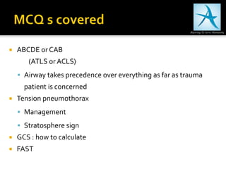
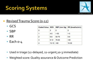
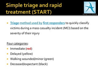
 Coordination with receiving hospital
 Emphasis is placed on
 Airway maintenance
 Control of external bleeding & Shock
 Immobilization of the patient
 Immediate transport to closest appropriate facility, preferably
a verified trauma center.
 Brief history on the way regarding time & mechanism of injury
 Nowadays, interventions at the scene have reduced to minimum
 Scoop & Run
Only Life
saving
Activities
 A: Airway with Cervical Spine control
 B: Breathing and ventilation
 C: Circulation with hemorrhage control
 D: Disability or neurological status
 E: Exposure & environment
 Inspect upper airway to detect obstruction
 Listen for gurgling, snoring or stridor
 Watch for chest movements & Feel airflow with volar aspect of
wrist & forearm
 Cervical Immobilization collar
 MILS when manipulating airway
 If intubated in prehospital setting, confirm position
 Airway & Breathing go hand-in-hand
 Look for central cyanosis
 Check symmetry of chest movements
 Watch for paradoxical chest movements
 Palpation : subcutaneous emphysema (snowball crepitus)
 Percussion : pneumothorax vs hemothorax
 Auscultation : absent breath sounds
 Supplemental oxygen
 Recognize airway obstruction
 Decreased LOC
 Direct trauma eg maxillofacial
Increasing neck sweling
 Maintain airway patency
 Simple airway manoeuvres
(chin lift or jaw thrust)
 Airway adjuncts
 Tracheal intubation
 Alternate airway devices
Indications for tracheal intubation and mechanical ventilation
other than to protect the airway are usually to manage
abnormalities of respiratory drive or mechanics and problems of
gas exchange e.g.:
 Severe traumatic brain injury
 Cervical spinal cord injury with related severe hypoventilation
 Severe disruption of the thorax (fractured ribs, flail segment)
 Hypoxaemia despite application of high-flow oxygen (lung
contusion, pulmonary haemorrhage, aspiration).
Open Pneumothorax
 If defect > 2/3 tracheal diameter; air is prefrentially drawn
through the defect
 Definitive management :
 Tube thoracostomy at separate side
 Cover the wound with occlusive dressing
Normal tracheal diameter 15-25 mm males & 10-21 mm females
Tension Pneumothorax
 Clinical Signs:Tachycardia, hypotension, deviated trachea, absent
breath sounds, hyperresonance on percussion
 Pushes mediastinum, compresses the oposite lung, kinking the great
veins & reducing venous return dramatically
 Emergency management : Needle Decompression
 Don’t wait for X ray confirmation
(14 G needle, mid-clavicular line,
 2nd intercostal space, Just above the rib)
 Definitive mgmt :Tube thoracostomy asap
 (5th intercostal space, Ant axillary line)
 Palpate carotid pulse (SBP approx 40-50 mm Hg)
 Look for obvious external hemmorhage
 Palpate peripheral pulses (rapid, faint)
 Look for jugular venous congestion (thoracic inflow
obstruction)
 Assess capillary refill, skin temperature & mottling
 Lab parameters: elevated lactate, increased base deficit,
reduced central/mixed venous oxygen saturation
 Short large bore iv cannula
 Monitoring (ECG, NIBP, Pulse oximetry, EtCO2)
 Emergency hemorrhage control
 External compression
 Tourniquet,as minimum as possible
 Pelvic compression
 Hemorrhagic shock resuscitation
 Crystalloids preferred
 If hemorrhage uncontrolled: permissive hypotension or
damage control resuscitation
Hagen-Poiseuille's
law, laminar flow
rate varies directly
with fourth power
of radius &
inversely with
length
Relief of obstructive shock
 Tension pneumothorax
 Cardiac tamponade
 Clinical signs suggestive
 Definitive diagnosis : transthoracic echocardiography
 Treatment: emergency clamshell thoracotomy in patients
with penetrating chest trauma & cardiac arrest
 Level of consciousness, GCS
 Lateralizing signs
 Brain stem reflexes
 Pupillary size and reaction
 U/l dilatation without light rxn: ipsilateral transtentorial
herniation
 B/l dilatation without light rxn: impending brain stem
compression or intoxication/severe hypothermia/ hypoxia
Flexion to painful stimuli: injury to upper midbrain (decortication)
Extension to painful stimuli: lower midbrain or pontine injury (decerebration)
Trauma patients, who do NOT fulfill all of the assessment criteria
listed below should receive C-spine control:
 No pain or tenderness around the vertebral column
 Awake, cooperative, & not under influence of drugs or alcohol
 No distracting pain from other injuries
 No neurological deficit.
Patient who is awake & can flex his neck so that his chin touches his
chest without pain
If signs of impending cerebral herniation
 Hypertonic osmotic solutions (HS, mannitol)
 Tracheal intubation & mechanical ventilation
 Deep sedation
 Normal to higher arterial pressures
 Urgent neurosurgical consultation
 Completely undress the patient
 Prevent hypothermia
 Warm blankets
 External warming devices
 Warm iv fluids
 Warm room temperature
 ECG
 Urinary output monitoring
 Gastric drainage monitoring
 Pulse oximetry
 EtCO2
 BP
 Radiological imaging (AP chest, AP pelvis)
Transurethral bladder catherization CI if
urethral injury is suspected
a) Blood at urethral meatus
b) Perineal echhymosis
c) High riding or non palpable prostrate
 Head toToe Examination
 AMPLE history
 Allergies
 Medication
 Past history
 Last meal
 Events and environment
Secondary survey is always after primary.
The patient may undergo an emergency laparotomy to stop
major intra-abdominal bleeding
 Head: GCS, look for periorbital or retroauricular hematomas
 Neurology: sensorimotor function
 Neck: cutaneous emphysema, palpate for pain, swelling &
dislocation
 Chest: palpate for sternum & rib #
 Abdomen: palpate
 Pelvis: compress horizontally & vertically to test stability
 Perineum: orifices
 Extremities: inspect, palpate & move joints, look for
compartment syndrome
 Back: log roll the patient, examine the spine
 Blood count
 Coagulation parameters
 KFT, LFT, muscle enzymes
 Cardiac biomarkers
 Toxicology
 TRAUMA Scan : head, neck, chest, abdomen, pelvis
CT scan
 Contrast can locate the ongoing hemorrhage
(min bleeding 0.5ml/min)
eFAST
1. Hepatorenal space
(Morison pouch)
2. Splenorenal space
(Koller pouch)
3. Pouch of Douglas
4. Pericardium
5. Pleural cavity left
& right
eFAST (Extended Focused Assessment with Sonography forTrauma
 M-mode
 depicts the glandular
echogenicity of the lung
abutted by the linear
appearance of the visceral
pleura.
 This sign is a normal
finding.
 In absence of a seashore
sign or presence of a
stratosphere sign,
pneumothorax is likely.
 eFAST
 prove presence of a pneumothorax.
 3.5-7.5 MHz ultrasound probe
 4th and 5th intercostal spaces
 Anterior clavicular line
 using the M-Mode of the machine.
 pleura and lung being indistinguishable
as linear hyperechogenic lines and is
fairly reliable for diagnosis of a
pneumothorax.
 M-mode finding
 presence of pleural effusion
 Due to the cyclical movement of the lung in inspiration and expiration, the
motion-time tracing (M-mode) ultrasound shows a sinusoid appearance
between the fluid and the line tissue.
 This finding indicates possibly but not with certainty, of pleural effusion,
empyema or blood in pleural space (Hemothorax)
 B-line is a discrete, laser-like, vertical, hyperechoic image, that
arises from the pleural line, extends to the bottom of the screen
without fading, and moves synchronously with respiration
 usually lost with any air
between the probe and the
lung tissue and therefore
whose presence with
seashore sign indicates
absence of a pneumothorax.
 ABCDE or CAB
(ATLS or ACLS)
 Airway takes precedence over everything as far as trauma
patient is concerned
 Tension pneumothorax
 Management
 Stratosphere sign
 GCS : how to calculate
 FAST
 Correct triage of patients to trauma centre
 Selecting adequate intensity of care
 Prognostication of short and long term outcomes
 Comparison of trauma centers
 RevisedTrauma Score (0-12)
 GCS
 SBP
 RR
 Each 0-4
 Used in triage (12-delayed, 11-urgent,10-3 immediate)
 Weighted score: Quality assurance & Outcome Prediction
 Abbreviated Injury Scale
 anatomically based system of grading injuries on an ordinal
scale ranging from 1 (minor injury) to 6 (lethal injury)
 Six body regions are defined, as follows:
▪ the thorax,
▪ abdomen and visceral pelvis,
▪ head and neck,
▪ face,
▪ bony pelvis and extremities,
▪ and external structures.
 Injury Severity Score (major trauma, ISS >15)
 Defined as the sum of squares of the highest AIS grade in
the 3 most severely injured body regions.
 Only one injury per body region is allowed.The ISS ranges
from 1-75, and an ISS of 75 is assigned to anyone with an AIS
of 6
 New or Modified ISS
 Squares of AIS scores of a patient’s 3 most severe injuries,
regardless of body region
 Triage method used by first responders to quickly classify
victims during a mass casualty incident (MCI) based on the
severity of their injury
Four categories:
 Immediate (red)
 Delayed (yellow)
 Walking wounded/minor (green)
 Deceased/expectant (black)
Trauma and critical care

Trauma and critical care

  • 1.
     Coordination withreceiving hospital  Emphasis is placed on  Airway maintenance  Control of external bleeding & Shock  Immobilization of the patient  Immediate transport to closest appropriate facility, preferably a verified trauma center.  Brief history on the way regarding time & mechanism of injury  Nowadays, interventions at the scene have reduced to minimum  Scoop & Run Only Life saving Activities
  • 3.
     A: Airwaywith Cervical Spine control  B: Breathing and ventilation  C: Circulation with hemorrhage control  D: Disability or neurological status  E: Exposure & environment
  • 4.
     Inspect upperairway to detect obstruction  Listen for gurgling, snoring or stridor  Watch for chest movements & Feel airflow with volar aspect of wrist & forearm  Cervical Immobilization collar  MILS when manipulating airway  If intubated in prehospital setting, confirm position
  • 5.
     Airway &Breathing go hand-in-hand  Look for central cyanosis  Check symmetry of chest movements  Watch for paradoxical chest movements  Palpation : subcutaneous emphysema (snowball crepitus)  Percussion : pneumothorax vs hemothorax  Auscultation : absent breath sounds
  • 6.
     Supplemental oxygen Recognize airway obstruction  Decreased LOC  Direct trauma eg maxillofacial Increasing neck sweling  Maintain airway patency  Simple airway manoeuvres (chin lift or jaw thrust)  Airway adjuncts  Tracheal intubation  Alternate airway devices
  • 7.
    Indications for trachealintubation and mechanical ventilation other than to protect the airway are usually to manage abnormalities of respiratory drive or mechanics and problems of gas exchange e.g.:  Severe traumatic brain injury  Cervical spinal cord injury with related severe hypoventilation  Severe disruption of the thorax (fractured ribs, flail segment)  Hypoxaemia despite application of high-flow oxygen (lung contusion, pulmonary haemorrhage, aspiration).
  • 8.
    Open Pneumothorax  Ifdefect > 2/3 tracheal diameter; air is prefrentially drawn through the defect  Definitive management :  Tube thoracostomy at separate side  Cover the wound with occlusive dressing Normal tracheal diameter 15-25 mm males & 10-21 mm females
  • 9.
    Tension Pneumothorax  ClinicalSigns:Tachycardia, hypotension, deviated trachea, absent breath sounds, hyperresonance on percussion  Pushes mediastinum, compresses the oposite lung, kinking the great veins & reducing venous return dramatically  Emergency management : Needle Decompression  Don’t wait for X ray confirmation (14 G needle, mid-clavicular line,  2nd intercostal space, Just above the rib)  Definitive mgmt :Tube thoracostomy asap  (5th intercostal space, Ant axillary line)
  • 10.
     Palpate carotidpulse (SBP approx 40-50 mm Hg)  Look for obvious external hemmorhage  Palpate peripheral pulses (rapid, faint)  Look for jugular venous congestion (thoracic inflow obstruction)  Assess capillary refill, skin temperature & mottling  Lab parameters: elevated lactate, increased base deficit, reduced central/mixed venous oxygen saturation
  • 11.
     Short largebore iv cannula  Monitoring (ECG, NIBP, Pulse oximetry, EtCO2)  Emergency hemorrhage control  External compression  Tourniquet,as minimum as possible  Pelvic compression  Hemorrhagic shock resuscitation  Crystalloids preferred  If hemorrhage uncontrolled: permissive hypotension or damage control resuscitation Hagen-Poiseuille's law, laminar flow rate varies directly with fourth power of radius & inversely with length
  • 12.
    Relief of obstructiveshock  Tension pneumothorax  Cardiac tamponade  Clinical signs suggestive  Definitive diagnosis : transthoracic echocardiography  Treatment: emergency clamshell thoracotomy in patients with penetrating chest trauma & cardiac arrest
  • 13.
     Level ofconsciousness, GCS  Lateralizing signs  Brain stem reflexes  Pupillary size and reaction  U/l dilatation without light rxn: ipsilateral transtentorial herniation  B/l dilatation without light rxn: impending brain stem compression or intoxication/severe hypothermia/ hypoxia
  • 14.
    Flexion to painfulstimuli: injury to upper midbrain (decortication) Extension to painful stimuli: lower midbrain or pontine injury (decerebration)
  • 15.
    Trauma patients, whodo NOT fulfill all of the assessment criteria listed below should receive C-spine control:  No pain or tenderness around the vertebral column  Awake, cooperative, & not under influence of drugs or alcohol  No distracting pain from other injuries  No neurological deficit. Patient who is awake & can flex his neck so that his chin touches his chest without pain
  • 16.
    If signs ofimpending cerebral herniation  Hypertonic osmotic solutions (HS, mannitol)  Tracheal intubation & mechanical ventilation  Deep sedation  Normal to higher arterial pressures  Urgent neurosurgical consultation
  • 17.
     Completely undressthe patient  Prevent hypothermia  Warm blankets  External warming devices  Warm iv fluids  Warm room temperature
  • 18.
     ECG  Urinaryoutput monitoring  Gastric drainage monitoring  Pulse oximetry  EtCO2  BP  Radiological imaging (AP chest, AP pelvis) Transurethral bladder catherization CI if urethral injury is suspected a) Blood at urethral meatus b) Perineal echhymosis c) High riding or non palpable prostrate
  • 19.
     Head toToeExamination  AMPLE history  Allergies  Medication  Past history  Last meal  Events and environment Secondary survey is always after primary. The patient may undergo an emergency laparotomy to stop major intra-abdominal bleeding
  • 20.
     Head: GCS,look for periorbital or retroauricular hematomas  Neurology: sensorimotor function  Neck: cutaneous emphysema, palpate for pain, swelling & dislocation  Chest: palpate for sternum & rib #  Abdomen: palpate  Pelvis: compress horizontally & vertically to test stability  Perineum: orifices  Extremities: inspect, palpate & move joints, look for compartment syndrome  Back: log roll the patient, examine the spine
  • 21.
     Blood count Coagulation parameters  KFT, LFT, muscle enzymes  Cardiac biomarkers  Toxicology  TRAUMA Scan : head, neck, chest, abdomen, pelvis CT scan  Contrast can locate the ongoing hemorrhage (min bleeding 0.5ml/min)
  • 22.
    eFAST 1. Hepatorenal space (Morisonpouch) 2. Splenorenal space (Koller pouch) 3. Pouch of Douglas 4. Pericardium 5. Pleural cavity left & right eFAST (Extended Focused Assessment with Sonography forTrauma
  • 23.
     M-mode  depictsthe glandular echogenicity of the lung abutted by the linear appearance of the visceral pleura.  This sign is a normal finding.  In absence of a seashore sign or presence of a stratosphere sign, pneumothorax is likely.
  • 24.
     eFAST  provepresence of a pneumothorax.  3.5-7.5 MHz ultrasound probe  4th and 5th intercostal spaces  Anterior clavicular line  using the M-Mode of the machine.  pleura and lung being indistinguishable as linear hyperechogenic lines and is fairly reliable for diagnosis of a pneumothorax.
  • 25.
     M-mode finding presence of pleural effusion  Due to the cyclical movement of the lung in inspiration and expiration, the motion-time tracing (M-mode) ultrasound shows a sinusoid appearance between the fluid and the line tissue.  This finding indicates possibly but not with certainty, of pleural effusion, empyema or blood in pleural space (Hemothorax)
  • 26.
     B-line isa discrete, laser-like, vertical, hyperechoic image, that arises from the pleural line, extends to the bottom of the screen without fading, and moves synchronously with respiration  usually lost with any air between the probe and the lung tissue and therefore whose presence with seashore sign indicates absence of a pneumothorax.
  • 27.
     ABCDE orCAB (ATLS or ACLS)  Airway takes precedence over everything as far as trauma patient is concerned  Tension pneumothorax  Management  Stratosphere sign  GCS : how to calculate  FAST
  • 28.
     Correct triageof patients to trauma centre  Selecting adequate intensity of care  Prognostication of short and long term outcomes  Comparison of trauma centers
  • 29.
     RevisedTrauma Score(0-12)  GCS  SBP  RR  Each 0-4  Used in triage (12-delayed, 11-urgent,10-3 immediate)  Weighted score: Quality assurance & Outcome Prediction
  • 30.
     Abbreviated InjuryScale  anatomically based system of grading injuries on an ordinal scale ranging from 1 (minor injury) to 6 (lethal injury)  Six body regions are defined, as follows: ▪ the thorax, ▪ abdomen and visceral pelvis, ▪ head and neck, ▪ face, ▪ bony pelvis and extremities, ▪ and external structures.
  • 31.
     Injury SeverityScore (major trauma, ISS >15)  Defined as the sum of squares of the highest AIS grade in the 3 most severely injured body regions.  Only one injury per body region is allowed.The ISS ranges from 1-75, and an ISS of 75 is assigned to anyone with an AIS of 6  New or Modified ISS  Squares of AIS scores of a patient’s 3 most severe injuries, regardless of body region
  • 32.
     Triage methodused by first responders to quickly classify victims during a mass casualty incident (MCI) based on the severity of their injury Four categories:  Immediate (red)  Delayed (yellow)  Walking wounded/minor (green)  Deceased/expectant (black)