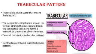
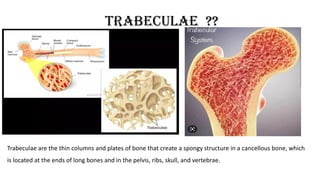
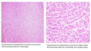
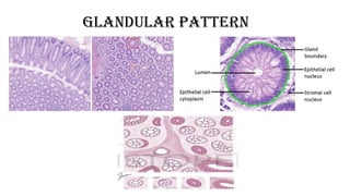
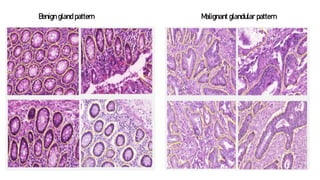
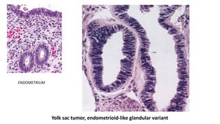
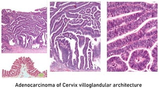
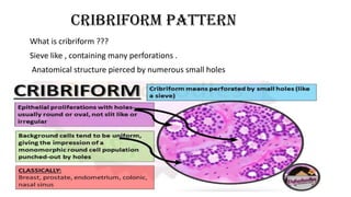
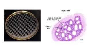
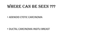
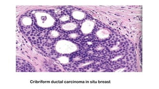
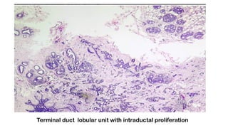
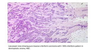
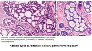
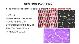
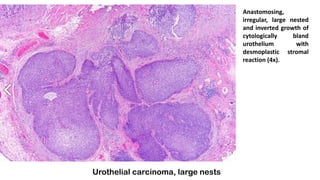
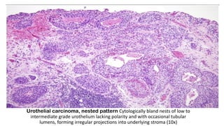
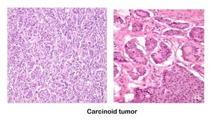
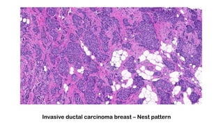
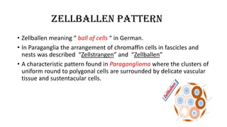
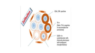
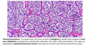
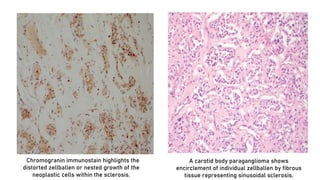
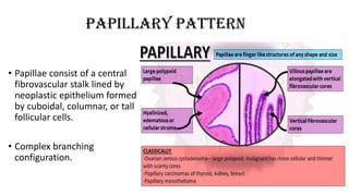
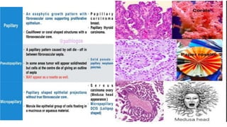
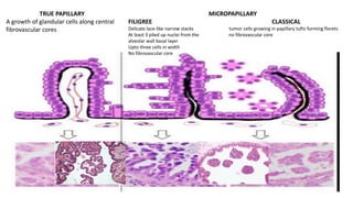
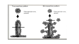
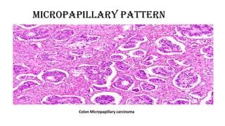
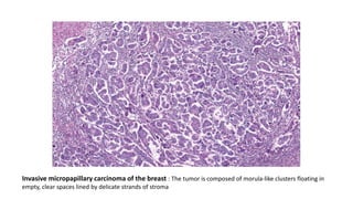
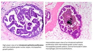
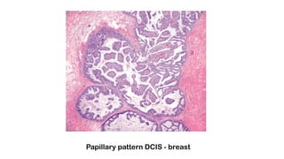
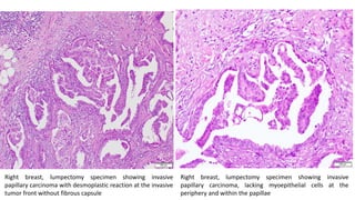
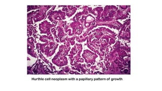
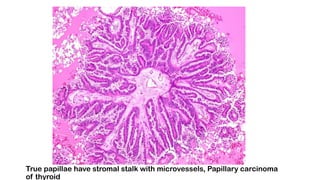
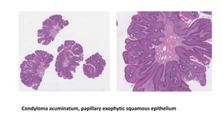
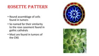
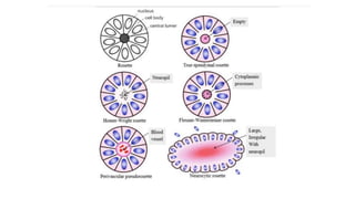
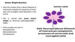
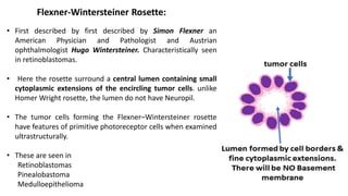
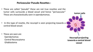
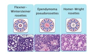
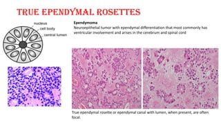
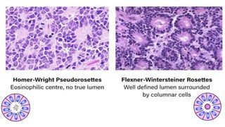
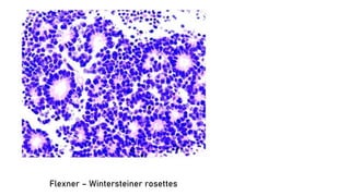
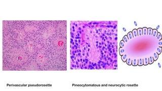
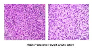
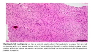
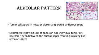
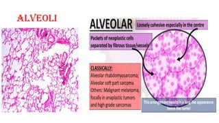
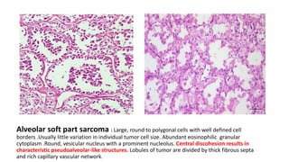
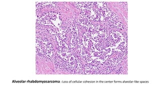
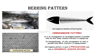
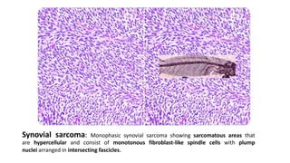
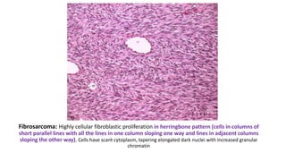
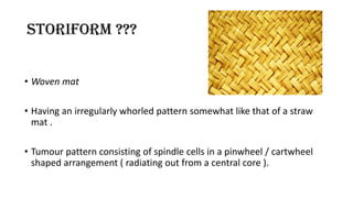
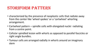
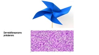
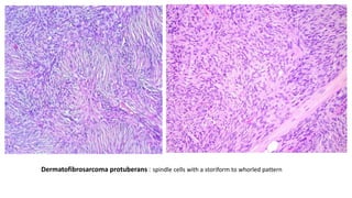
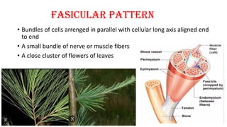
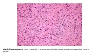
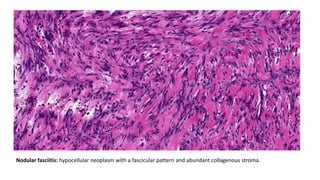
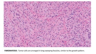
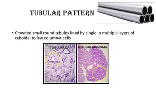
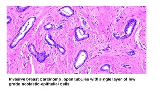
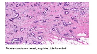
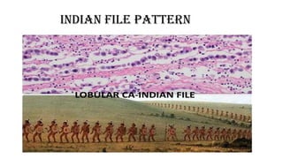
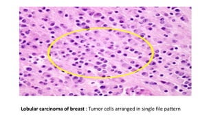
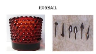
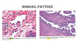
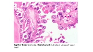
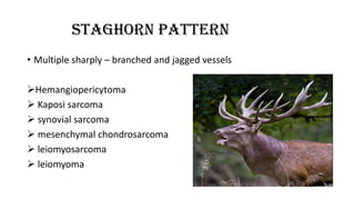
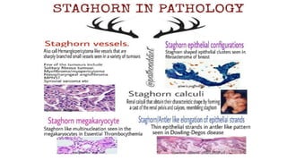
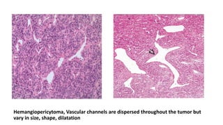
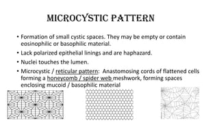
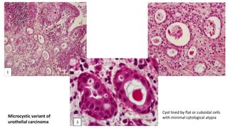
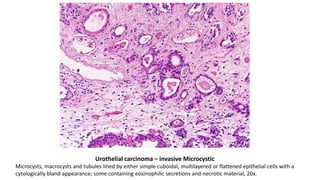
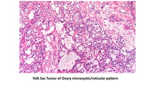
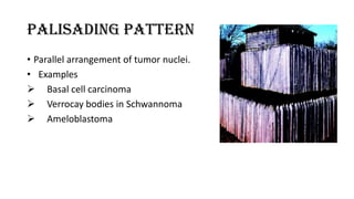
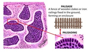
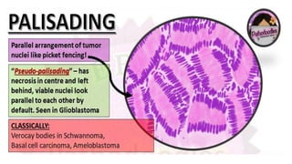
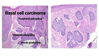
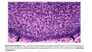
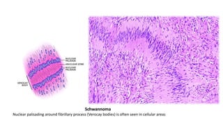
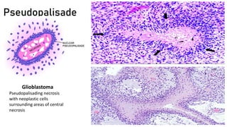
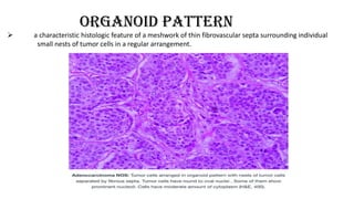
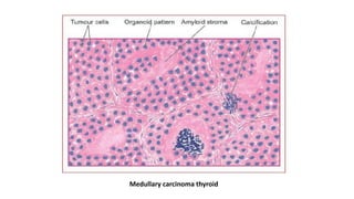
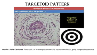
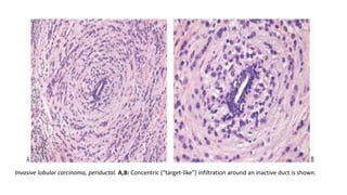
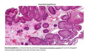
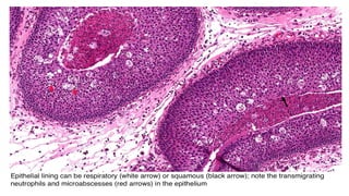
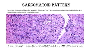
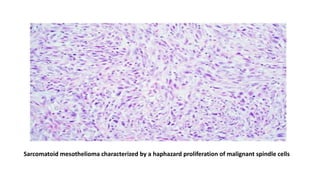
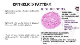
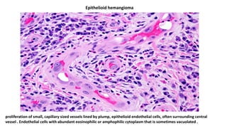
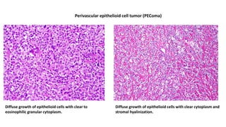
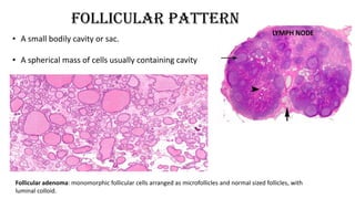
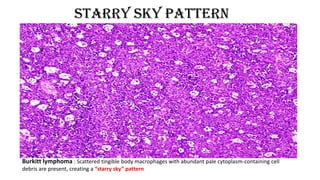
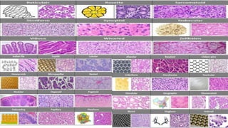
ARCHITECTURAL
PATTERNS IN
HISTOPATHOLOGY
Dr Dharmaraj Vilasrao Pawar
Junior Resident 2
objectives
 By the end of this session you should be able to differentiate
between most of the patterns in histopathology
TRABECULAr pattern
• Trabecula is a Latin word that means
'little beam'.
• The neoplastic epithelium is seen in the
form of strands that is separated from
the connective tissue and forms a
network or trabeculae of variable sizes.
• Two cell thick (microtrabecular pattern)
• Eight to ten cell thick ( macrotrabecular
pattern)
TRABECULAE ??
Trabeculae are the thin columns and plates of bone that create a spongy structure in a cancellous bone, which
is located at the ends of long bones and in the pelvis, ribs, skull, and vertebrae.
WHERE CAN BE SEEN ??
• CARCINOID TUMORS (Most common)
• HEPATOCELLULAR CARCINOMA
• GRANULOSA CELL TUMOR
Pulmonary carcinoid tumor showing trabecular pattern
CARCINOID TUMOR
Poorly differentiated carcinoma of thyroid
Hepatectomy for hepatocellular carcinoma at low power
demonstrating trabecular morphology
Hepatectomy for hepatocellular carcinoma at higher power
demonstrating trabecular morphology and cytologic atypia
GLANDULAR PATTERN
Benign gland pattern Malignant glandular pattern
Yolk sac tumor, endometrioid-like glandular variant
ENDOMETRIUM
Adenocarcinoma of Cervix villoglandular architecture
CRIBRIFORM PATTERN
What is cribriform ???
Sieve like , containing many perforations .
Anatomical structure pierced by numerous small holes
Where can be seen ???
• ADENOID CYSTIC CARCINOMA
• DUCTAL CARCINOMA INSITU BREAST
Cribriform ductal carcinoma in situ breast
Terminal duct lobular unit with intraductal proliferation
Low power view showing pure invasive cribriform carcinoma with > 90% cribriform pattern in
desmoplastic stroma, H&E.
Adenoid cystic carcinoma of salivary gland cribriform pattern
Cribriform pattern is composed predominantly of myoepithelial cells admixed
with hyalinized or myxoid globules. Scattered ductal elements (arrows) may
also been present.
NESTING PATTERN
• The proliferating epithelial cells are present in groups or small nests
• SEEN IN
 UROTHELIAL CARCINOMA
CARCINOID TUMOR
NEURO ENDOCRINE TUMOR
PHEOCROMOCYTOMA
PARAGANGLIOMA
Urothelial carcinoma, large nests
Anastomosing,
irregular, large nested
and inverted growth of
cytologically bland
urothelium with
desmoplastic stromal
reaction (4x).
Urothelial carcinoma, nested pattern Cytologically bland nests of low to
intermediate grade urothelium lacking polarity and with occasional tubular
lumens, forming irregular projections into underlying stroma (10x)
Carcinoid tumor
Invasive ductal carcinoma breast – Nest pattern
ZELLBALLEN PATTERN
• Zellballen meaning “ ball of cells “ in German.
• In Paraganglia the arrangement of chromaffin cells in fascicles and
nests was described “Zellstrangen” and “Zellballen”
• A characteristic pattern found in Paraganglioma where the clusters of
uniform round to polygonal cells are surrounded by delicate vascular
tissue and sustentacular cells.
Pheochromocytomas : In a typical case, the tumor consists of polygonal or spindle cells arranged in small
nests (Zellballen) surrounded by sustentacular cells (best seen with S-100 immunostain). The nests are
separated by a delicate fibrovascular stroma. The nuclei are round or oval and have prominent nucleoli.
Chromogranin immunostain highlights the
distorted zellballen or nested growth of the
neoplastic cells within the sclerosis.
A carotid body paraganglioma shows
encirclement of individual zellballen by fibrous
tissue representing sinusoidal sclerosis.
PAPILLARY PATTERN
• Papillae consist of a central
fibrovascular stalk lined by
neoplastic epithelium formed
by cuboidal, columnar, or tall
follicular cells.
• Complex branching
configuration.
TRUE PAPILLARY
A growth of glandular cells along central
fibrovascular cores
MICROPAPILLARY
FILIGREE CLASSICAL
Delicate lace-like narrow stacks tumor cells growing in papillary tufts forming florets
At least 3 piled up nuclei from the no fibrovascular core
alveolar wall basal layer
Upto three cells in width
No fibrovascular core
MICROPAPILLARY PATTERN
Colon Micropapillary carcinoma
Invasive micropapillary carcinoma of the breast : The tumor is composed of morula-like clusters floating in
empty, clear spaces lined by delicate strands of stroma
High power view of an intraductal epithelial proliferation
with intermediate grade nuclear atypia, micropapillary
growth pattern.
Intermediate power view of an intraductal epithelial
proliferation with intermediate grade nuclear atypia,
micropapillary growth pattern, luminal hemosiderin laden
macrophages and microcalcifications.
Papillary pattern DCIS - breast
Right breast, lumpectomy specimen showing invasive
papillary carcinoma with desmoplastic reaction at the invasive
tumor front without fibrous capsule
Right breast, lumpectomy specimen showing invasive
papillary carcinoma, lacking myoepithelial cells at the
periphery and within the papillae
Hurthle cell neoplasm with a papillary pattern of growth
True papillae have stromal stalk with microvessels, Papillary carcinoma
of thyroid
Condyloma acuminatum, papillary exophytic squamous epithelium
ROSETTE PATTERN
• Round assemblage of cells
found in tumors
• So named for their similarity
to the rose casement found in
gothic cathdrals
• Most are found in tumors of
the CNS
Homer Wright Rosettes:
• Here the rosettes have a dense feltwork of
interwoven cytoplasmic processes of nerve
cells and neuroglial cells( Neuropil) in the
centre.
• This is named after James Homer
Wright an American pathologist(1869 –
1928 )
• These rosettes are seen in
Neuroblastoma
Medulloblastoma
Pineoblastoma
Flexner-Wintersteiner Rosette:
• First described by first described by Simon Flexner an
American Physician and Pathologist and Austrian
ophthalmologist Hugo Wintersteiner. Characteristically seen
in retinoblastomas.
• Here the rosette surround a central lumen containing small
cytoplasmic extensions of the encircling tumor cells. unlike
Homer Wright rosette, the lumen do not have Neuropil.
• The tumor cells forming the Flexner–Wintersteiner rosette
have features of primitive photoreceptor cells when examined
ultrastructurally.
• These are seen in
Retinoblastomas
Pinealobastoma
Medulloepithelioma
Perivascular Pseudo Rosettes :
• These are called “pseudo” these are not true rosettes and the
tumor cells surrounds a blood vessel and hence “perivascular”
These are charactestically seen in ependymomas.
• In this type of rosette, the neuropil is seen projecting towards a
central blood vessel.
• These are seen are
Ependymomas
Central Neurocytoma
Glioblastoma
True ependymal rosettes
True ependymal rosette or ependymal canal with lumen, when present, are often
focal.
Ependymoma
Neuroepithelial tumor with ependymal differentiation that most commonly has
ventricular involvement and arises in the cerebrum and spinal cord
Flexner – Wintersteiner rosettes
Perivascular pseudorosette Pineocytomatous and neurocytic rosette
SYNCiTIAL PATTERN
• Single cell or cytoplasmic mass containing several nuclei, formed by
fusion of cells .
• Having cytoplasmic continuity between the constituent cells
Medullary carcinoma of thyroid, syncytial pattern
Meningothelial meningioma can have a syncytial growth pattern that needs to be separated from sheeted
architecture, which is an atypical feature. Uniform, bland nuclei and abundant cytoplasm support syncytial growth
pattern, while other atypical features such as mitoses, hypercellularity, macronuclei and small cell change support
sheeted architecture.
ALVEOLAR PATTERN
• Tumor cells grow in nests or clusters separated by fibrous septa
• Central cells showing loss of cohesion and individual tumor cell
necrosis is seen between the fibrous septa resulting in a lung like
alveolar spaces
ALVEOLI
Alveolar soft part sarcoma : Large, round to polygonal cells with well defined cell
borders .Usually little variation in individual tumor cell size. Abundant eosinophilic granular
cytoplasm .Round, vesicular nucleus with a prominent nucleolus. Central discohesion results in
characteristic pseudoalveolar-like structures. Lobules of tumor are divided by thick fibrous septa
and rich capillary vascular network.
Alveolar rhabdomyosarcoma : Loss of cellular cohesion in the center forms alveolar-like spaces
HERRING PATTERN
Synovial sarcoma: Monophasic synovial sarcoma showing sarcomatous areas that
are hypercellular and consist of monotonous fibroblast-like spindle cells with plump
nuclei arranged in intersecting fascicles.
Fibrosarcoma: Highly cellular fibroblastic proliferation in herringbone pattern (cells in columns of
short parallel lines with all the lines in one column sloping one way and lines in adjacent columns
sloping the other way). Cells have scant cytoplasm, tapering elongated dark nuclei with increased granular
chromatin
STORIFORM ???
• Woven mat
• Having an irregularly whorled pattern somewhat like that of a straw
mat .
• Tumour pattern consisting of spindle cells in a pinwheel / cartwheel
shaped arrangement ( radiating out from a central core ).
STORIFORM PATTERN
• characterized by the presence of neoplastic cells that radiate away
from the center like 'wheel spokes' or a 'cartwheel' whorling
arrangement.
• Cartwheel pattern – spindle cells with elongated nuclei radiating
from a centre point.
• Cellular spindled lesion with whorls as opposed to parallel fascicles or
right angle bundles.
• Tumour cells are arranged radially in whorls around an imaginary
stem
Dermatofibrosarcoma
protuberans
Dermatofibrosarcoma protuberans : spindle cells with a storiform to whorled pattern
FASICULAR PATTERN
• Bundles of cells arrenged in parallel with cellular long axis aligned end
to end
• A small bundle of nerve or muscle fibers
• A close cluster of flowers of leaves
Classic leiomyosarcoma: Tumor cells are set in long intersecting fascicles parallel and perpendicular to the plane of
section.
Nodular fasciitis: hypocellular neoplasm with a fascicular pattern and abundant collagenous stroma.
FIBROMATOSIS : Tumor cells are arranged in long sweeping fascicles, similar to the growth pattern.
TUBULAR PATTERN
• Crowded small round tubules lined by single to multiple layers of
cuboidal to low columnar cells
Invasive breast carcinoma, open tubules with single layer of low
grade neolastic epithelial cells
Tubular carcinoma breast, angulated tubules noted
INDIAN FILE PATTERN
Lobular carcinoma of breast : Tumor cells arranged in single file pattern
HOBNAIL
HOBNAIL PATTERN
Papillary thyroid carcinoma , Hobnail varient : Hobnail cells with apically placed
nuclei
STAGHORN PATTERN
• Multiple sharply – branched and jagged vessels
Hemangiopericytoma
 Kaposi sarcoma
 synovial sarcoma
 mesenchymal chondrosarcoma
 leiomyosarcoma
 leiomyoma
Hemangiopericytoma, Vascular channels are dispersed throughout the tumor but
vary in size, shape, dilatation
Solitary fibrous tumor of breast
MICROCYSTIC PATTERN
• Formation of small cystic spaces. They may be empty or contain
eosinophilic or basophilic material.
• Lack polarized epithelial linings and are haphazard.
• Nuclei touches the lumen.
• Microcystic / reticular pattern: Anastomosing cords of flattened cells
forming a honeycomb / spider web meshwork, forming spaces
enclosing mucoid / basophilic material
Cyst lined by flat or cuboidal cells
with minimal cytological atypia
Microcystic variant of
urothelial carcinoma
Urothelial carcinoma – invasive Microcystic
Microcysts, macrocysts and tubules lined by either simple cuboidal, multilayered or flattened epithelial cells with a
cytologically bland appearance; some containing eosinophilic secretions and necrotic material, 20x.
Yolk Sac Tumor of Ovary microcystic/reticular pattern
PALISADING PATTERN
• Parallel arrangement of tumor nuclei.
• Examples
 Basal cell carcinoma
 Verrocay bodies in Schwannoma
 Ameloblastoma
Basal cell carcinoma: these cells have scanty cytoplasm, elongated hyperchromatic nuclei. At the periphery of
these nests, the cells are radially arranged with long axis parallel to each other. This is referred to as “ palisading
arrangement”
Schwannoma
Nuclear palisading around fibrillary process (Verocay bodies) is often seen in cellular areas
Pseudopalisade
Glioblastoma
Pseudopalisading necrosis
with neoplastic cells
surrounding areas of central
necrosis
ORGANOID PATTERN
 a characteristic histologic feature of a meshwork of thin fibrovascular septa surrounding individual
small nests of tumor cells in a regular arrangement.
Medullary carcinoma thyroid
Targetoid pattern
Invasive Lobular Carcinoma: Tumor cells can be arranged concentrically around normal ducts, giving a targetoid appearance
Invasive lobular carcinoma, periductal. A,B: Concentric (“target-like”) infiltration around an inactive duct is shown.
Comedo pattern
Ductal carcinoma in situ : Intermediate power view of an intraductal epithelial proliferation with
intermediate grade nuclear atypia, cribriform growth pattern, comedo necrosis and microcalcifications.
Comedo : Central expansile necrosis containing cellular debris, generally associated with high grade DCIS,
frequently associated with coarse microcalcifications
Ductal carcinoma in situ : Medium power view of an intraductal epithelial proliferation with high grade
nuclear atypia , flat growth pattern and central necrosis.
Salivary duct carcinoma
Inverted papilloma is characterized by prominent downward endophytic growth of round to elongated
interconnected epithelial nests with smooth outer contour
Inverted papilloma
Sarcomatoid pattern
comprised of spindle-shaped cells arranged in sheets or fascicles that form nonspecific architectural patterns
that resemble those seen in various sarcomas
10x photomicrograph of sarcomatoid spindle cell dedifferentiation in a RCC with fascicular growth.
Sarcomatoid mesothelioma characterized by a haphazard proliferation of malignant spindle cells
Epithelioid pattern
 Epithelioid morphology refers to resemblance to
epithelial cells.
 Epithelioid cells usually feature a polygonal
shape and exhibit abundant cytoplasm.
 These can show variable growth patterns as
solid, nesting, sheet-like, cord-like, or glandular-
like.
proliferation of small, capillary sized vessels lined by plump, epithelioid endothelial cells, often surrounding central
vessel . Endothelial cells with abundant eosinophilic or amphophilic cytoplasm that is sometimes vacuolated .
Epithelioid hemangioma
Perivascular epithelioid cell tumor (PEComa)
Diffuse growth of epithelioid cells with clear to
eosinophilic granular cytoplasm.
Diffuse growth of epithelioid cells with clear cytoplasm and
stromal hyalinization.
Follicular pattern
• A small bodily cavity or sac.
• A spherical mass of cells usually containing cavity
Follicular adenoma: monomorphic follicular cells arranged as microfollicles and normal sized follicles, with
luminal colloid.
Burkitt lymphoma : Scattered tingible body macrophages with abundant pale cytoplasm-containing cell
debris are present, creating a “starry sky” pattern
Starry sky pattern
REFERENCES
• Robbins and Cotron: Pathologic Basis of Disease, Tenth edition
• Mills and Sternbergs: Diagnostic surgical Pathology, Seventh edition
• Rosai and Ackerman’s: Surgical pathology, eleventh edition
• WHO classification of tumors, Female genital tumors, Fifth edition
• WHO classification of tumors, Breast tumors, Fifth edition

ARCHITECTURAL PATTERNS IN HISTOPATHOLOGY pdf- [Autosaved].pdf

  • 1.
  • 2.
    objectives  By theend of this session you should be able to differentiate between most of the patterns in histopathology
  • 3.
    TRABECULAr pattern • Trabeculais a Latin word that means 'little beam'. • The neoplastic epithelium is seen in the form of strands that is separated from the connective tissue and forms a network or trabeculae of variable sizes. • Two cell thick (microtrabecular pattern) • Eight to ten cell thick ( macrotrabecular pattern)
  • 4.
    TRABECULAE ?? Trabeculae arethe thin columns and plates of bone that create a spongy structure in a cancellous bone, which is located at the ends of long bones and in the pelvis, ribs, skull, and vertebrae.
  • 5.
    WHERE CAN BESEEN ?? • CARCINOID TUMORS (Most common) • HEPATOCELLULAR CARCINOMA • GRANULOSA CELL TUMOR
  • 6.
    Pulmonary carcinoid tumorshowing trabecular pattern
  • 7.
  • 8.
  • 9.
    Hepatectomy for hepatocellularcarcinoma at low power demonstrating trabecular morphology Hepatectomy for hepatocellular carcinoma at higher power demonstrating trabecular morphology and cytologic atypia
  • 10.
  • 11.
    Benign gland patternMalignant glandular pattern
  • 12.
    Yolk sac tumor,endometrioid-like glandular variant ENDOMETRIUM
  • 13.
    Adenocarcinoma of Cervixvilloglandular architecture
  • 14.
    CRIBRIFORM PATTERN What iscribriform ??? Sieve like , containing many perforations . Anatomical structure pierced by numerous small holes
  • 16.
    Where can beseen ??? • ADENOID CYSTIC CARCINOMA • DUCTAL CARCINOMA INSITU BREAST
  • 17.
  • 18.
    Terminal duct lobularunit with intraductal proliferation
  • 19.
    Low power viewshowing pure invasive cribriform carcinoma with > 90% cribriform pattern in desmoplastic stroma, H&E.
  • 20.
    Adenoid cystic carcinomaof salivary gland cribriform pattern Cribriform pattern is composed predominantly of myoepithelial cells admixed with hyalinized or myxoid globules. Scattered ductal elements (arrows) may also been present.
  • 21.
    NESTING PATTERN • Theproliferating epithelial cells are present in groups or small nests • SEEN IN  UROTHELIAL CARCINOMA CARCINOID TUMOR NEURO ENDOCRINE TUMOR PHEOCROMOCYTOMA PARAGANGLIOMA
  • 22.
    Urothelial carcinoma, largenests Anastomosing, irregular, large nested and inverted growth of cytologically bland urothelium with desmoplastic stromal reaction (4x).
  • 23.
    Urothelial carcinoma, nestedpattern Cytologically bland nests of low to intermediate grade urothelium lacking polarity and with occasional tubular lumens, forming irregular projections into underlying stroma (10x)
  • 24.
  • 25.
    Invasive ductal carcinomabreast – Nest pattern
  • 26.
    ZELLBALLEN PATTERN • Zellballenmeaning “ ball of cells “ in German. • In Paraganglia the arrangement of chromaffin cells in fascicles and nests was described “Zellstrangen” and “Zellballen” • A characteristic pattern found in Paraganglioma where the clusters of uniform round to polygonal cells are surrounded by delicate vascular tissue and sustentacular cells.
  • 28.
    Pheochromocytomas : Ina typical case, the tumor consists of polygonal or spindle cells arranged in small nests (Zellballen) surrounded by sustentacular cells (best seen with S-100 immunostain). The nests are separated by a delicate fibrovascular stroma. The nuclei are round or oval and have prominent nucleoli.
  • 29.
    Chromogranin immunostain highlightsthe distorted zellballen or nested growth of the neoplastic cells within the sclerosis. A carotid body paraganglioma shows encirclement of individual zellballen by fibrous tissue representing sinusoidal sclerosis.
  • 30.
    PAPILLARY PATTERN • Papillaeconsist of a central fibrovascular stalk lined by neoplastic epithelium formed by cuboidal, columnar, or tall follicular cells. • Complex branching configuration.
  • 32.
    TRUE PAPILLARY A growthof glandular cells along central fibrovascular cores MICROPAPILLARY FILIGREE CLASSICAL Delicate lace-like narrow stacks tumor cells growing in papillary tufts forming florets At least 3 piled up nuclei from the no fibrovascular core alveolar wall basal layer Upto three cells in width No fibrovascular core
  • 34.
  • 35.
    Invasive micropapillary carcinomaof the breast : The tumor is composed of morula-like clusters floating in empty, clear spaces lined by delicate strands of stroma
  • 36.
    High power viewof an intraductal epithelial proliferation with intermediate grade nuclear atypia, micropapillary growth pattern. Intermediate power view of an intraductal epithelial proliferation with intermediate grade nuclear atypia, micropapillary growth pattern, luminal hemosiderin laden macrophages and microcalcifications.
  • 37.
  • 38.
    Right breast, lumpectomyspecimen showing invasive papillary carcinoma with desmoplastic reaction at the invasive tumor front without fibrous capsule Right breast, lumpectomy specimen showing invasive papillary carcinoma, lacking myoepithelial cells at the periphery and within the papillae
  • 39.
    Hurthle cell neoplasmwith a papillary pattern of growth
  • 40.
    True papillae havestromal stalk with microvessels, Papillary carcinoma of thyroid
  • 41.
    Condyloma acuminatum, papillaryexophytic squamous epithelium
  • 42.
    ROSETTE PATTERN • Roundassemblage of cells found in tumors • So named for their similarity to the rose casement found in gothic cathdrals • Most are found in tumors of the CNS
  • 44.
    Homer Wright Rosettes: •Here the rosettes have a dense feltwork of interwoven cytoplasmic processes of nerve cells and neuroglial cells( Neuropil) in the centre. • This is named after James Homer Wright an American pathologist(1869 – 1928 ) • These rosettes are seen in Neuroblastoma Medulloblastoma Pineoblastoma
  • 45.
    Flexner-Wintersteiner Rosette: • Firstdescribed by first described by Simon Flexner an American Physician and Pathologist and Austrian ophthalmologist Hugo Wintersteiner. Characteristically seen in retinoblastomas. • Here the rosette surround a central lumen containing small cytoplasmic extensions of the encircling tumor cells. unlike Homer Wright rosette, the lumen do not have Neuropil. • The tumor cells forming the Flexner–Wintersteiner rosette have features of primitive photoreceptor cells when examined ultrastructurally. • These are seen in Retinoblastomas Pinealobastoma Medulloepithelioma
  • 46.
    Perivascular Pseudo Rosettes: • These are called “pseudo” these are not true rosettes and the tumor cells surrounds a blood vessel and hence “perivascular” These are charactestically seen in ependymomas. • In this type of rosette, the neuropil is seen projecting towards a central blood vessel. • These are seen are Ependymomas Central Neurocytoma Glioblastoma
  • 48.
    True ependymal rosettes Trueependymal rosette or ependymal canal with lumen, when present, are often focal. Ependymoma Neuroepithelial tumor with ependymal differentiation that most commonly has ventricular involvement and arises in the cerebrum and spinal cord
  • 50.
  • 51.
  • 52.
    SYNCiTIAL PATTERN • Singlecell or cytoplasmic mass containing several nuclei, formed by fusion of cells . • Having cytoplasmic continuity between the constituent cells
  • 53.
    Medullary carcinoma ofthyroid, syncytial pattern
  • 54.
    Meningothelial meningioma canhave a syncytial growth pattern that needs to be separated from sheeted architecture, which is an atypical feature. Uniform, bland nuclei and abundant cytoplasm support syncytial growth pattern, while other atypical features such as mitoses, hypercellularity, macronuclei and small cell change support sheeted architecture.
  • 55.
    ALVEOLAR PATTERN • Tumorcells grow in nests or clusters separated by fibrous septa • Central cells showing loss of cohesion and individual tumor cell necrosis is seen between the fibrous septa resulting in a lung like alveolar spaces
  • 56.
  • 57.
    Alveolar soft partsarcoma : Large, round to polygonal cells with well defined cell borders .Usually little variation in individual tumor cell size. Abundant eosinophilic granular cytoplasm .Round, vesicular nucleus with a prominent nucleolus. Central discohesion results in characteristic pseudoalveolar-like structures. Lobules of tumor are divided by thick fibrous septa and rich capillary vascular network.
  • 58.
    Alveolar rhabdomyosarcoma :Loss of cellular cohesion in the center forms alveolar-like spaces
  • 59.
  • 60.
    Synovial sarcoma: Monophasicsynovial sarcoma showing sarcomatous areas that are hypercellular and consist of monotonous fibroblast-like spindle cells with plump nuclei arranged in intersecting fascicles.
  • 61.
    Fibrosarcoma: Highly cellularfibroblastic proliferation in herringbone pattern (cells in columns of short parallel lines with all the lines in one column sloping one way and lines in adjacent columns sloping the other way). Cells have scant cytoplasm, tapering elongated dark nuclei with increased granular chromatin
  • 62.
    STORIFORM ??? • Wovenmat • Having an irregularly whorled pattern somewhat like that of a straw mat . • Tumour pattern consisting of spindle cells in a pinwheel / cartwheel shaped arrangement ( radiating out from a central core ).
  • 63.
    STORIFORM PATTERN • characterizedby the presence of neoplastic cells that radiate away from the center like 'wheel spokes' or a 'cartwheel' whorling arrangement. • Cartwheel pattern – spindle cells with elongated nuclei radiating from a centre point. • Cellular spindled lesion with whorls as opposed to parallel fascicles or right angle bundles. • Tumour cells are arranged radially in whorls around an imaginary stem
  • 64.
  • 65.
    Dermatofibrosarcoma protuberans :spindle cells with a storiform to whorled pattern
  • 66.
    FASICULAR PATTERN • Bundlesof cells arrenged in parallel with cellular long axis aligned end to end • A small bundle of nerve or muscle fibers • A close cluster of flowers of leaves
  • 67.
    Classic leiomyosarcoma: Tumorcells are set in long intersecting fascicles parallel and perpendicular to the plane of section.
  • 68.
    Nodular fasciitis: hypocellularneoplasm with a fascicular pattern and abundant collagenous stroma.
  • 69.
    FIBROMATOSIS : Tumorcells are arranged in long sweeping fascicles, similar to the growth pattern.
  • 70.
    TUBULAR PATTERN • Crowdedsmall round tubules lined by single to multiple layers of cuboidal to low columnar cells
  • 71.
    Invasive breast carcinoma,open tubules with single layer of low grade neolastic epithelial cells
  • 72.
    Tubular carcinoma breast,angulated tubules noted
  • 73.
  • 74.
    Lobular carcinoma ofbreast : Tumor cells arranged in single file pattern
  • 75.
  • 76.
  • 77.
    Papillary thyroid carcinoma, Hobnail varient : Hobnail cells with apically placed nuclei
  • 78.
    STAGHORN PATTERN • Multiplesharply – branched and jagged vessels Hemangiopericytoma  Kaposi sarcoma  synovial sarcoma  mesenchymal chondrosarcoma  leiomyosarcoma  leiomyoma
  • 80.
    Hemangiopericytoma, Vascular channelsare dispersed throughout the tumor but vary in size, shape, dilatation
  • 81.
  • 82.
    MICROCYSTIC PATTERN • Formationof small cystic spaces. They may be empty or contain eosinophilic or basophilic material. • Lack polarized epithelial linings and are haphazard. • Nuclei touches the lumen. • Microcystic / reticular pattern: Anastomosing cords of flattened cells forming a honeycomb / spider web meshwork, forming spaces enclosing mucoid / basophilic material
  • 83.
    Cyst lined byflat or cuboidal cells with minimal cytological atypia Microcystic variant of urothelial carcinoma
  • 84.
    Urothelial carcinoma –invasive Microcystic Microcysts, macrocysts and tubules lined by either simple cuboidal, multilayered or flattened epithelial cells with a cytologically bland appearance; some containing eosinophilic secretions and necrotic material, 20x.
  • 85.
    Yolk Sac Tumorof Ovary microcystic/reticular pattern
  • 86.
    PALISADING PATTERN • Parallelarrangement of tumor nuclei. • Examples  Basal cell carcinoma  Verrocay bodies in Schwannoma  Ameloblastoma
  • 90.
    Basal cell carcinoma:these cells have scanty cytoplasm, elongated hyperchromatic nuclei. At the periphery of these nests, the cells are radially arranged with long axis parallel to each other. This is referred to as “ palisading arrangement”
  • 91.
    Schwannoma Nuclear palisading aroundfibrillary process (Verocay bodies) is often seen in cellular areas
  • 92.
  • 93.
    ORGANOID PATTERN  acharacteristic histologic feature of a meshwork of thin fibrovascular septa surrounding individual small nests of tumor cells in a regular arrangement.
  • 94.
  • 95.
    Targetoid pattern Invasive LobularCarcinoma: Tumor cells can be arranged concentrically around normal ducts, giving a targetoid appearance
  • 96.
    Invasive lobular carcinoma,periductal. A,B: Concentric (“target-like”) infiltration around an inactive duct is shown.
  • 97.
    Comedo pattern Ductal carcinomain situ : Intermediate power view of an intraductal epithelial proliferation with intermediate grade nuclear atypia, cribriform growth pattern, comedo necrosis and microcalcifications. Comedo : Central expansile necrosis containing cellular debris, generally associated with high grade DCIS, frequently associated with coarse microcalcifications
  • 98.
    Ductal carcinoma insitu : Medium power view of an intraductal epithelial proliferation with high grade nuclear atypia , flat growth pattern and central necrosis.
  • 100.
  • 101.
    Inverted papilloma ischaracterized by prominent downward endophytic growth of round to elongated interconnected epithelial nests with smooth outer contour Inverted papilloma
  • 103.
    Sarcomatoid pattern comprised ofspindle-shaped cells arranged in sheets or fascicles that form nonspecific architectural patterns that resemble those seen in various sarcomas 10x photomicrograph of sarcomatoid spindle cell dedifferentiation in a RCC with fascicular growth.
  • 104.
    Sarcomatoid mesothelioma characterizedby a haphazard proliferation of malignant spindle cells
  • 105.
    Epithelioid pattern  Epithelioidmorphology refers to resemblance to epithelial cells.  Epithelioid cells usually feature a polygonal shape and exhibit abundant cytoplasm.  These can show variable growth patterns as solid, nesting, sheet-like, cord-like, or glandular- like.
  • 106.
    proliferation of small,capillary sized vessels lined by plump, epithelioid endothelial cells, often surrounding central vessel . Endothelial cells with abundant eosinophilic or amphophilic cytoplasm that is sometimes vacuolated . Epithelioid hemangioma
  • 107.
    Perivascular epithelioid celltumor (PEComa) Diffuse growth of epithelioid cells with clear to eosinophilic granular cytoplasm. Diffuse growth of epithelioid cells with clear cytoplasm and stromal hyalinization.
  • 108.
    Follicular pattern • Asmall bodily cavity or sac. • A spherical mass of cells usually containing cavity Follicular adenoma: monomorphic follicular cells arranged as microfollicles and normal sized follicles, with luminal colloid.
  • 109.
    Burkitt lymphoma :Scattered tingible body macrophages with abundant pale cytoplasm-containing cell debris are present, creating a “starry sky” pattern Starry sky pattern
  • 112.
    REFERENCES • Robbins andCotron: Pathologic Basis of Disease, Tenth edition • Mills and Sternbergs: Diagnostic surgical Pathology, Seventh edition • Rosai and Ackerman’s: Surgical pathology, eleventh edition • WHO classification of tumors, Female genital tumors, Fifth edition • WHO classification of tumors, Breast tumors, Fifth edition