
Download to read offline

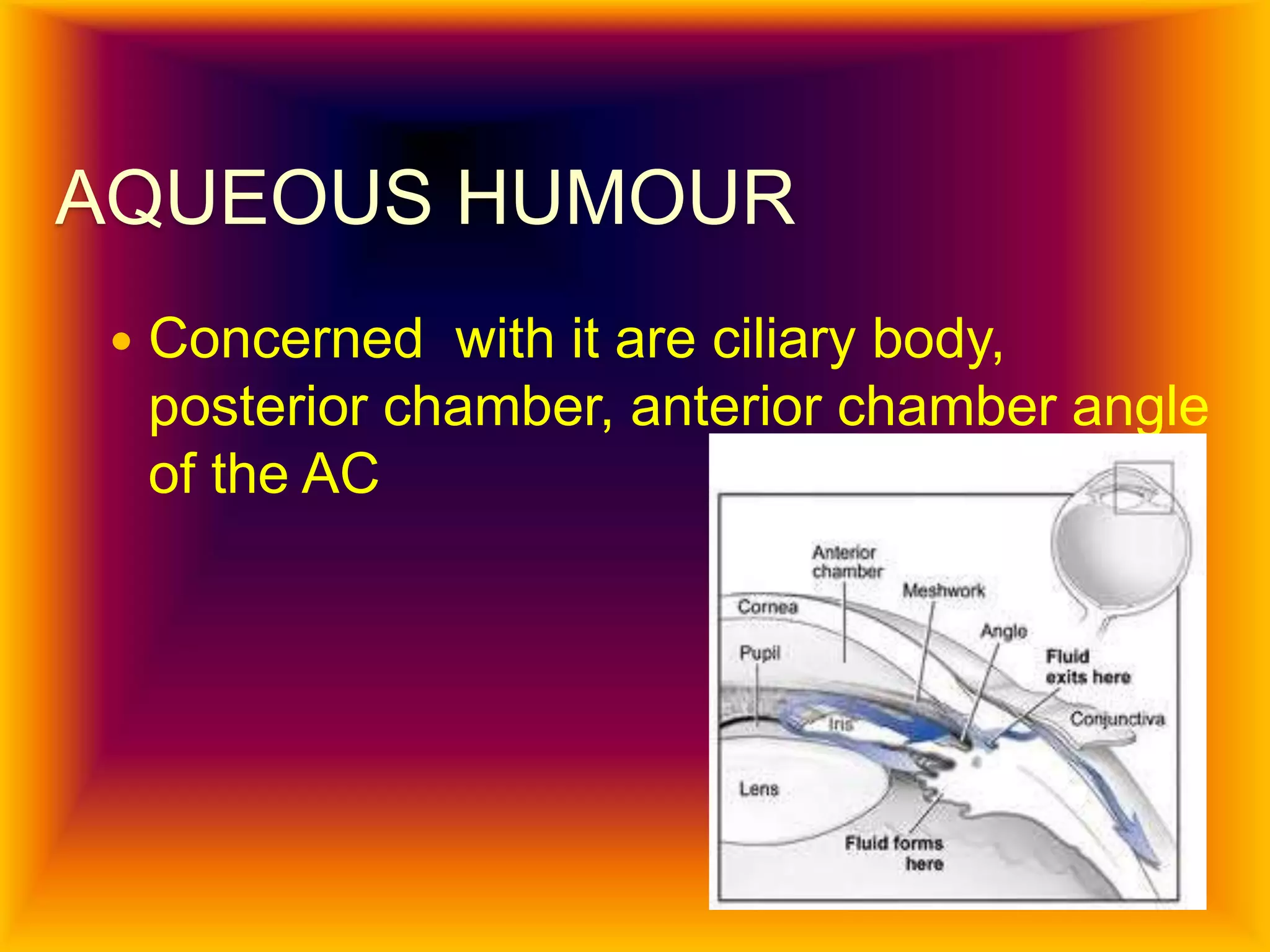


The document summarizes the anatomy and physiology of the aqueous humour in the eye. It describes how the ciliary body produces aqueous humour which flows into the posterior chamber. From there it passes through the pupil into the anterior chamber and exits through the trabecular meshwork into Schlemm's canal and collector channels for drainage into the episcleral veins. It maintains intraocular pressure and the optical and metabolic functions of the eye.


