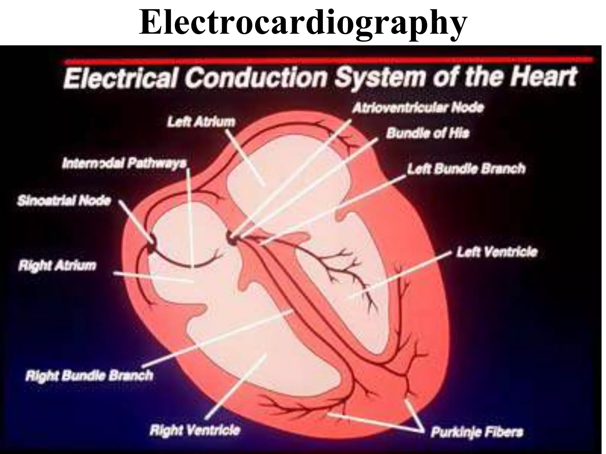
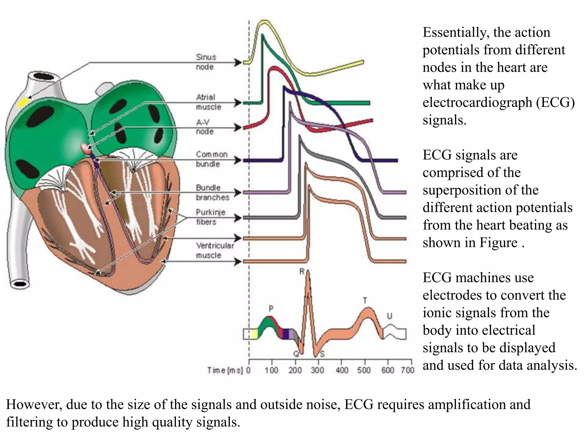
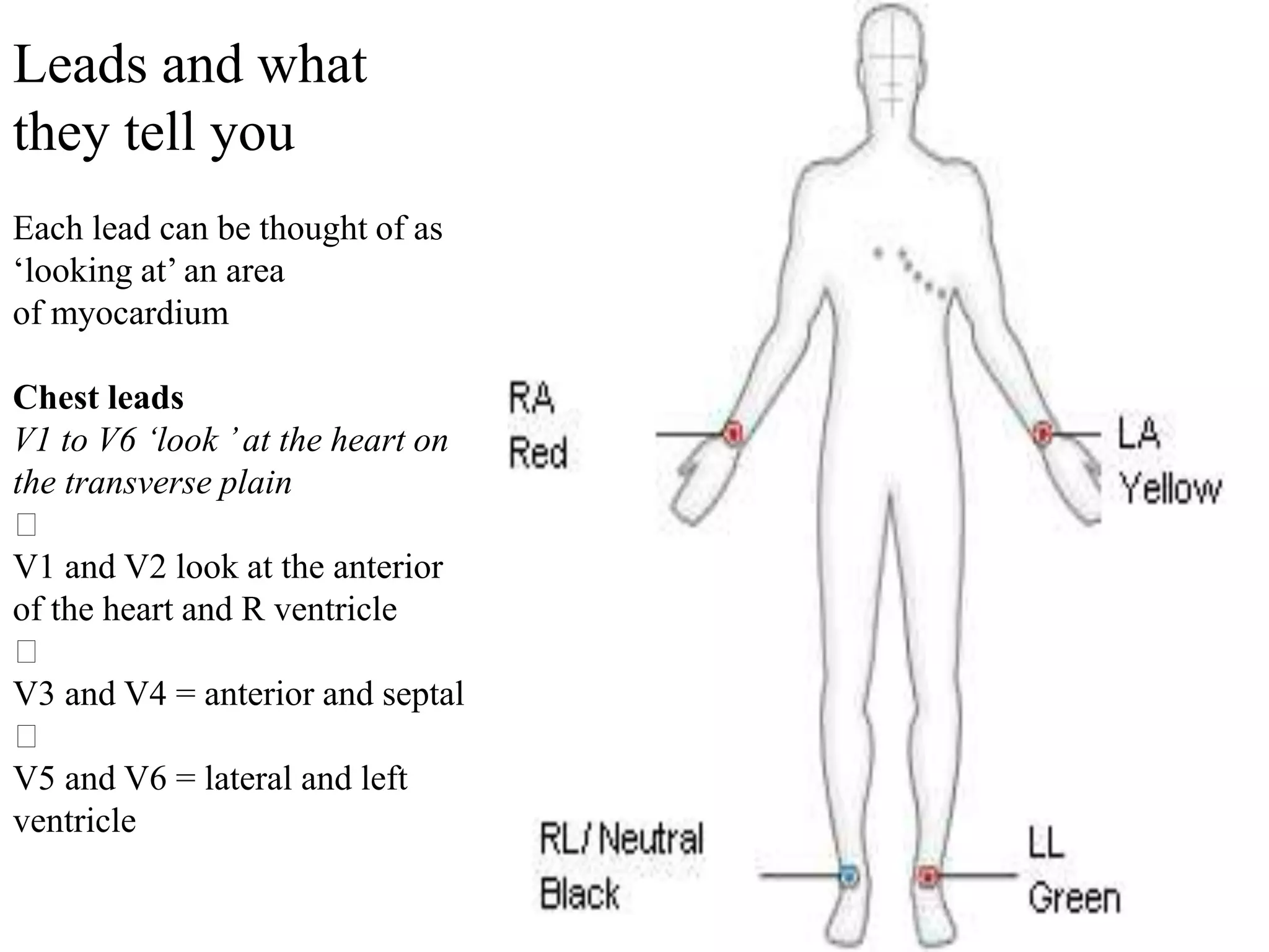
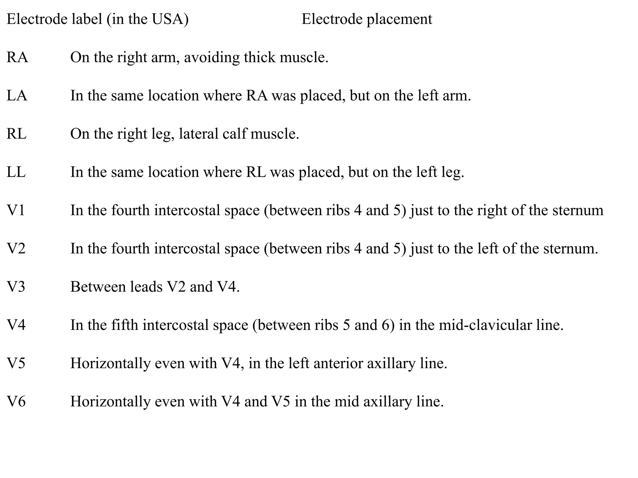
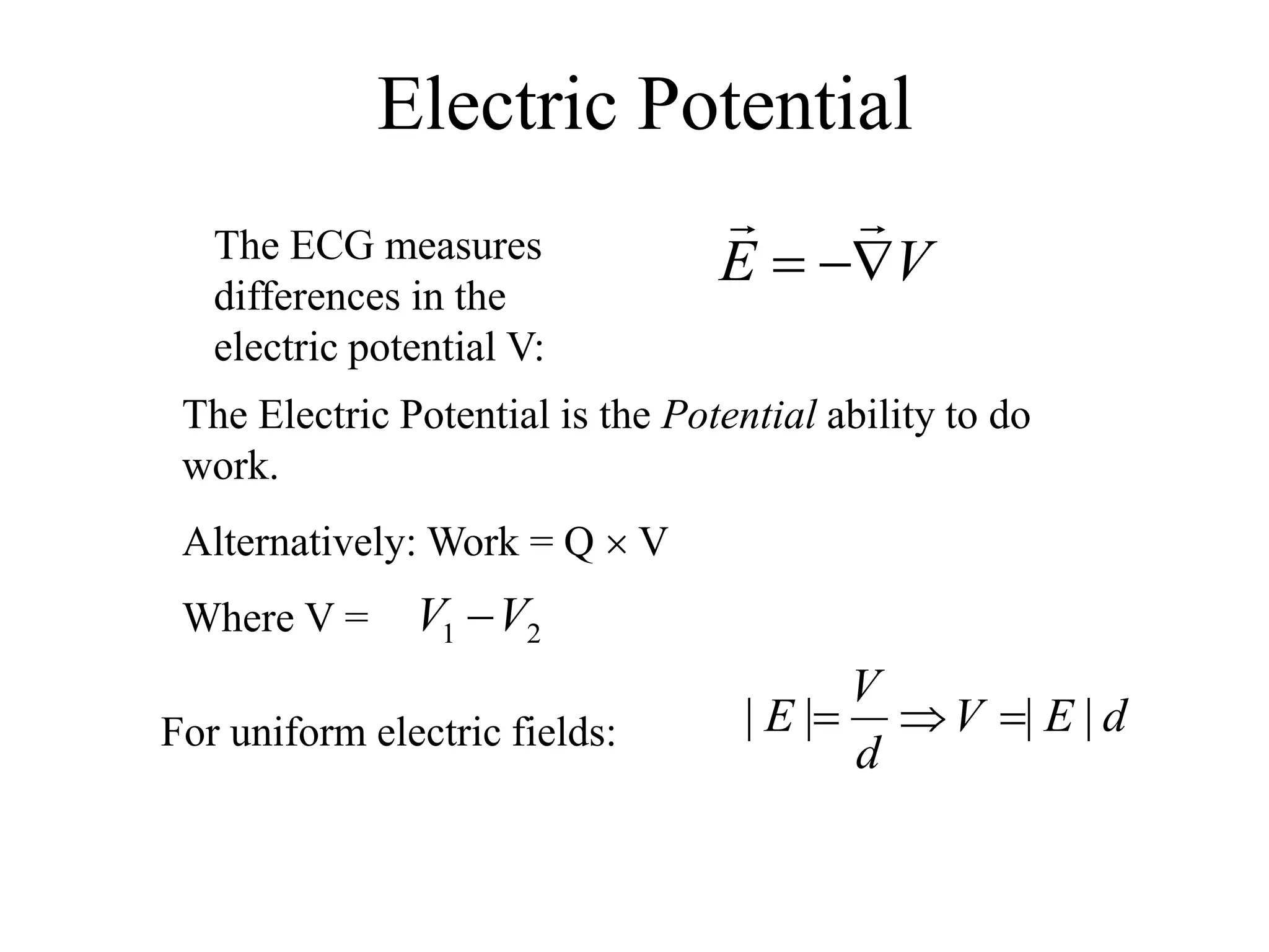
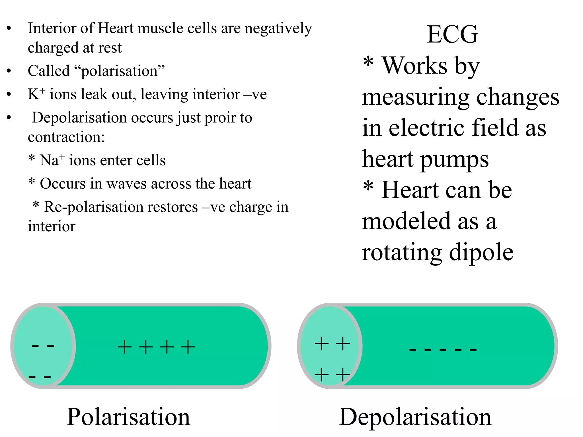
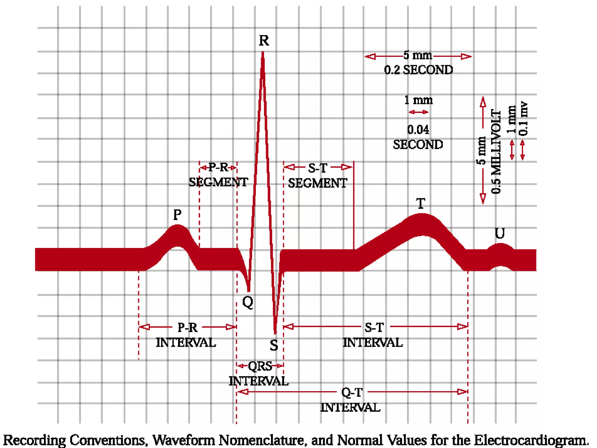
Electrocardiography
Cells in humans act like little batteries. These cells have different ion
concentrations inside and outside of their membranes which create
small electric potentials called biopotentials. When there is a
disturbance in a biopotential this gives rise to an action potential which
is the depolarization and repolarization of the cell
Essentially, the action
potentials from different
nodes in the heart are
what make up
electrocardiograph (ECG)
signals.
ECG signals are
comprised of the
superposition of the
different action potentials
from the heart beating as
shown in Figure .
ECG machines use
electrodes to convert the
ionic signals from the
body into electrical
signals to be displayed
and used for data analysis.
However, due to the size of the signals and outside noise, ECG requires amplification and
filtering to produce high quality signals.
Leads and what
they tell you
Each lead can be thought of as
‘looking at’ an area
of myocardium
Chest leads
V1 to V6 ‘look ’at the heart on
the transverse plain
􀂃
V1 and V2 look at the anterior
of the heart and R ventricle
􀂃
V3 and V4 = anterior and septal
􀂃
V5 and V6 = lateral and left
ventricle
Electrode label (in the USA) Electrode placement
RA On the right arm, avoiding thick muscle.
LA In the same location where RA was placed, but on the left arm.
RL On the right leg, lateral calf muscle.
LL In the same location where RL was placed, but on the left leg.
V1 In the fourth intercostal space (between ribs 4 and 5) just to the right of the sternum
V2 In the fourth intercostal space (between ribs 4 and 5) just to the left of the sternum.
V3 Between leads V2 and V4.
V4 In the fifth intercostal space (between ribs 5 and 6) in the mid-clavicular line.
V5 Horizontally even with V4, in the left anterior axillary line.
V6 Horizontally even with V4 and V5 in the mid axillary line.
The ECG measures
differences in the
electric potential V:
VE 

The Electric Potential is the Potential ability to do
work.
Alternatively: Work = Q  V
Where V = 21 VV 
For uniform electric fields: dEV
d
V
E |||| 
Electric Potential
• Interior of Heart muscle cells are negatively
charged at rest
• Called “polarisation”
• K+ ions leak out, leaving interior –ve
• Depolarisation occurs just proir to
contraction:
* Na+ ions enter cells
* Occurs in waves across the heart
* Re-polarisation restores –ve charge in
interior
+ + + + - - - - -+ +
+ +
- -
- -
Polarisation Depolarisation
ECG
* Works by
measuring changes
in electric field as
heart pumps
* Heart can be
modeled as a
rotating dipole
Depolarisation begins at the SA node
The wave of depolarisation spreads across the atria
It reaches the AV node and the accessory bundle
Conduction is delayed as usual by the in-built delay in the AV node
However, the accessory bundle has no such delay and depolarisation begins
early in the part of the ventricle served by the bundle
As the depolarisation in this part of the ventricle does not travel in the high
speed conduction pathway, the spread of depolarisation across the ventricle is
slow, causing a slow rising delta wave
Until rapid depolarisation resumes via the normal pathway and a more
normal complex follows
How does the ECG work?
􀂃
Electrical impulse (wave of depolarisation) picked
up by placing electrodes on patient
􀂃
The voltage change is sensed by measuring the
current change across 2 electrodes – a positive
electrode and a negative electrode
􀂃If the electrical impulse travels towards the
positive electrode this results in a positive
deflection
􀂃
If the impulse travels away from the positive
electrode this results in a negative deflection
Apps of electrostatistics

Apps of electrostatistics

  • 2.
  • 3.
    Cells in humansact like little batteries. These cells have different ion concentrations inside and outside of their membranes which create small electric potentials called biopotentials. When there is a disturbance in a biopotential this gives rise to an action potential which is the depolarization and repolarization of the cell
  • 4.
    Essentially, the action potentialsfrom different nodes in the heart are what make up electrocardiograph (ECG) signals. ECG signals are comprised of the superposition of the different action potentials from the heart beating as shown in Figure . ECG machines use electrodes to convert the ionic signals from the body into electrical signals to be displayed and used for data analysis. However, due to the size of the signals and outside noise, ECG requires amplification and filtering to produce high quality signals.
  • 5.
    Leads and what theytell you Each lead can be thought of as ‘looking at’ an area of myocardium Chest leads V1 to V6 ‘look ’at the heart on the transverse plain 􀂃 V1 and V2 look at the anterior of the heart and R ventricle 􀂃 V3 and V4 = anterior and septal 􀂃 V5 and V6 = lateral and left ventricle
  • 6.
    Electrode label (inthe USA) Electrode placement RA On the right arm, avoiding thick muscle. LA In the same location where RA was placed, but on the left arm. RL On the right leg, lateral calf muscle. LL In the same location where RL was placed, but on the left leg. V1 In the fourth intercostal space (between ribs 4 and 5) just to the right of the sternum V2 In the fourth intercostal space (between ribs 4 and 5) just to the left of the sternum. V3 Between leads V2 and V4. V4 In the fifth intercostal space (between ribs 5 and 6) in the mid-clavicular line. V5 Horizontally even with V4, in the left anterior axillary line. V6 Horizontally even with V4 and V5 in the mid axillary line.
  • 7.
    The ECG measures differencesin the electric potential V: VE   The Electric Potential is the Potential ability to do work. Alternatively: Work = Q  V Where V = 21 VV  For uniform electric fields: dEV d V E ||||  Electric Potential
  • 8.
    • Interior ofHeart muscle cells are negatively charged at rest • Called “polarisation” • K+ ions leak out, leaving interior –ve • Depolarisation occurs just proir to contraction: * Na+ ions enter cells * Occurs in waves across the heart * Re-polarisation restores –ve charge in interior + + + + - - - - -+ + + + - - - - Polarisation Depolarisation ECG * Works by measuring changes in electric field as heart pumps * Heart can be modeled as a rotating dipole
  • 9.
    Depolarisation begins atthe SA node The wave of depolarisation spreads across the atria It reaches the AV node and the accessory bundle Conduction is delayed as usual by the in-built delay in the AV node However, the accessory bundle has no such delay and depolarisation begins early in the part of the ventricle served by the bundle As the depolarisation in this part of the ventricle does not travel in the high speed conduction pathway, the spread of depolarisation across the ventricle is slow, causing a slow rising delta wave Until rapid depolarisation resumes via the normal pathway and a more normal complex follows
  • 11.
    How does theECG work? 􀂃 Electrical impulse (wave of depolarisation) picked up by placing electrodes on patient 􀂃 The voltage change is sensed by measuring the current change across 2 electrodes – a positive electrode and a negative electrode 􀂃If the electrical impulse travels towards the positive electrode this results in a positive deflection 􀂃 If the impulse travels away from the positive electrode this results in a negative deflection