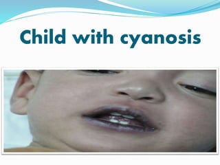
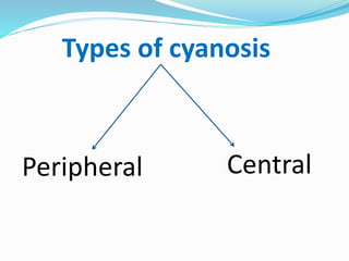
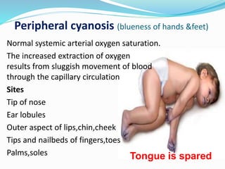
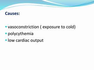
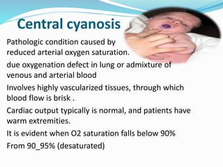
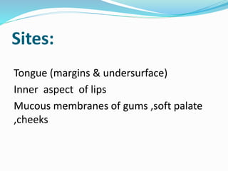
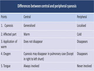
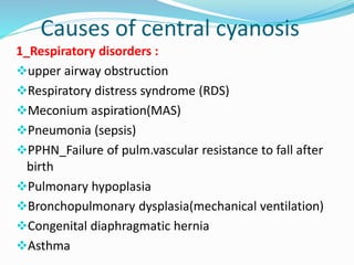
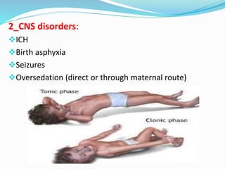
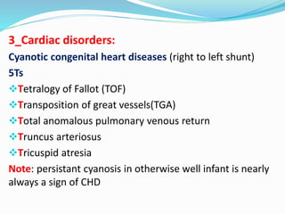
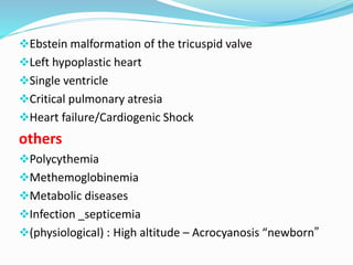
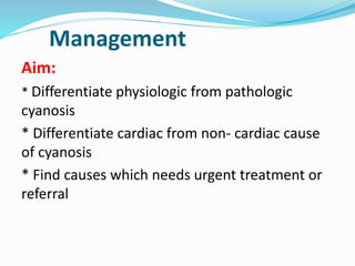
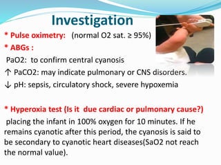
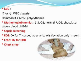
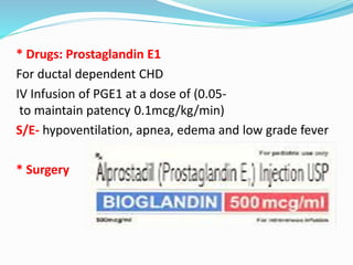
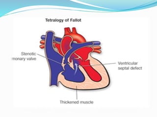
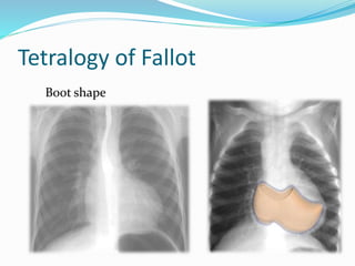
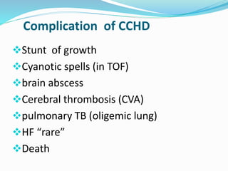
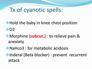
Child with cyanosis
Presented by : 
Group C4 
5th year medical students 
Tripoli university 
Pediatric
Objectives 
 Definition of cyanosis 
 Types of cyanosis 
 Causes of cyanosis 
Complications 
Management
Cyanosis is derived from the colour ‘cyan’, which comes 
from ‘kyanous’, the Greek word for blue . 
Definition: 
It is Bluish discoloration of skin and mucous membrane 
caused by increase concentration of reduced 
hemoglobin > 5g/dl 
so its not less pronounced if the child is anemic.
Types of cyanosis 
Peripheral Central
Peripheral cyanosis (blueness of hands &feet) 
Normal systemic arterial oxygen saturation. 
The increased extraction of oxygen 
results from sluggish movement of blood 
through the capillary circulation 
Sites 
Tip of nose 
Ear lobules 
Outer aspect of lips,chin,cheek 
Tips and nailbeds of fingers,toes 
Palms,soles Tongue is spared
Causes: 
 vasoconstriction ( exposure to cold) 
 polycythemia 
 low cardiac output
Central cyanosis 
Pathologic condition caused by 
reduced arterial oxygen saturation. 
due oxygenation defect in lung or admixture of 
venous and arterial blood 
Involves highly vascularized tissues, through which 
blood flow is brisk . 
Cardiac output typically is normal, and patients have 
warm extremities. 
It is evident when O2 saturation falls below 90% 
From 90_95% (desaturated)
Sites: 
Tongue (margins & undersurface) 
Inner aspect of lips 
Mucous membranes of gums ,soft palate 
,cheeks
Causes of central cyanosis 
1_Respiratory disorders : 
upper airway obstruction 
Respiratory distress syndrome (RDS) 
Meconium aspiration(MAS) 
Pneumonia (sepsis) 
PPHN_Failure of pulm.vascular resistance to fall after 
birth 
Pulmonary hypoplasia 
Bronchopulmonary dysplasia(mechanical ventilation) 
Congenital diaphragmatic hernia 
Asthma
2_CNS disorders: 
ICH 
Birth asphyxia 
Seizures 
Oversedation (direct or through maternal route)
3_Cardiac disorders: 
Cyanotic congenital heart diseases (right to left shunt) 
5Ts 
Tetralogy of Fallot (TOF) 
Transposition of great vessels(TGA) 
Total anomalous pulmonary venous return 
Truncus arteriosus 
Tricuspid atresia 
Note: persistant cyanosis in otherwise well infant is nearly 
always a sign of CHD
Ebstein malformation of the tricuspid valve 
Left hypoplastic heart 
Single ventricle 
Critical pulmonary atresia 
Heart failure/Cardiogenic Shock 
others 
Polycythemia 
Methemoglobinemia 
Metabolic diseases 
Infection _septicemia 
(physiological) : High altitude – Acrocyanosis “newborn”
cause example 
Cardiovascular disorders congenital cyanotic heart disease 
Respiratory disorders - Pneumonia 
- Bronchiolitis 
- RDS & meconium aspiration 
- Pneumothorax & pleural effusion 
0CNS disorders - intra cranial hemorrhage0 
- Tonic clonic seizure 
hematological disorders - Methemoglobinemia 
- Polycythemia 
- Congenital cyanosis 
other disorders - High Altitude 
- hypothermia 
- obstructive sleep apnea
Differential Cyanosis 
Hands red (less blue) and feet blue seen in PDA with 
reversal of shunt (Differential Cyanosis) Requires 
pulmonary vascular resistance elevated to a systemic level 
and a patent ductus arteriosus 
Left to right sunt pulmonary HT reversed shunt 
(Rt Lt shunt) 
Desaturated blood from the ductus enters the aorta distal 
to the left subclavian artery, sparing the brachiocephalic 
circulation.
Management 
Aim: 
* Differentiate physiologic from pathologic 
cyanosis 
* Differentiate cardiac from non- cardiac cause 
of cyanosis 
* Find causes which needs urgent treatment or 
referral
Do : 
1_complete maternal and newborn history 
2_perform a full physical examination 
3_ Investigation
Investigation 
* Pulse oximetry: (normal O2 sat. ≥ 95%) 
* ABGs : 
PaO2: to confirm central cyanosis 
↑ PaCO2: may indicate pulmonary or CNS disorders. 
↓ pH: sepsis, circulatory shock, severe hypoxemia 
* Hyperoxia test (Is it due cardiac or pulmonary cause?) 
placing the infant in 100% oxygen for 10 minutes. If he 
remains cyanotic after this period, the cyanosis is said to 
be secondary to cyanotic heart diseases(SaO2 not reach 
the normal value).
* CBC : 
↑ or ↓ WBC : sepsis 
Hematocrit > 65% : polycythemia 
* Methemoglobinemia : ↓ SaO2, normal PaO2, chocolate-brown 
blood , HB-M 
* Sepsis screening 
* ECG: Dx for Tricusped atresia (Lt axis deviation only is seen) 
* Echo: Dx for CHD 
* Chest x-ray
Treatment 
* Warming of the affected area: in peripheral cyanosis 
* Oxygenation & adequate ventilation 
(PaO2 normalizes completely during artificial ventilation 
in infant with CNS disorder) 
* IV fluids 
Children who have difficulty in feeding due to cyanosis 
need fluids to be administrated. 
* If sepsis is suspected or another specific cause is not 
identified, start on broad spectrum antibiotics then obtain 
a full septic screening
* Drugs: Prostaglandin E1 
For ductal dependent CHD 
IV Infusion of PGE1 at a dose of (0.05- 
to maintain patency 0.1mcg/kg/min) 
S/E- hypoventilation, apnea, edema and low grade fever 
* Surgery
CCHD
Tetralogy of Fallot 
Boot shape
Total Anomalous Pulmonary 
Venous Return(TAPVR) 
Snowman
Transposition of Great Arteries 
Egg on a string
Truncus arteriosus
Tricusped atresia
Complication of CCHD 
Stunt of growth 
Cyanotic spells (in TOF) 
brain abscess 
Cerebral thrombosis (CVA) 
pulmonary TB (oligemic lung) 
HF “rare” 
Death
Tx of cyanotic spells: 
Hold the baby in knee chest position 
O2 
Morphine (subcut.) : to relieve pain & 
anexiety 
NaHco3 : for metabolic acidosis 
Inderal (Beta blocker) : prevent recurrent 
attack
Thank you

Child with cyanosis

  • 1.
  • 2.
    Presented by : Group C4 5th year medical students Tripoli university Pediatric
  • 3.
    Objectives  Definitionof cyanosis  Types of cyanosis  Causes of cyanosis Complications Management
  • 4.
    Cyanosis is derivedfrom the colour ‘cyan’, which comes from ‘kyanous’, the Greek word for blue . Definition: It is Bluish discoloration of skin and mucous membrane caused by increase concentration of reduced hemoglobin > 5g/dl so its not less pronounced if the child is anemic.
  • 5.
    Types of cyanosis Peripheral Central
  • 6.
    Peripheral cyanosis (bluenessof hands &feet) Normal systemic arterial oxygen saturation. The increased extraction of oxygen results from sluggish movement of blood through the capillary circulation Sites Tip of nose Ear lobules Outer aspect of lips,chin,cheek Tips and nailbeds of fingers,toes Palms,soles Tongue is spared
  • 7.
    Causes:  vasoconstriction( exposure to cold)  polycythemia  low cardiac output
  • 8.
    Central cyanosis Pathologiccondition caused by reduced arterial oxygen saturation. due oxygenation defect in lung or admixture of venous and arterial blood Involves highly vascularized tissues, through which blood flow is brisk . Cardiac output typically is normal, and patients have warm extremities. It is evident when O2 saturation falls below 90% From 90_95% (desaturated)
  • 9.
    Sites: Tongue (margins& undersurface) Inner aspect of lips Mucous membranes of gums ,soft palate ,cheeks
  • 11.
    Causes of centralcyanosis 1_Respiratory disorders : upper airway obstruction Respiratory distress syndrome (RDS) Meconium aspiration(MAS) Pneumonia (sepsis) PPHN_Failure of pulm.vascular resistance to fall after birth Pulmonary hypoplasia Bronchopulmonary dysplasia(mechanical ventilation) Congenital diaphragmatic hernia Asthma
  • 12.
    2_CNS disorders: ICH Birth asphyxia Seizures Oversedation (direct or through maternal route)
  • 13.
    3_Cardiac disorders: Cyanoticcongenital heart diseases (right to left shunt) 5Ts Tetralogy of Fallot (TOF) Transposition of great vessels(TGA) Total anomalous pulmonary venous return Truncus arteriosus Tricuspid atresia Note: persistant cyanosis in otherwise well infant is nearly always a sign of CHD
  • 14.
    Ebstein malformation ofthe tricuspid valve Left hypoplastic heart Single ventricle Critical pulmonary atresia Heart failure/Cardiogenic Shock others Polycythemia Methemoglobinemia Metabolic diseases Infection _septicemia (physiological) : High altitude – Acrocyanosis “newborn”
  • 15.
    cause example Cardiovasculardisorders congenital cyanotic heart disease Respiratory disorders - Pneumonia - Bronchiolitis - RDS & meconium aspiration - Pneumothorax & pleural effusion 0CNS disorders - intra cranial hemorrhage0 - Tonic clonic seizure hematological disorders - Methemoglobinemia - Polycythemia - Congenital cyanosis other disorders - High Altitude - hypothermia - obstructive sleep apnea
  • 16.
    Differential Cyanosis Handsred (less blue) and feet blue seen in PDA with reversal of shunt (Differential Cyanosis) Requires pulmonary vascular resistance elevated to a systemic level and a patent ductus arteriosus Left to right sunt pulmonary HT reversed shunt (Rt Lt shunt) Desaturated blood from the ductus enters the aorta distal to the left subclavian artery, sparing the brachiocephalic circulation.
  • 17.
    Management Aim: *Differentiate physiologic from pathologic cyanosis * Differentiate cardiac from non- cardiac cause of cyanosis * Find causes which needs urgent treatment or referral
  • 18.
    Do : 1_completematernal and newborn history 2_perform a full physical examination 3_ Investigation
  • 19.
    Investigation * Pulseoximetry: (normal O2 sat. ≥ 95%) * ABGs : PaO2: to confirm central cyanosis ↑ PaCO2: may indicate pulmonary or CNS disorders. ↓ pH: sepsis, circulatory shock, severe hypoxemia * Hyperoxia test (Is it due cardiac or pulmonary cause?) placing the infant in 100% oxygen for 10 minutes. If he remains cyanotic after this period, the cyanosis is said to be secondary to cyanotic heart diseases(SaO2 not reach the normal value).
  • 20.
    * CBC : ↑ or ↓ WBC : sepsis Hematocrit > 65% : polycythemia * Methemoglobinemia : ↓ SaO2, normal PaO2, chocolate-brown blood , HB-M * Sepsis screening * ECG: Dx for Tricusped atresia (Lt axis deviation only is seen) * Echo: Dx for CHD * Chest x-ray
  • 21.
    Treatment * Warmingof the affected area: in peripheral cyanosis * Oxygenation & adequate ventilation (PaO2 normalizes completely during artificial ventilation in infant with CNS disorder) * IV fluids Children who have difficulty in feeding due to cyanosis need fluids to be administrated. * If sepsis is suspected or another specific cause is not identified, start on broad spectrum antibiotics then obtain a full septic screening
  • 22.
    * Drugs: ProstaglandinE1 For ductal dependent CHD IV Infusion of PGE1 at a dose of (0.05- to maintain patency 0.1mcg/kg/min) S/E- hypoventilation, apnea, edema and low grade fever * Surgery
  • 23.
  • 25.
  • 26.
    Total Anomalous Pulmonary Venous Return(TAPVR) Snowman
  • 27.
    Transposition of GreatArteries Egg on a string
  • 28.
  • 29.
  • 30.
    Complication of CCHD Stunt of growth Cyanotic spells (in TOF) brain abscess Cerebral thrombosis (CVA) pulmonary TB (oligemic lung) HF “rare” Death
  • 31.
    Tx of cyanoticspells: Hold the baby in knee chest position O2 Morphine (subcut.) : to relieve pain & anexiety NaHco3 : for metabolic acidosis Inderal (Beta blocker) : prevent recurrent attack
  • 32.

Editor's Notes

  • #13 Tonic clonic type of convulsion
  • #15 Newborn cyanotic at birth when transfer from intrauterine to extrauterine life , so need quick warming &dryness
  • #18  As : physiological Cyanosis soon after birth-normal transition from intrauterine to extrauterine life History of convulsion & general depression ,shallow, irregular respirations and periods of apnea strongly suggest CNS problem
  • #19 Pneumonia/ sepsis- PROM Foul smelling liquor Maternal pyrexia Maternal GBS TTN – Birth by cesarean section with or without labor Male sex Family history of asthma (especially in mother) Macrosomia Maternal diabetes Polycythemia- small-for-gestational age MAS- Post maturity Small for gestational age Placental dysfunction Fetal distress Meconium stained liquor Pneumothorax- Aggressive resucitation IPPV Meconiun aspiration HMD Hypoplastic lung Staph pneumonia Hyaline membrane disease- Premature infant Infant of diabetic mother
  • #21 CHD= Cyanotic heart diseases SaO2= oxgen saturation
  • #31 Brain abscess becos. Blood from Rt to Lt without pass to lung which has a phagocytic activity (filter bld) so go to the brain & cause abscess if contain organisms