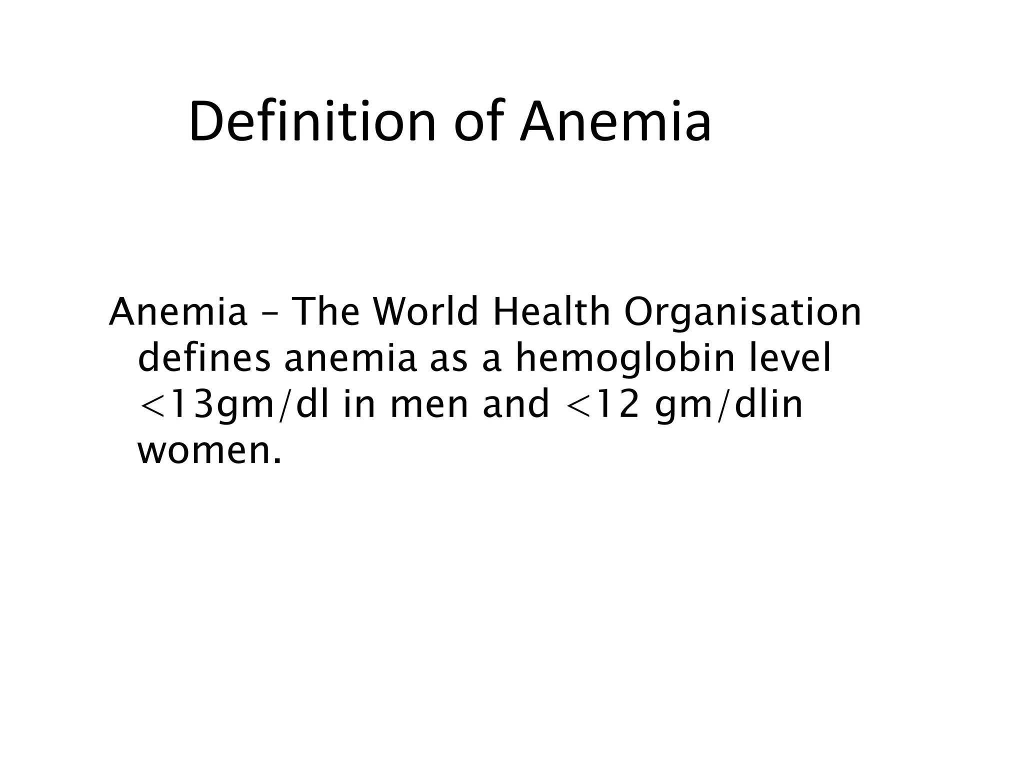
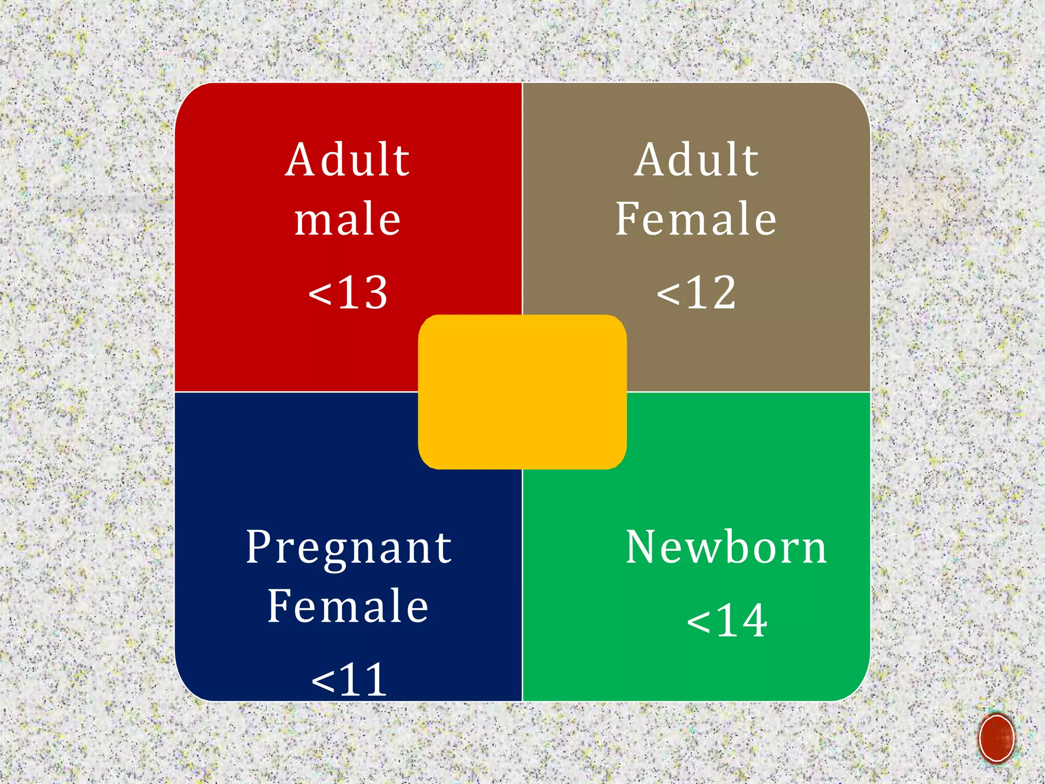
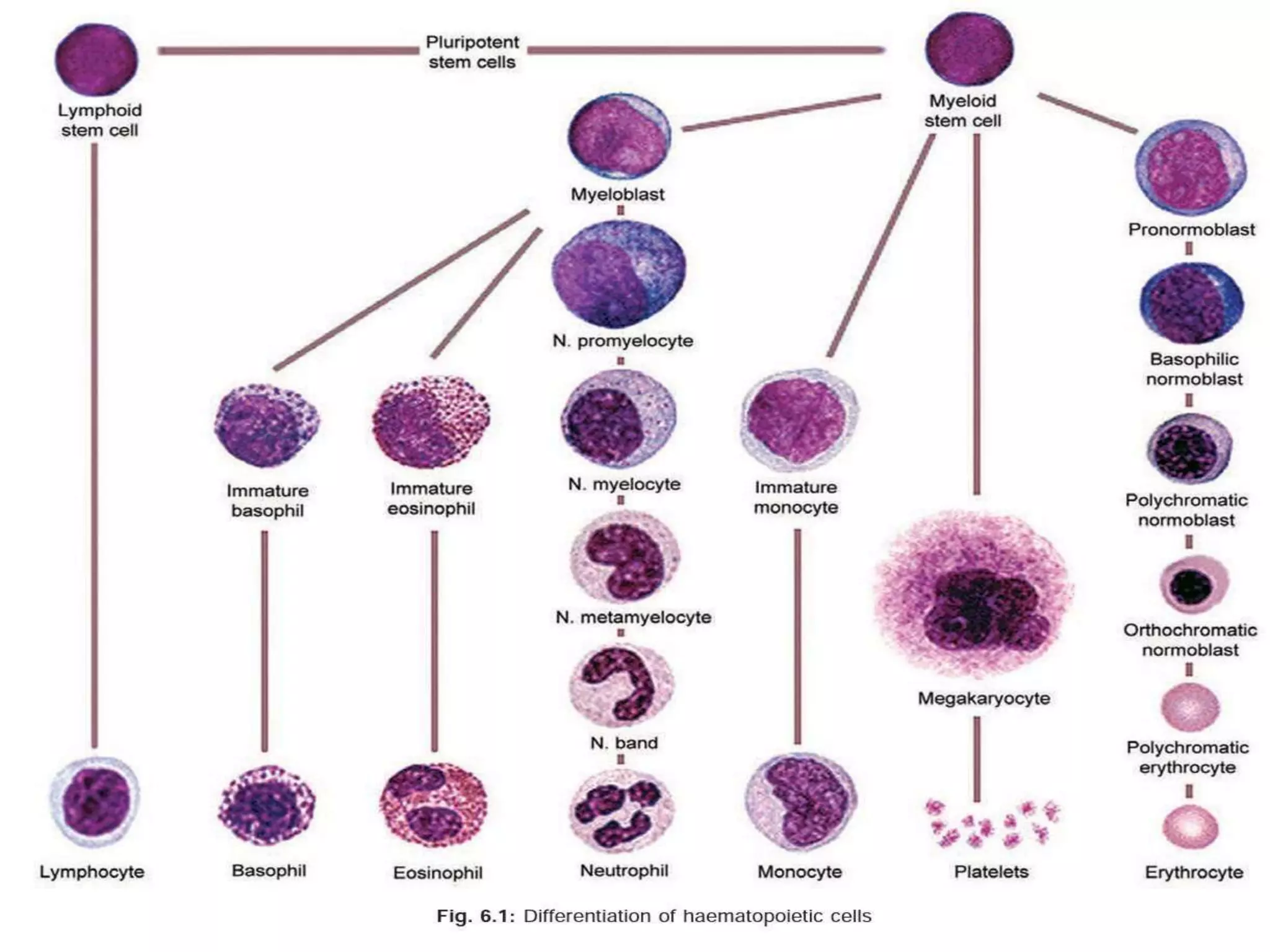
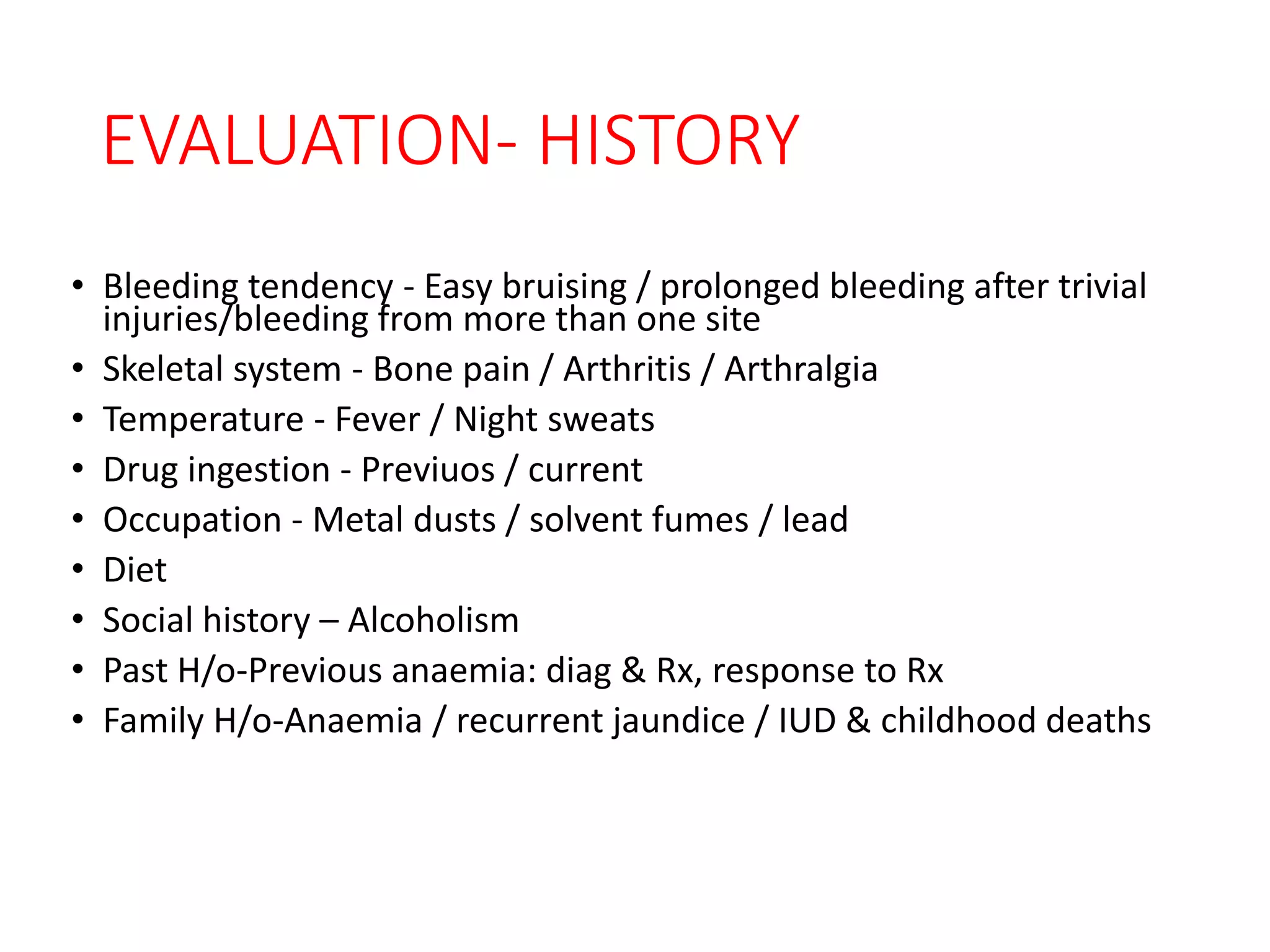
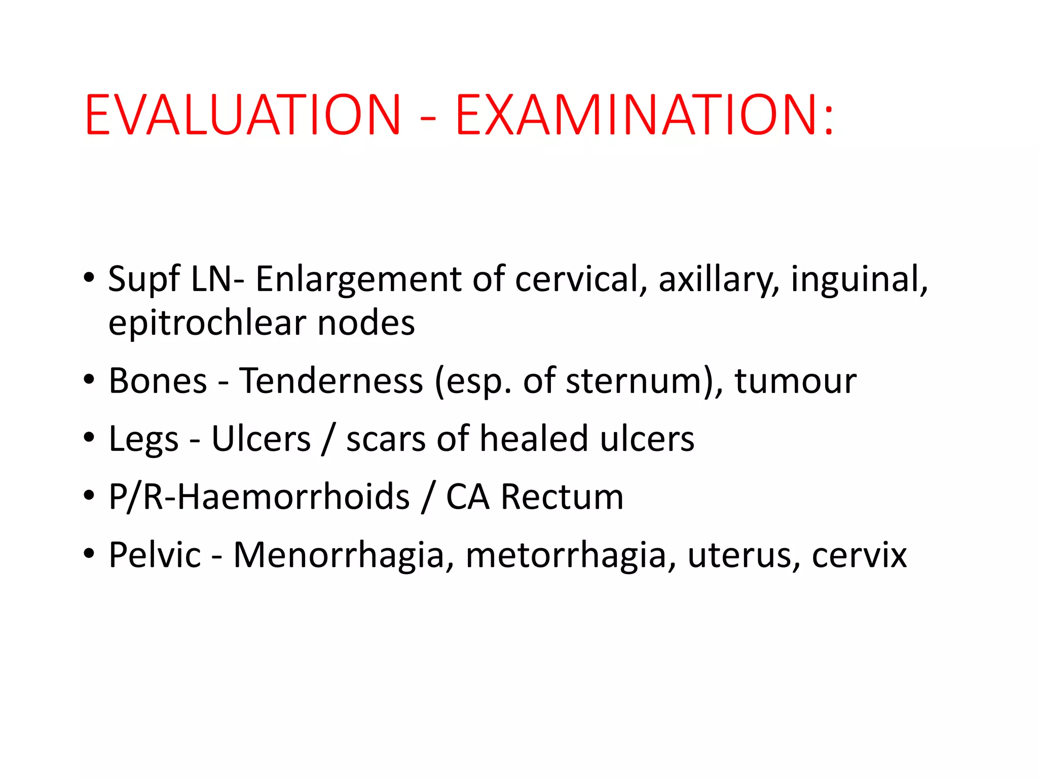
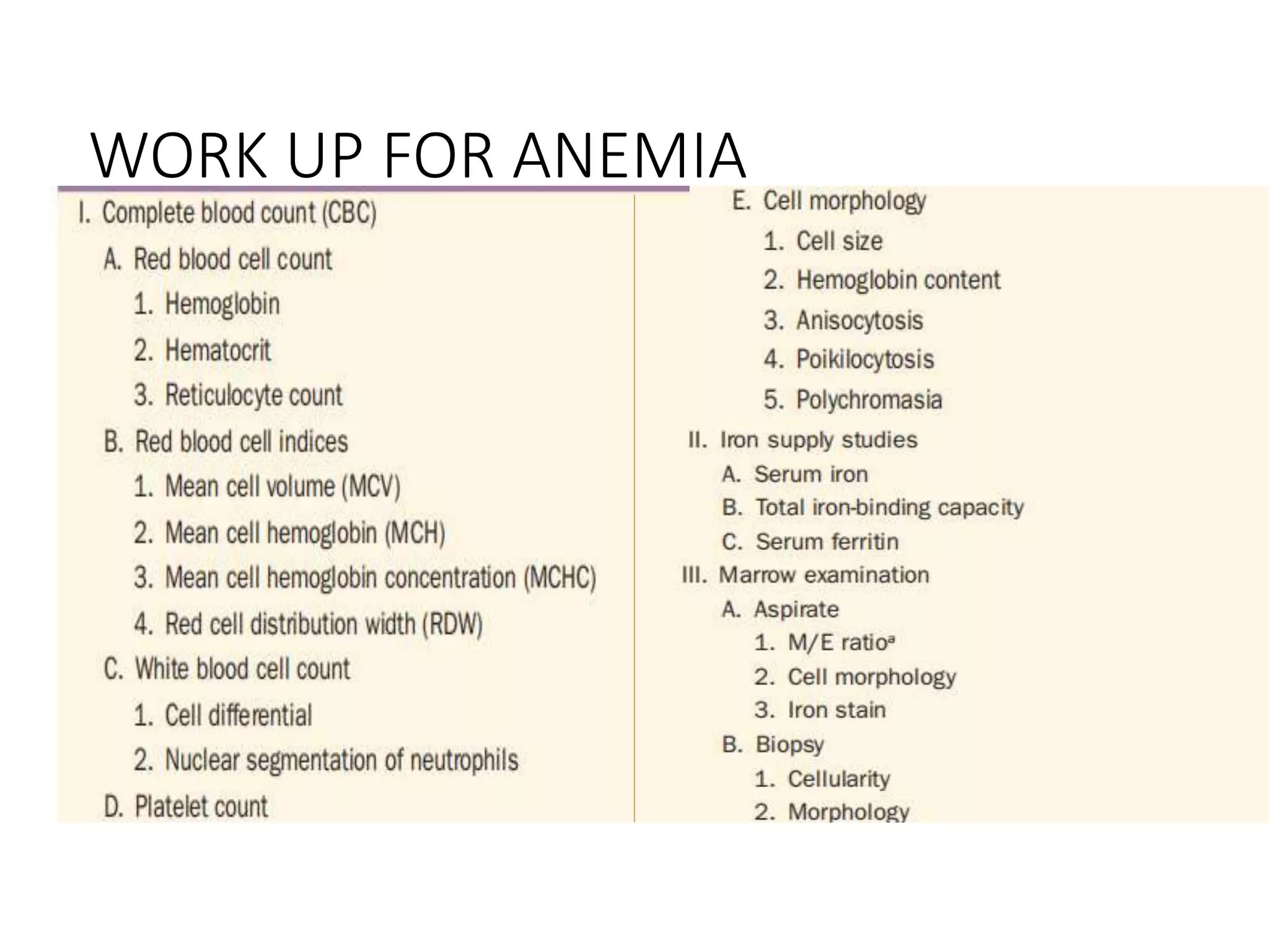
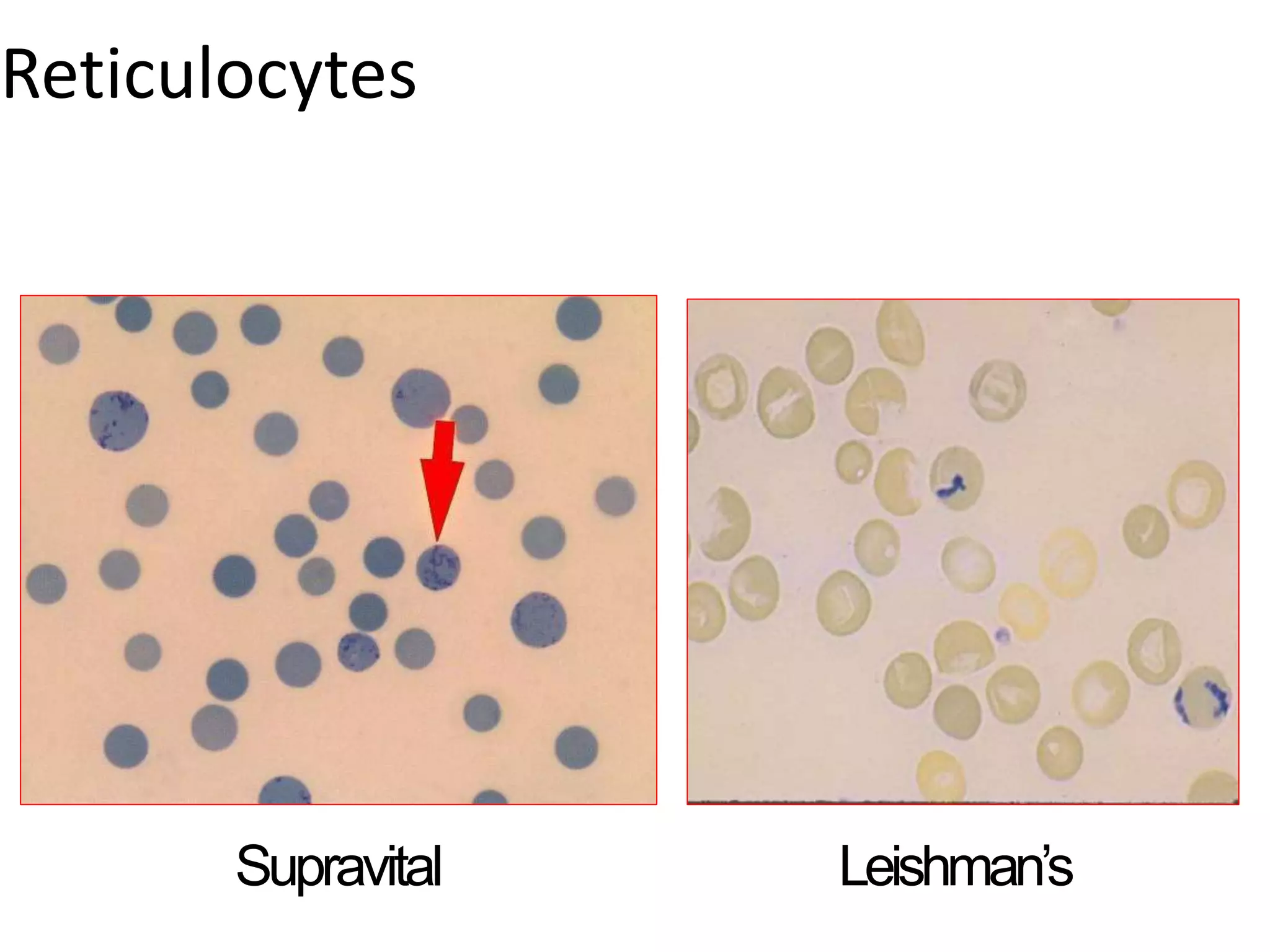
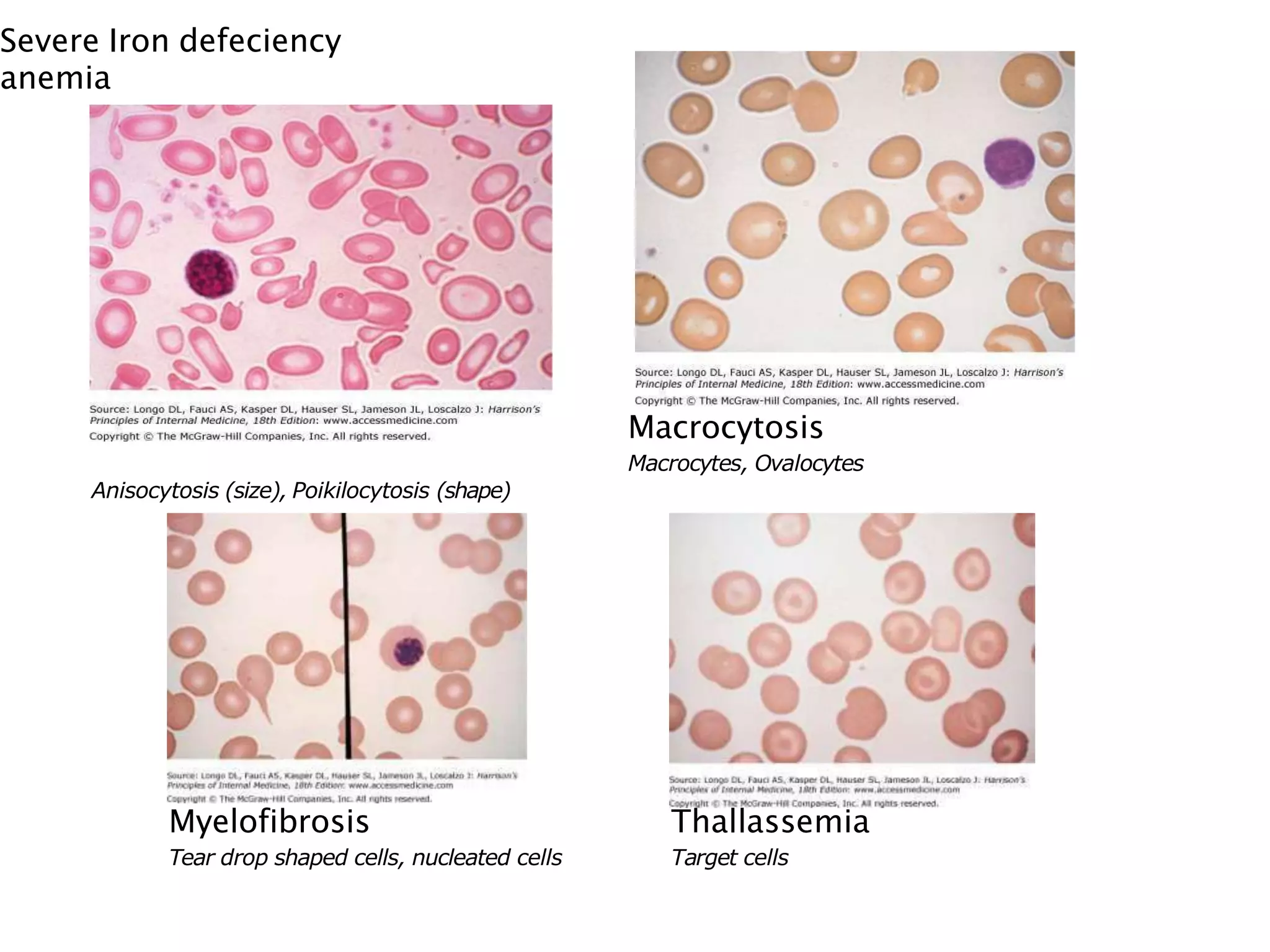
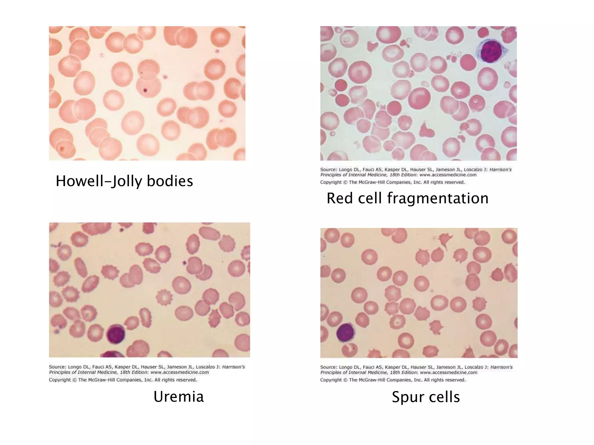
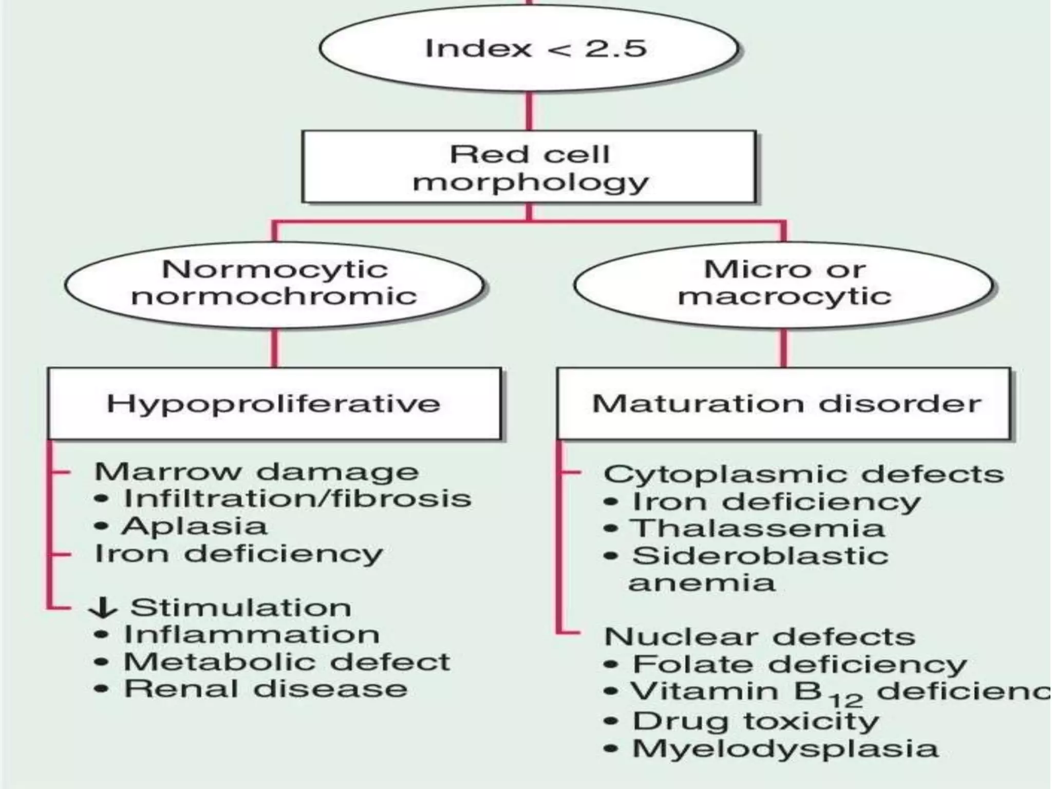
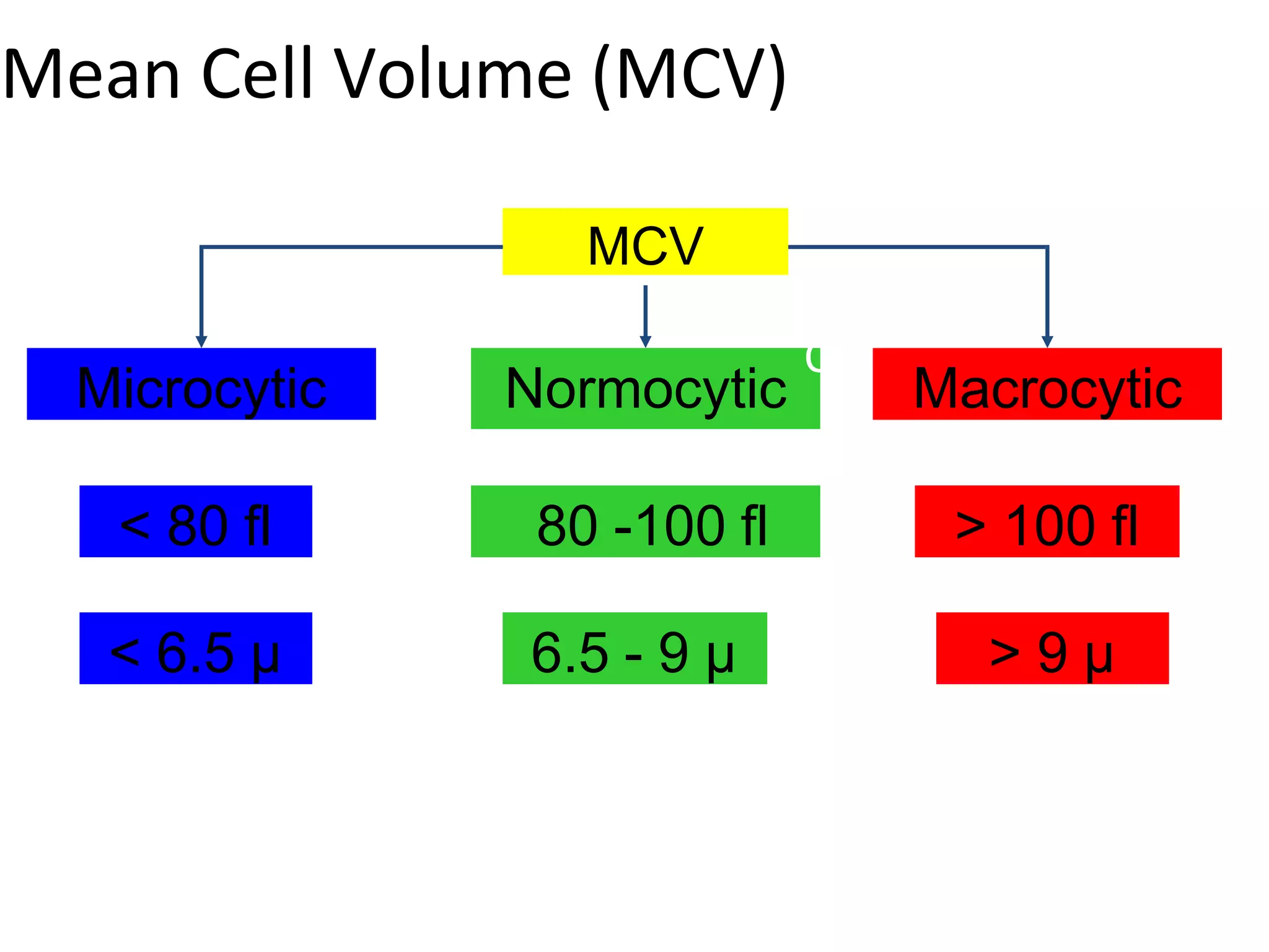
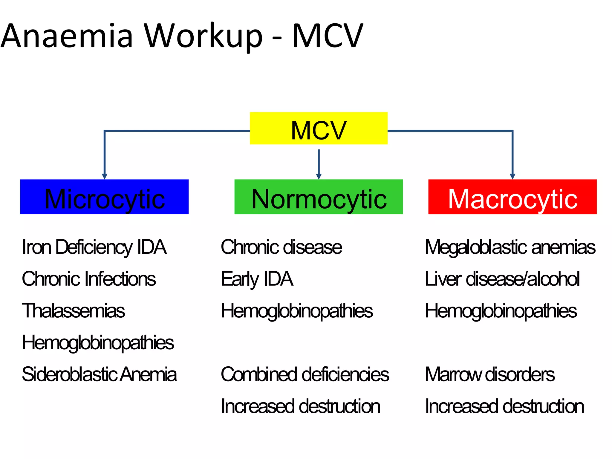
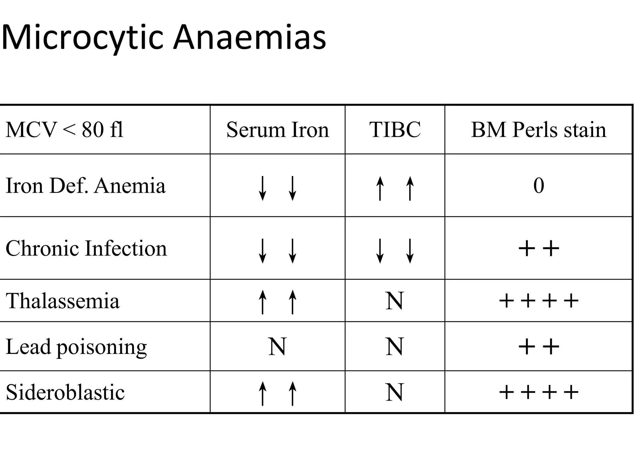
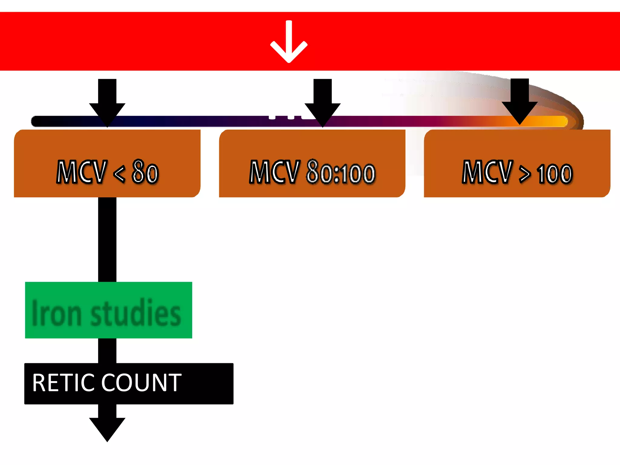
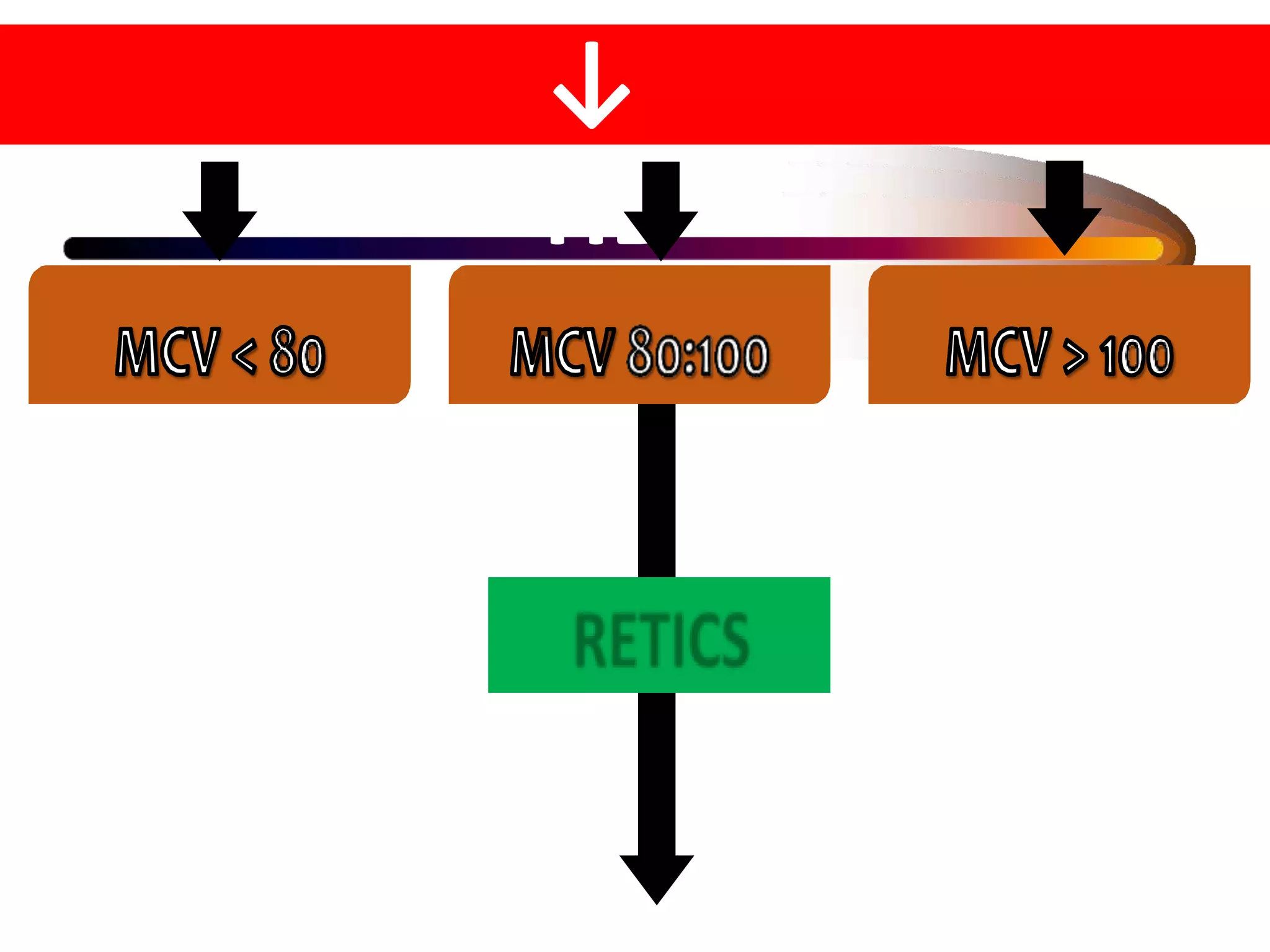
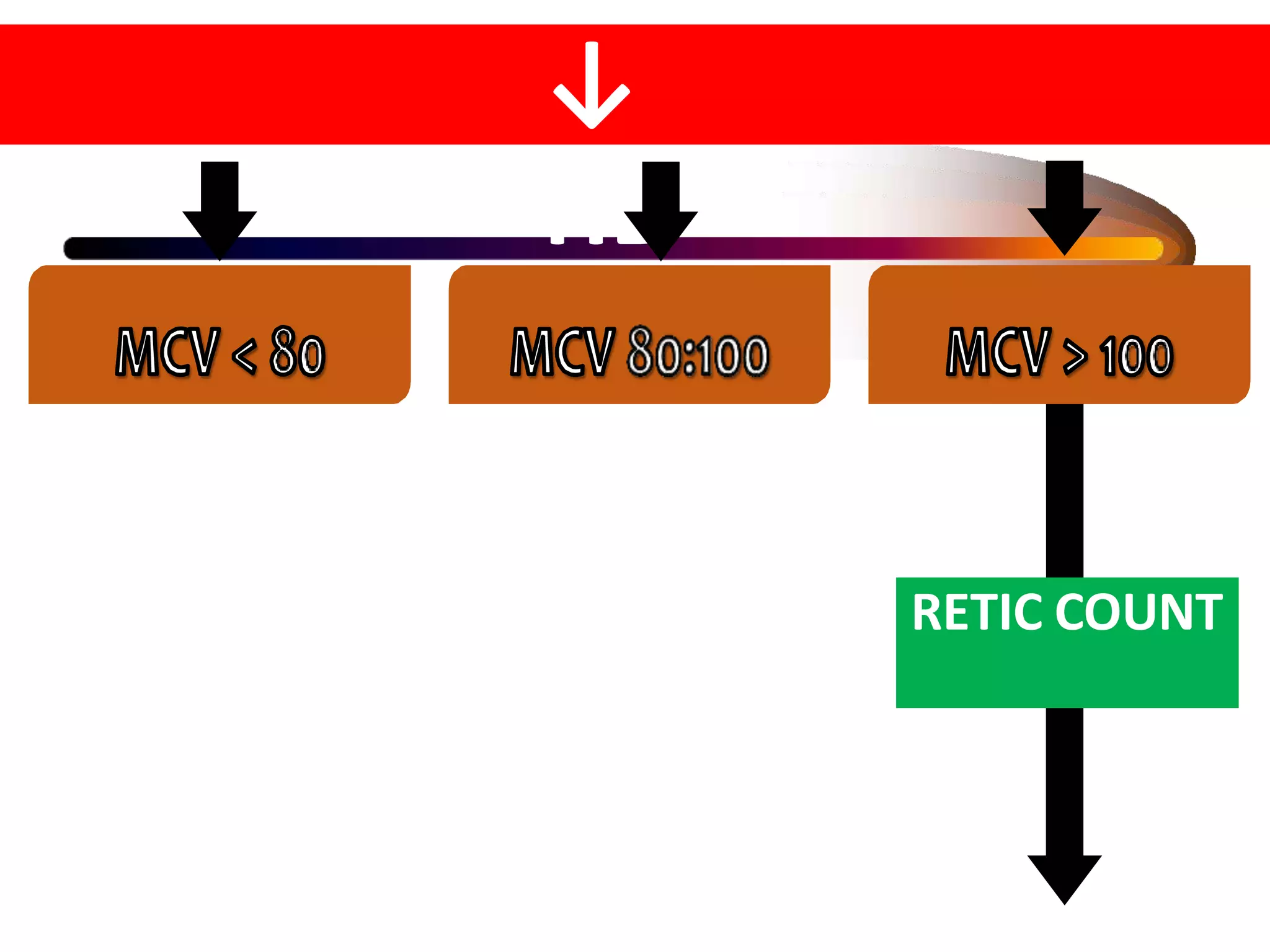
APPROACH TO ANEMIA
Guided by Dr SHIDRAM K
sir
Presented by -Dr Gadeppa
Definition of Anemia
Anemia – The World Health Organisation
defines anemia as a hemoglobin level
<13gm/dl in men and <12 gm/dlin
women.
Adult
male
<13
Adult
Female
<12
Pregnant
Female
<11
Newborn
<14
Erythron
•
•
Erythron is the machinery of RBC production
EPO, IL, Growth factors, Cytokines – stimulate it
• Hypoxia is strong stimulus for the Erythron
• Its functioning is influenced by
1. Normal renal production of EPO
2. A functioning Erythroid marrow
3. An adequate supply of substrates for Hb production
ERYTHROPOIETIN
• Glycoprotein hormone
• Produced by peritubular capillary lining of
cells in kidney
• Small amount in liver
• EPO gene regulation is by Hypoxia inducible
factor 1α
• Normal levels 10 – 25 U/l
• T1/2 – 6-9 hrs
Normal Red Cells
Nonucleus, Enzymepackets
Biconcavediscs–Haem+Gl
Center1/3pallor
Pinkcytoplasm(Hbfilled)
Cellsize7- 8µm
Hemoglobin (Hb)
EVALUATION- HISTORY
• Age/Sex
• Rate of onset - Rapid/Slow
• Blood loss - Haematemesis/malena/bleeding piles/menorrhagia/
metorrhagia/epistaxis/hematuria /haemoptysis
• Abdomen - Appetitie/weight
loss/dysphagia/regurgitation/dyspepsia/ abd
pain/diarrhoea/constipation/jaundice/soreness of tongue/previous
abd surgeries
• Reproductive - Menstrual history in detail/number & interval
between pregnancies/miscarriages
• Urinary system - Nocturnal polyuria
• CNS-Parasthesiae / difficulty in walking
EVALUATION- HISTORY
• Bleeding tendency - Easy bruising / prolonged bleeding after trivial
injuries/bleeding from more than one site
• Skeletal system - Bone pain / Arthritis / Arthralgia
• Temperature - Fever / Night sweats
• Drug ingestion - Previuos / current
• Occupation - Metal dusts / solvent fumes / lead
• Diet
• Social history – Alcoholism
• Past H/o-Previous anaemia: diag & Rx, response to Rx
• Family H/o-Anaemia / recurrent jaundice / IUD & childhood deaths
EVALUATION - EXAMINATION:
• Skin - Colour, texture, petechiae, ecchymoses, scratch marks.
• Nails - Brittleness, longitudinal ridging, koilonychia
• Conjunctiva/Sclera - Pallor, icterus, haemorrhages
• Retina - Haemorrhages, s/o HTN/renal failure, other changes
• Mouth - Mucous membrane: Pallor, petechiae
• Gums: Bleeding, hypertrophy
• Tongue: Redness, atrophy of papillae
• Abdomen - HSM, either HM or SM, tenderness, mass, ascites
• CVS-BP, valvular, vascular prosthesis
• CNS- Peripheral neuritis, s/s/o SADSC
EVALUATION - EXAMINATION:
• Supf LN- Enlargement of cervical, axillary, inguinal,
epitrochlear nodes
• Bones - Tenderness (esp. of sternum), tumour
• Legs - Ulcers / scars of healed ulcers
• P/R-Haemorrhoids / CA Rectum
• Pelvic - Menorrhagia, metorrhagia, uterus, cervix
WORK UP FOR ANEMIA
The Three Basic Measures
Measurement Normal Range
A. RBC count 5 million 4 to 6
B. Hemoglobin 15 g% 12 to 17
C. Hematocrit 45 38 to 50
The Three Derived Indicies
Measurement
A. RBC count
Normal
5 million
Range
4 to 6
B. Hemoglobin 15 g% 12 to 17
C. Hematocrit 45 38 to 50
MCV C ÷ A x 10 = 90 fl
MCH B ÷ A x 10 = 30 pg
MCHC B ÷ C x 100 = 33%
RETICULOCYTE COUNT %
• Reticulocytes are immature RBC
• ‘RBC to be’ or Apprentice RBC
• Fragments of nuclear material
• RNA strands which stain blue
• Normal Less than 2%
Reticulocytes
Leishman’s
Supravital
Reticulocyte correction
• Correction 1 - in presence of anemia, reticulocyte count spuriously elevated when it is
related to reduced number of RBCs in anemic patient. (relatively more in number due to
reduced mature RBC)
• Correction 2 - An additional correction needs to be made because reticulocytes released
under intense EPO stimulation remain in the peripheral blood for more than usual 1 day
(prematurely released Reticulocyte remains longer in circulation)
Reticulocyte count %of reticulocytes in RBC
population
Corrected reticulocyte count %reticulocytes ×(patient Hct/45)
Reticulocyte production index Corrected reticulocyte count ÷
maturation time in peripheral
blood in days
Absolute reticulocyte count %reticulocytes ×RBC count/L
CORRECTED RETICULOCYTE COUNT
Red cell distribution width
• RDW measures range of variation of red cell
volume
• Normal range is 11.5 to 14.5 %
• It is measure of anisocytosis
• Usually elevated in deficiency of Iron, Folate,
B12
• Usually normal in Hemoglobinopathy
RBC Size – Anisocytosis
Different sizes of RBC
MENTZERS INDEX
• MCV/RBC
• >13 - S/O IRON DEFICIENCY ANEMIA
• 11-13 - INDETERMINATE
• <11 - THALASSEMIA TRAIT
Peripheral Smear Study
• Are all RBC of the same size ?
• Are all RBC of the same normal discoid shape ?
• How is the colour (Hb content) saturation ?
• Are all the RBC of same colour/ multi coloured ?
• Are there any RBC inclusions ?
• Are intra RBC there any hemo-parasites ?
• Are leucocytes normal in number and D.C ?
• Is platelet distribution adequate ?
Cell size
Hb content
Anisocytosis
Poikilocytosis
Polychromasia
Gives clues to
specific disorders
Normal peripheral smear
Severe Iron defeciency
anemia
Anisocytosis (size), Poikilocytosis (shape)
Macrocytosis
Macrocytes, Ovalocytes
Myelofibrosis
Tear drop shaped cells, nucleated cells
Thallassemia
Target cells
Howell-Jolly bodies
Red cell fragmentation
Uremia Spur cells
Target Cells
1. Liver Disease
2. Thalassemia
3. HbDDisease
4. Post splenectomy
Shistocytes
Fragmented, Helmet or triangle shaped
RBC
1. MAHA
2. Prosthetic
valves
3. Uremia
4. Malignant HT
Tear Drop Cells
1. Myelofibosis
2. Infiltration of
BM
3. Tumours of BM
4. Thalassemia
Types of Anaemia
Mean Cell Volume (MCV)
measured by
M and RDW
Microcytic
< 80 fl
MCV
Normocytic
CV
Macrocytic
80 -100 fl > 100 fl
< 6.5 µ 6.5 - 9 µ > 9 µ
Anaemia Workup - MCV
Microcytic
MCV
Normocytic Macrocytic
IronDeficiency IDA
Chronic Infections
Thalassemias
Hemoglobinopathies
SideroblasticAnemia
Chronic disease
Early IDA
Hemoglobinopathies
Combined deficiencies
Increaseddestruction
Megaloblastic anemias
Liver disease/alcohol
Hemoglobinopathies
Marrowdisorders
Increaseddestruction
Hypoproliferative Anaemias
Failureofcell
maturation
Nuclear
breakdown
Cytoplasmi
c
breakdown
MegaloblasticAnaemia
DefectiveDNAsynthesis
FolateorB12 deficiency Haemdefect Globindefect
Thalassemia
SicklecellA
Fe Phorph
IDA, SA
IDA – Special Tests
Iron related tests Normal IDA
Serum Ferritin
(pmo/L)
33-270 < 33
TIBC (µg/dL) 300-340 > 400
Serum Iron (µg/dL) 50-150 < 30
Saturation % 30-50 < 10
Bone marrow Iron ++ Absent
Microcytic Anaemias
MCV < 80 fl Serum Iron TIBC BM Perls stain
Iron Def. Anemia ↓↓ ↑↑ 0
Chronic Infection ↓↓ ↓↓ + +
Thalassemia ↑↑ N + + + +
Lead poisoning N N + +
Sideroblastic ↑↑ N + + + +
IDA
• Microcytic
• RDW
• Hypochromic
• RPI
• Serum ferritin
• TIBC
• Serum Iron
• BM Fe Stain
• Response to Fe Rx.
MCV < 80 fl, RBC < 6 µ
Widened and shift to left MCH <
27 pg, MCHC < 30%
< 2
Very low < 30 (p mols/L)
Increased > 400 (µg/dL)
Very low < 30 (µg/dL)
Absent Fe
Excellent
IDA
• Look for occult blood loss – 2 days non veg. free
• Pica and Pagophagia
• Absorption of Haem Iron > Fe++
• Food, Phytates, Ca, Phosphate, antacids ↓absorption
• Ascorbic acid ↑absorption
• Oral iron Rx. always is the best
• Oral iron therapy.
• Packed cell transfusion in emergency
• Continue Fe Rx at least 2 months after normal Hb
• 1 gram ↑in Hb every week can be expected
• Always supplement protein for the Globin component
Microcytic Hypochromic - IDA
Severe Hypochromia
Ringed Sideroblasts in BM
PrussianBlueStain
Macrocytic Anaemias
• Megaloblastic Macrocytic –B12 and Folate↓
• Non Megaloblastic Macrocytic Anaemias
1. Liver disease/alcohol
2. Hemoglobinopathies
3. Metabolic disorders, Hypothyroidism
4. Myelodystrophy, BM infiltration
5. Accelerated Erythropoesis -↑destruction
6. Drugs (cytotoxics, immunosuppressants, AZT,
anticonvulsants)
Anemia - Macrocytic (MCV > 100)
– Macrocytic anemias may be asymptomatic
until the Hb is as low as 6 grams
– MCV 100-110 fl must look for other causes of
macrocytosis
– MCV > 110 fl almost always folate or B12
deficiency
Macrocytosis of Alcoholism
• MCV elevation usually slight (100-110 fl)
• Minimal or no anemia
• Macrocytes round (not oval)
• Neutrophil hyper segmentation absent
• Folate stores normal
DRUGS CAUSING MEGALOBLASTICANEMIA
 Folate antagonists (e.g., methotrexate)
 Purine antagonists (e.g., 6-mercaptopurine)
 Pyrimidine antagonists (e.g., cytosine arabinoside)
 Alkylating agents (e.g., cyclophosphamide)
 Zidovudine (AZT
, Retrovir)
 Trimethoprim
 Oral contraceptives
 Nitrous oxide
 Arsenic
Megaloblastic Hematopoiesis
Marrow failure due to
• Disrupted DNA synth. & ineffective erythropoesis
• Giant precursors (Megaloblasts)
• Nuclear : Cytoplasmic dyssynchrony in marrow
• Neutrophil hyper segmentation & macro ovalocytes
• Anemia (and often leukopenia & thrombocytopenia)
• Almost always due to B12 or folate deficiency
Pernicious Anaemia - Tongue
Bald, smooth, lemon
yellowish red tongue
Megaloblastic anemia
Anisocytosis - Macrocytic Anaemia
Normocytic Anaemias
• Chronic disease
• Early IDA
• Hemoglobinopathies
• Primary marrow disorders
• Combined deficiencies
Anaemia of Chronic Disease
• Thyroid diseases
• Malignancy
• Collagen Vascular Disease
– Rheumatoid Arthritis
– SLE
– Polymyositis
– Polyarteritis Nodosa
• IBD
– Ulcerative Colitis
– Crohn’s Disease
• Chronic Infections
– HIV, Osteomyelitis
– Tuberculosis
• Renal Failure
• Anaemia is usually mild and non-progressive, rarely less than
9 gm/dL.
• Anaemia is never severe
• Anaemia resolves when underlying cause is treated.
• Anaemia of chronic inflammation is not responsive to
haematinics like iron, folate, vitamin B12.
• Transfusion is rarely indicated.
• Higher doses of Epo is required to treat ACD than for the
therapy of renal anaemia.
‘Dimorphic’ Anaemia
• Folate & Fe deficiency (pregnancy, alcoholism)
• B12 & Fe deficiency (PA with atrophic gastritis)
• Thalassemia minor & B12 or folate deficiency
• Fe deficiency & hemolysis (prosthetic valve)
• Folate deficiency & hemolysis (Hb SS disease)
• Peripheral smear exam is critical to assess these
• RDW is increased very much
Hemolytic Anaemia
Anemia of increased RBC destruction
– Normochromic, normocytic anemia
– Shortened RBC survival
– Reticulocytosis – due to ↑ RBC destruction
Will not be symptomatic until the RBC life span is
reduced to 20 days – BM compensates 6 times
Tests Used to Diagnose Hemolysis
1. Reticulocyte count
2. Combined with serial Hb
3. Serum LDH
4. Serum bilirubin
5. Haptoglobin
6. Urine hemosiderin
7. Hemoglobinuria
Findings in Hemolytic Anaemia
Reticulocyte count and RPI Increased
Serum Unconjugated Bilirubin Increased
Serum LDH 1: LDH 2 Increased
Serum Haptoglobin Decreased
Urine Hemoglobin Present
Urine Hemosiderin Present
Urine Urobilinogen Increased
Cr 51 labeled RBC life span Decreased
Tests to define
the cause of hemolysis
1. Hemoglobin electrophoresis
2. Hemoglobin A2 (βeta-Thalassemia trait)
3. RBC enzymes (G6PD, PK, etc)
4. Direct & indirect antiglobulin tests (immune)
5. Cold agglutinins
6. Osmotic fragility (spherocytosis)
7. Acid hemolysis test (PNH)
8. Clotting profile (DIC)
Spherocytosis
Elliptocytes
Hereditary Elliptocytosis, B12 or Folate↓
Sickle Cell Anaemia
Micro Angiopathic Hemolytic Anaemia
ANEMIA DUE TO IMPAIRED BONE MARROW
RESPONSE
Red blood cell aplasia
Aplastic anemia
Myelodysplasia
Leukemias
Myelophthisic anemia
Marrow infiltration
Myeloma
Congenital Dyserythropoietic Anemias
Aplastic Anaemia
1. Chloramphenicol
2. Carbimazole
3. Sulfonamides
4. Phenytoin
5. Anti Ca drugs
BM - Aplastic Anaemia
↓
HB
RETIC COUNT
CONDITION SERUM
IRON
TIBC FERRITIN COMMENT
Iron deficiency ↓ ↑ ↓ Responsive to iron
Chronic
inflammation
↓ ↓  Unresponsive to iron
Thalassemia major ↑ N N
Reticulocytosis and
indirect
bilirubinemia
Lead poisoning N N N Basophilic stippling of
RBCs
Sideroblastic anemia ↑ N  Ring sideroblasts in
marrow
↓
HB
RPI <2.5 RPI >2.5
• Anemia of
chronic
disease
• CKD
• Acute
Bloodloss
• Leukemia
• Approach to
Pancytopeni
a
• Approach to
Hemolytic
anemia
↓
HB
RETIC COUNT
REFERENCES
• Harrison’s Principles of Internal Medicine 20th
edition
• The Washington’s Manual of Medical
Therapeutics 34th edition
• Robbins & Cotran Pathologic Basis of Disease, 8th
edition
approach to anemia.pptx

approach to anemia.pptx

  • 1.
    APPROACH TO ANEMIA Guidedby Dr SHIDRAM K sir Presented by -Dr Gadeppa
  • 2.
    Definition of Anemia Anemia– The World Health Organisation defines anemia as a hemoglobin level <13gm/dl in men and <12 gm/dlin women.
  • 3.
  • 4.
    Erythron • • Erythron is themachinery of RBC production EPO, IL, Growth factors, Cytokines – stimulate it • Hypoxia is strong stimulus for the Erythron • Its functioning is influenced by 1. Normal renal production of EPO 2. A functioning Erythroid marrow 3. An adequate supply of substrates for Hb production
  • 6.
    ERYTHROPOIETIN • Glycoprotein hormone •Produced by peritubular capillary lining of cells in kidney • Small amount in liver • EPO gene regulation is by Hypoxia inducible factor 1α • Normal levels 10 – 25 U/l • T1/2 – 6-9 hrs
  • 7.
    Normal Red Cells Nonucleus,Enzymepackets Biconcavediscs–Haem+Gl Center1/3pallor Pinkcytoplasm(Hbfilled) Cellsize7- 8µm
  • 8.
  • 10.
    EVALUATION- HISTORY • Age/Sex •Rate of onset - Rapid/Slow • Blood loss - Haematemesis/malena/bleeding piles/menorrhagia/ metorrhagia/epistaxis/hematuria /haemoptysis • Abdomen - Appetitie/weight loss/dysphagia/regurgitation/dyspepsia/ abd pain/diarrhoea/constipation/jaundice/soreness of tongue/previous abd surgeries • Reproductive - Menstrual history in detail/number & interval between pregnancies/miscarriages • Urinary system - Nocturnal polyuria • CNS-Parasthesiae / difficulty in walking
  • 11.
    EVALUATION- HISTORY • Bleedingtendency - Easy bruising / prolonged bleeding after trivial injuries/bleeding from more than one site • Skeletal system - Bone pain / Arthritis / Arthralgia • Temperature - Fever / Night sweats • Drug ingestion - Previuos / current • Occupation - Metal dusts / solvent fumes / lead • Diet • Social history – Alcoholism • Past H/o-Previous anaemia: diag & Rx, response to Rx • Family H/o-Anaemia / recurrent jaundice / IUD & childhood deaths
  • 12.
    EVALUATION - EXAMINATION: •Skin - Colour, texture, petechiae, ecchymoses, scratch marks. • Nails - Brittleness, longitudinal ridging, koilonychia • Conjunctiva/Sclera - Pallor, icterus, haemorrhages • Retina - Haemorrhages, s/o HTN/renal failure, other changes • Mouth - Mucous membrane: Pallor, petechiae • Gums: Bleeding, hypertrophy • Tongue: Redness, atrophy of papillae • Abdomen - HSM, either HM or SM, tenderness, mass, ascites • CVS-BP, valvular, vascular prosthesis • CNS- Peripheral neuritis, s/s/o SADSC
  • 13.
    EVALUATION - EXAMINATION: •Supf LN- Enlargement of cervical, axillary, inguinal, epitrochlear nodes • Bones - Tenderness (esp. of sternum), tumour • Legs - Ulcers / scars of healed ulcers • P/R-Haemorrhoids / CA Rectum • Pelvic - Menorrhagia, metorrhagia, uterus, cervix
  • 14.
  • 15.
    The Three BasicMeasures Measurement Normal Range A. RBC count 5 million 4 to 6 B. Hemoglobin 15 g% 12 to 17 C. Hematocrit 45 38 to 50
  • 16.
    The Three DerivedIndicies Measurement A. RBC count Normal 5 million Range 4 to 6 B. Hemoglobin 15 g% 12 to 17 C. Hematocrit 45 38 to 50 MCV C ÷ A x 10 = 90 fl MCH B ÷ A x 10 = 30 pg MCHC B ÷ C x 100 = 33%
  • 17.
    RETICULOCYTE COUNT % •Reticulocytes are immature RBC • ‘RBC to be’ or Apprentice RBC • Fragments of nuclear material • RNA strands which stain blue • Normal Less than 2%
  • 18.
  • 19.
    Reticulocyte correction • Correction1 - in presence of anemia, reticulocyte count spuriously elevated when it is related to reduced number of RBCs in anemic patient. (relatively more in number due to reduced mature RBC) • Correction 2 - An additional correction needs to be made because reticulocytes released under intense EPO stimulation remain in the peripheral blood for more than usual 1 day (prematurely released Reticulocyte remains longer in circulation) Reticulocyte count %of reticulocytes in RBC population Corrected reticulocyte count %reticulocytes ×(patient Hct/45) Reticulocyte production index Corrected reticulocyte count ÷ maturation time in peripheral blood in days Absolute reticulocyte count %reticulocytes ×RBC count/L
  • 20.
  • 21.
    Red cell distributionwidth • RDW measures range of variation of red cell volume • Normal range is 11.5 to 14.5 % • It is measure of anisocytosis • Usually elevated in deficiency of Iron, Folate, B12 • Usually normal in Hemoglobinopathy
  • 22.
    RBC Size –Anisocytosis Different sizes of RBC
  • 23.
    MENTZERS INDEX • MCV/RBC •>13 - S/O IRON DEFICIENCY ANEMIA • 11-13 - INDETERMINATE • <11 - THALASSEMIA TRAIT
  • 24.
    Peripheral Smear Study •Are all RBC of the same size ? • Are all RBC of the same normal discoid shape ? • How is the colour (Hb content) saturation ? • Are all the RBC of same colour/ multi coloured ? • Are there any RBC inclusions ? • Are intra RBC there any hemo-parasites ? • Are leucocytes normal in number and D.C ? • Is platelet distribution adequate ?
  • 25.
    Cell size Hb content Anisocytosis Poikilocytosis Polychromasia Givesclues to specific disorders Normal peripheral smear
  • 26.
    Severe Iron defeciency anemia Anisocytosis(size), Poikilocytosis (shape) Macrocytosis Macrocytes, Ovalocytes Myelofibrosis Tear drop shaped cells, nucleated cells Thallassemia Target cells
  • 27.
    Howell-Jolly bodies Red cellfragmentation Uremia Spur cells
  • 28.
    Target Cells 1. LiverDisease 2. Thalassemia 3. HbDDisease 4. Post splenectomy
  • 29.
    Shistocytes Fragmented, Helmet ortriangle shaped RBC 1. MAHA 2. Prosthetic valves 3. Uremia 4. Malignant HT
  • 30.
    Tear Drop Cells 1.Myelofibosis 2. Infiltration of BM 3. Tumours of BM 4. Thalassemia
  • 34.
  • 35.
    Mean Cell Volume(MCV) measured by M and RDW Microcytic < 80 fl MCV Normocytic CV Macrocytic 80 -100 fl > 100 fl < 6.5 µ 6.5 - 9 µ > 9 µ
  • 36.
    Anaemia Workup -MCV Microcytic MCV Normocytic Macrocytic IronDeficiency IDA Chronic Infections Thalassemias Hemoglobinopathies SideroblasticAnemia Chronic disease Early IDA Hemoglobinopathies Combined deficiencies Increaseddestruction Megaloblastic anemias Liver disease/alcohol Hemoglobinopathies Marrowdisorders Increaseddestruction
  • 37.
  • 38.
    IDA – SpecialTests Iron related tests Normal IDA Serum Ferritin (pmo/L) 33-270 < 33 TIBC (µg/dL) 300-340 > 400 Serum Iron (µg/dL) 50-150 < 30 Saturation % 30-50 < 10 Bone marrow Iron ++ Absent
  • 39.
    Microcytic Anaemias MCV <80 fl Serum Iron TIBC BM Perls stain Iron Def. Anemia ↓↓ ↑↑ 0 Chronic Infection ↓↓ ↓↓ + + Thalassemia ↑↑ N + + + + Lead poisoning N N + + Sideroblastic ↑↑ N + + + +
  • 40.
    IDA • Microcytic • RDW •Hypochromic • RPI • Serum ferritin • TIBC • Serum Iron • BM Fe Stain • Response to Fe Rx. MCV < 80 fl, RBC < 6 µ Widened and shift to left MCH < 27 pg, MCHC < 30% < 2 Very low < 30 (p mols/L) Increased > 400 (µg/dL) Very low < 30 (µg/dL) Absent Fe Excellent
  • 41.
    IDA • Look foroccult blood loss – 2 days non veg. free • Pica and Pagophagia • Absorption of Haem Iron > Fe++ • Food, Phytates, Ca, Phosphate, antacids ↓absorption • Ascorbic acid ↑absorption • Oral iron Rx. always is the best
  • 42.
    • Oral irontherapy. • Packed cell transfusion in emergency • Continue Fe Rx at least 2 months after normal Hb • 1 gram ↑in Hb every week can be expected • Always supplement protein for the Globin component
  • 43.
  • 44.
  • 45.
    Ringed Sideroblasts inBM PrussianBlueStain
  • 46.
    Macrocytic Anaemias • MegaloblasticMacrocytic –B12 and Folate↓ • Non Megaloblastic Macrocytic Anaemias 1. Liver disease/alcohol 2. Hemoglobinopathies 3. Metabolic disorders, Hypothyroidism 4. Myelodystrophy, BM infiltration 5. Accelerated Erythropoesis -↑destruction 6. Drugs (cytotoxics, immunosuppressants, AZT, anticonvulsants)
  • 47.
    Anemia - Macrocytic(MCV > 100) – Macrocytic anemias may be asymptomatic until the Hb is as low as 6 grams – MCV 100-110 fl must look for other causes of macrocytosis – MCV > 110 fl almost always folate or B12 deficiency
  • 48.
    Macrocytosis of Alcoholism •MCV elevation usually slight (100-110 fl) • Minimal or no anemia • Macrocytes round (not oval) • Neutrophil hyper segmentation absent • Folate stores normal
  • 49.
    DRUGS CAUSING MEGALOBLASTICANEMIA Folate antagonists (e.g., methotrexate)  Purine antagonists (e.g., 6-mercaptopurine)  Pyrimidine antagonists (e.g., cytosine arabinoside)  Alkylating agents (e.g., cyclophosphamide)  Zidovudine (AZT , Retrovir)  Trimethoprim  Oral contraceptives  Nitrous oxide  Arsenic
  • 50.
    Megaloblastic Hematopoiesis Marrow failuredue to • Disrupted DNA synth. & ineffective erythropoesis • Giant precursors (Megaloblasts) • Nuclear : Cytoplasmic dyssynchrony in marrow • Neutrophil hyper segmentation & macro ovalocytes • Anemia (and often leukopenia & thrombocytopenia) • Almost always due to B12 or folate deficiency
  • 51.
    Pernicious Anaemia -Tongue Bald, smooth, lemon yellowish red tongue
  • 52.
  • 53.
  • 54.
    Normocytic Anaemias • Chronicdisease • Early IDA • Hemoglobinopathies • Primary marrow disorders • Combined deficiencies
  • 55.
    Anaemia of ChronicDisease • Thyroid diseases • Malignancy • Collagen Vascular Disease – Rheumatoid Arthritis – SLE – Polymyositis – Polyarteritis Nodosa • IBD – Ulcerative Colitis – Crohn’s Disease • Chronic Infections – HIV, Osteomyelitis – Tuberculosis • Renal Failure
  • 57.
    • Anaemia isusually mild and non-progressive, rarely less than 9 gm/dL. • Anaemia is never severe • Anaemia resolves when underlying cause is treated. • Anaemia of chronic inflammation is not responsive to haematinics like iron, folate, vitamin B12. • Transfusion is rarely indicated. • Higher doses of Epo is required to treat ACD than for the therapy of renal anaemia.
  • 58.
    ‘Dimorphic’ Anaemia • Folate& Fe deficiency (pregnancy, alcoholism) • B12 & Fe deficiency (PA with atrophic gastritis) • Thalassemia minor & B12 or folate deficiency • Fe deficiency & hemolysis (prosthetic valve) • Folate deficiency & hemolysis (Hb SS disease) • Peripheral smear exam is critical to assess these • RDW is increased very much
  • 59.
    Hemolytic Anaemia Anemia ofincreased RBC destruction – Normochromic, normocytic anemia – Shortened RBC survival – Reticulocytosis – due to ↑ RBC destruction Will not be symptomatic until the RBC life span is reduced to 20 days – BM compensates 6 times
  • 60.
    Tests Used toDiagnose Hemolysis 1. Reticulocyte count 2. Combined with serial Hb 3. Serum LDH 4. Serum bilirubin 5. Haptoglobin 6. Urine hemosiderin 7. Hemoglobinuria
  • 61.
    Findings in HemolyticAnaemia Reticulocyte count and RPI Increased Serum Unconjugated Bilirubin Increased Serum LDH 1: LDH 2 Increased Serum Haptoglobin Decreased Urine Hemoglobin Present Urine Hemosiderin Present Urine Urobilinogen Increased Cr 51 labeled RBC life span Decreased
  • 62.
    Tests to define thecause of hemolysis 1. Hemoglobin electrophoresis 2. Hemoglobin A2 (βeta-Thalassemia trait) 3. RBC enzymes (G6PD, PK, etc) 4. Direct & indirect antiglobulin tests (immune) 5. Cold agglutinins 6. Osmotic fragility (spherocytosis) 7. Acid hemolysis test (PNH) 8. Clotting profile (DIC)
  • 63.
  • 64.
  • 65.
  • 66.
  • 68.
    ANEMIA DUE TOIMPAIRED BONE MARROW RESPONSE Red blood cell aplasia Aplastic anemia Myelodysplasia Leukemias Myelophthisic anemia Marrow infiltration Myeloma Congenital Dyserythropoietic Anemias
  • 69.
    Aplastic Anaemia 1. Chloramphenicol 2.Carbimazole 3. Sulfonamides 4. Phenytoin 5. Anti Ca drugs
  • 70.
  • 71.
  • 76.
    CONDITION SERUM IRON TIBC FERRITINCOMMENT Iron deficiency ↓ ↑ ↓ Responsive to iron Chronic inflammation ↓ ↓  Unresponsive to iron Thalassemia major ↑ N N Reticulocytosis and indirect bilirubinemia Lead poisoning N N N Basophilic stippling of RBCs Sideroblastic anemia ↑ N  Ring sideroblasts in marrow
  • 77.
  • 78.
    RPI <2.5 RPI>2.5 • Anemia of chronic disease • CKD • Acute Bloodloss • Leukemia • Approach to Pancytopeni a • Approach to Hemolytic anemia
  • 79.
  • 83.
    REFERENCES • Harrison’s Principlesof Internal Medicine 20th edition • The Washington’s Manual of Medical Therapeutics 34th edition • Robbins & Cotran Pathologic Basis of Disease, 8th edition