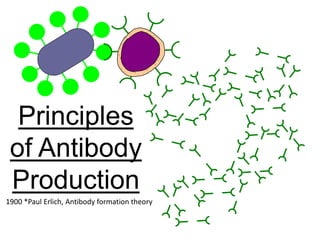
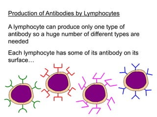
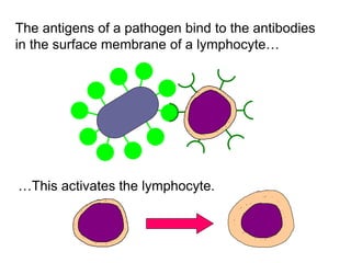
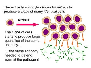
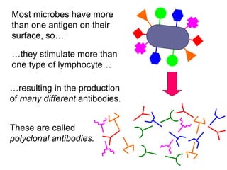
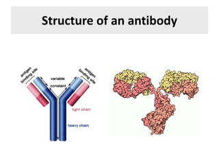
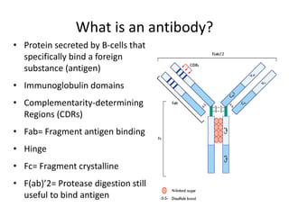
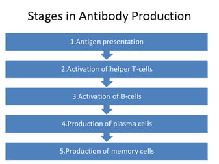
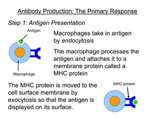
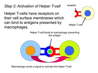
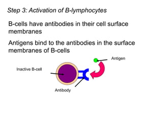
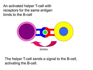
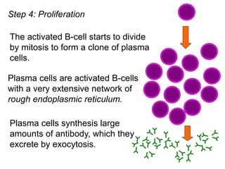
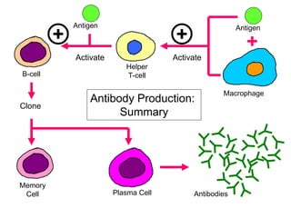
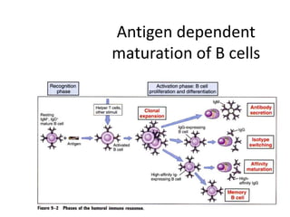
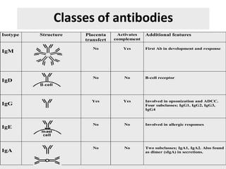
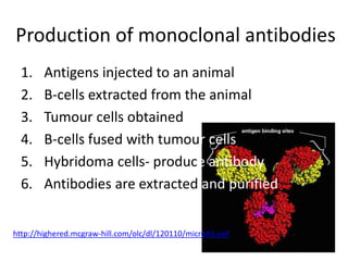
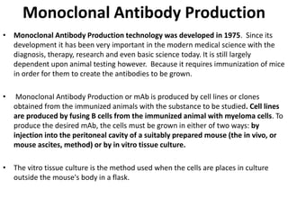
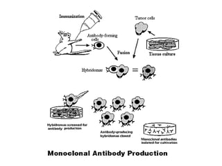
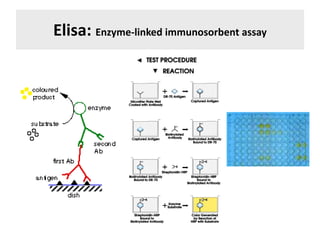
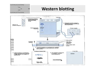
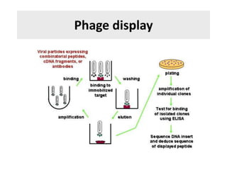
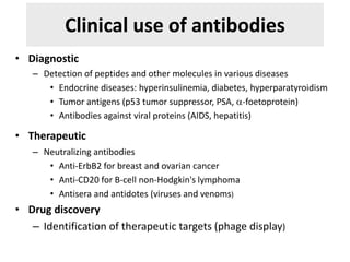
SUBMITTED TO:
Dr. NARAYAN MOGER
DEPARTMENT OF
BIOTECHNOLOGY
SUBMITTED BY:
Balaji Rathod
ID NO. PGS16AGR7165
Principles
of Antibody
Production
1900 *Paul Erlich, Antibody formation theory
The blood contains two types of white blood
cell or leukocyte
Phagocytes ingest bacteria by endocytosis
Lymphocytes produce antibodies
Antibodies
• Antibodies are proteins secreted by B cells
that specifically bind to a foreign substance-
Antigen
• Antigens are foreign substances that
stimulate the production of antibodies
• Many of the molecules on the surface of
viruses and bacteria are antigens
Antibodies are specific – they usually bind to
only one specific antigen.
Antigen
Antibodies
Microbe
Production of Antibodies by Lymphocytes
A lymphocyte can produce only one type of
antibody so a huge number of different types are
needed
Each lymphocyte has some of its antibody on its
surface…
The antigens of a pathogen bind to the antibodies
in the surface membrane of a lymphocyte…
…This activates the lymphocyte.
The active lymphocyte divides by mitosis to
produce a clone of many identical cells
MITOSIS
The clone of cells
starts to produce large
quantities of the same
antibody…
… the same antibody
needed to defend
against the pathogen!
Most microbes have more
than one antigen on their
surface, so…
…they stimulate more than
one type of lymphocyte…
…resulting in the production
of many different antibodies.
These are called
polyclonal antibodies.
Structure of an antibody
What is an antibody?
• Protein secreted by B-cells that
specifically bind a foreign
substance (antigen)
• Immunoglobulin domains
• Complementarity-determining
Regions (CDRs)
• Fab= Fragment antigen binding
• Hinge
• Fc= Fragment crystalline
• F(ab)’2= Protease digestion still
useful to bind antigen
Stages in Antibody Production
5.Production of memory cells
4.Production of plasma cells
3.Activation of B-cells
2.Activation of helper T-cells
1.Antigen presentation
Antibody Production: The Primary Response
Macrophages take in antigen
by endocytosis
Antigen
Macrophage
The macrophage processes the
antigen and attaches it to a
membrane protein called a
MHC protein
The MHC protein is moved to the
cell surface membrane by
exocytosis so that the antigen is
displayed on its surface.
MHC protein
Step 1: Antigen Presentation
Helper T-cell binds to macrophage presenting
the antigen
Step 2: Activation of Helper T-cell
Helper T-cells have receptors on
their cell surface membranes which
can bind to antigens presented by
macrophages.
receptor
Helper T-cell
Macrophage sends a signal to activate the helper T-cell
Step 3: Activation of B-lymphocytes
B-cells have antibodies in their cell surface
membranes
Inactive B-cell
Antibody
Antigens bind to the antibodies in the surface
membranes of B-cells
Antigen
An activated helper T-cell with
receptors for the same antigen
binds to the B-cell
SIGNAL
The helper T-cell sends a signal to the B-cell,
activating the B-cell.
Step 4: Proliferation
Plasma cells are activated B-cells
with a very extensive network of
rough endoplasmic reticulum.
Plasma cells synthesis large
amounts of antibody, which they
excrete by exocytosis.
The activated B-cell starts to divide
by mitosis to form a clone of plasma
cells.
The Secondary Response: Memory Cells
• If an antigen invades your body a second time, a
much faster response occurs which produces
much larger quantity of the required antibody.
•When activated B-cells are dividing during the
primary response, some cells stop dividing and
secreting antibody and become memory cells.
• Large numbers of memory cells remain in the
body for a long time…
• …they are capable of producing large amounts of
antibody very quickly when stimulated.
B-cell
Macrophage
Antigen
Activate
Antigen
Helper
T-cell
Activate
Clone
Memory
Cell Plasma Cell Antibodies
Antibody Production:
Summary
Principles of antibody production
• Clonal selection
– > 1015 antibodies
– B-cells clone themselves
• Challenge and response
– Immunity developed only when disease
challenges the immune system
Antigen dependent
maturation of B cells
Classes of antibodies
Isotype Structure Placenta
transfert
Activates
complement
Additional features
IgM
No Yes First Ab in development and response
IgD
No No B-cell receptor
IgG
Yes Yes Involved in opsonization and ADCC.
Four subclasses; IgG1, IgG2, IgG3,
IgG4
IgE
No No Involved in allergic responses
IgA
No No Two subclasses; IgA1, IgA2. Also found
as dimer (sIgA) in secretions.
Production of monoclonal antibodies
1. Antigens injected to an animal
2. B-cells extracted from the animal
3. Tumour cells obtained
4. B-cells fused with tumour cells
5. Hybridoma cells- produce antibody
6. Antibodies are extracted and purified
http://highered.mcgraw-hill.com/olc/dl/120110/micro43.swf
Monoclonal Antibody Production
• Monoclonal Antibody Production technology was developed in 1975. Since its
development it has been very important in the modern medical science with the
diagnosis, therapy, research and even basic science today. It is still largely
dependent upon animal testing however. Because it requires immunization of mice
in order for them to create the antibodies to be grown.
• Monoclonal Antibody Production or mAb is produced by cell lines or clones
obtained from the immunized animals with the substance to be studied. Cell lines
are produced by fusing B cells from the immunized animal with myeloma cells. To
produce the desired mAb, the cells must be grown in either of two ways: by
injection into the peritoneal cavity of a suitably prepared mouse (the in vivo, or
mouse ascites, method) or by in vitro tissue culture.
• The vitro tissue culture is the method used when the cells are places in culture
outside the mouse's body in a flask.
Why this method is used!!
• This method is used because antibodies must be formed from
the immunization of the substance being studied. So
antibodies must be produced. Once the antibodies are
produced the animal aspect of the study can be eliminated
and tissue culture can then be used.
• When using live mice researchers have found that it is the
better option because in vitro doesn’t always produce
adequate cell lines that are adaptive to tissue culture. Protein
denaturation can occur from purification techniques and
antibody activity is decreased with normal activity not
represented. Also cell lines could possibly become
contaminated when using in vitro technique.
Polyclonal antibodies:
If an animal is immunized with a protein, a wide array of B
cells will be stimulated to produce anti-protein antibodies.
Antibodies may be made to a number of different epitopes
of the protein.
Even antibodies that bind to the same epitope may have
different antigen-binding sites and bind the epitope with
different affinity.
The mixture of antibodies produced in response to an
antigen are referred to as polyclonal antibodies (they are
produced by many different clones of B cells).
Producing polyclonal antibodies
MONOCLONAL VS POLYCLONAL
ANTIBODIES
MONOCLONAL ANTIBODIES POLYCLONAL ANTIBODIES
Expensive to produce Inexpensive to produce
Training is required for the
technology used
Skills required are low
Time scale is long for hybridization. Time scale is short
Can produce large amount of
specific antibodies.
Produces large amount of non
specific antibodies.
Recognizes only 1 epitope on an
antigen
Recognizes multiple epitopes on
anyone antigen
Once hybridoma is made it is a
constant and renewable source.
No or low batch to batch variability.
Can have batch to batch variability.
Active and passive immunity
• Active immunity: antibodies by the organism
itself
• Passive immunity: antibodies received from
another organism
– During pregnancy antibodies passed to the fetus
Antigenic determinants
• An antibody will recognize
– Epitope: defined segment of an antigen
– Immunoreactivity of epitopes may depend on primary,
secondary, tertiary or quaternary structure of an
antigen
– Define the possible applications
– Variability of epitopes depends on the species
• Antibodies are antigen themselves
Commercial production of antibodies:
polyclonal vs monoclonal
• Slected clones from a polyclonal each recognizing a
single epitope can be fused to a tumor cell
(hybridoma) to proliferate indefinitely
• Host animals ca be used to raise antibodies against
a given antigen
Laboratory use of antibodies
• Quantitation of an antigen
– RIA, Elisa
• Identification and characterization of protein antigens
– Immunoprecipitation
– Western blotting
• Cell surface labelling and separation
• Localisation of antigens within tissues or cells
• Expression librairies
• Phage display
Elisa: Enzyme-linked immunosorbent assay
Western blotting
Phage display
Clinical use of antibodies
• Diagnostic
– Detection of peptides and other molecules in various diseases
• Endocrine diseases: hyperinsulinemia, diabetes, hyperparatyroidism
• Tumor antigens (p53 tumor suppressor, PSA, a-foetoprotein)
• Antibodies against viral proteins (AIDS, hepatitis)
• Therapeutic
– Neutralizing antibodies
• Anti-ErbB2 for breast and ovarian cancer
• Anti-CD20 for B-cell non-Hodgkin's lymphoma
• Antisera and antidotes (viruses and venoms)
• Drug discovery
– Identification of therapeutic targets (phage display)
Physiological roles of antibodies
• Protect against
– Viral infections
– Bacterial infections
– Foreign bodies
• Antigens
• Deleterious in
– Autoimmune diseases
• Reumathoid arthritis Lupus
• Type 1 diabetes Croh’n disease
– Graft rejection and hypersensitivity
responses
Lacking an antibody for your
protein or antigen of interest is
limiting the progression of your
research!

Antibody

  • 1.
    SUBMITTED TO: Dr. NARAYANMOGER DEPARTMENT OF BIOTECHNOLOGY SUBMITTED BY: Balaji Rathod ID NO. PGS16AGR7165
  • 2.
    Principles of Antibody Production 1900 *PaulErlich, Antibody formation theory
  • 3.
    The blood containstwo types of white blood cell or leukocyte Phagocytes ingest bacteria by endocytosis Lymphocytes produce antibodies
  • 4.
    Antibodies • Antibodies areproteins secreted by B cells that specifically bind to a foreign substance- Antigen • Antigens are foreign substances that stimulate the production of antibodies • Many of the molecules on the surface of viruses and bacteria are antigens
  • 5.
    Antibodies are specific– they usually bind to only one specific antigen. Antigen Antibodies Microbe
  • 6.
    Production of Antibodiesby Lymphocytes A lymphocyte can produce only one type of antibody so a huge number of different types are needed Each lymphocyte has some of its antibody on its surface…
  • 7.
    The antigens ofa pathogen bind to the antibodies in the surface membrane of a lymphocyte… …This activates the lymphocyte.
  • 8.
    The active lymphocytedivides by mitosis to produce a clone of many identical cells MITOSIS The clone of cells starts to produce large quantities of the same antibody… … the same antibody needed to defend against the pathogen!
  • 9.
    Most microbes havemore than one antigen on their surface, so… …they stimulate more than one type of lymphocyte… …resulting in the production of many different antibodies. These are called polyclonal antibodies.
  • 10.
  • 11.
    What is anantibody? • Protein secreted by B-cells that specifically bind a foreign substance (antigen) • Immunoglobulin domains • Complementarity-determining Regions (CDRs) • Fab= Fragment antigen binding • Hinge • Fc= Fragment crystalline • F(ab)’2= Protease digestion still useful to bind antigen
  • 12.
    Stages in AntibodyProduction 5.Production of memory cells 4.Production of plasma cells 3.Activation of B-cells 2.Activation of helper T-cells 1.Antigen presentation
  • 13.
    Antibody Production: ThePrimary Response Macrophages take in antigen by endocytosis Antigen Macrophage The macrophage processes the antigen and attaches it to a membrane protein called a MHC protein The MHC protein is moved to the cell surface membrane by exocytosis so that the antigen is displayed on its surface. MHC protein Step 1: Antigen Presentation
  • 14.
    Helper T-cell bindsto macrophage presenting the antigen Step 2: Activation of Helper T-cell Helper T-cells have receptors on their cell surface membranes which can bind to antigens presented by macrophages. receptor Helper T-cell Macrophage sends a signal to activate the helper T-cell
  • 15.
    Step 3: Activationof B-lymphocytes B-cells have antibodies in their cell surface membranes Inactive B-cell Antibody Antigens bind to the antibodies in the surface membranes of B-cells Antigen
  • 16.
    An activated helperT-cell with receptors for the same antigen binds to the B-cell SIGNAL The helper T-cell sends a signal to the B-cell, activating the B-cell.
  • 17.
    Step 4: Proliferation Plasmacells are activated B-cells with a very extensive network of rough endoplasmic reticulum. Plasma cells synthesis large amounts of antibody, which they excrete by exocytosis. The activated B-cell starts to divide by mitosis to form a clone of plasma cells.
  • 18.
    The Secondary Response:Memory Cells • If an antigen invades your body a second time, a much faster response occurs which produces much larger quantity of the required antibody. •When activated B-cells are dividing during the primary response, some cells stop dividing and secreting antibody and become memory cells. • Large numbers of memory cells remain in the body for a long time… • …they are capable of producing large amounts of antibody very quickly when stimulated.
  • 19.
  • 20.
    Principles of antibodyproduction • Clonal selection – > 1015 antibodies – B-cells clone themselves • Challenge and response – Immunity developed only when disease challenges the immune system
  • 21.
  • 22.
    Classes of antibodies IsotypeStructure Placenta transfert Activates complement Additional features IgM No Yes First Ab in development and response IgD No No B-cell receptor IgG Yes Yes Involved in opsonization and ADCC. Four subclasses; IgG1, IgG2, IgG3, IgG4 IgE No No Involved in allergic responses IgA No No Two subclasses; IgA1, IgA2. Also found as dimer (sIgA) in secretions.
  • 23.
    Production of monoclonalantibodies 1. Antigens injected to an animal 2. B-cells extracted from the animal 3. Tumour cells obtained 4. B-cells fused with tumour cells 5. Hybridoma cells- produce antibody 6. Antibodies are extracted and purified http://highered.mcgraw-hill.com/olc/dl/120110/micro43.swf
  • 24.
    Monoclonal Antibody Production •Monoclonal Antibody Production technology was developed in 1975. Since its development it has been very important in the modern medical science with the diagnosis, therapy, research and even basic science today. It is still largely dependent upon animal testing however. Because it requires immunization of mice in order for them to create the antibodies to be grown. • Monoclonal Antibody Production or mAb is produced by cell lines or clones obtained from the immunized animals with the substance to be studied. Cell lines are produced by fusing B cells from the immunized animal with myeloma cells. To produce the desired mAb, the cells must be grown in either of two ways: by injection into the peritoneal cavity of a suitably prepared mouse (the in vivo, or mouse ascites, method) or by in vitro tissue culture. • The vitro tissue culture is the method used when the cells are places in culture outside the mouse's body in a flask.
  • 26.
    Why this methodis used!! • This method is used because antibodies must be formed from the immunization of the substance being studied. So antibodies must be produced. Once the antibodies are produced the animal aspect of the study can be eliminated and tissue culture can then be used. • When using live mice researchers have found that it is the better option because in vitro doesn’t always produce adequate cell lines that are adaptive to tissue culture. Protein denaturation can occur from purification techniques and antibody activity is decreased with normal activity not represented. Also cell lines could possibly become contaminated when using in vitro technique.
  • 27.
    Polyclonal antibodies: If ananimal is immunized with a protein, a wide array of B cells will be stimulated to produce anti-protein antibodies. Antibodies may be made to a number of different epitopes of the protein. Even antibodies that bind to the same epitope may have different antigen-binding sites and bind the epitope with different affinity. The mixture of antibodies produced in response to an antigen are referred to as polyclonal antibodies (they are produced by many different clones of B cells).
  • 28.
  • 29.
    MONOCLONAL VS POLYCLONAL ANTIBODIES MONOCLONALANTIBODIES POLYCLONAL ANTIBODIES Expensive to produce Inexpensive to produce Training is required for the technology used Skills required are low Time scale is long for hybridization. Time scale is short Can produce large amount of specific antibodies. Produces large amount of non specific antibodies. Recognizes only 1 epitope on an antigen Recognizes multiple epitopes on anyone antigen Once hybridoma is made it is a constant and renewable source. No or low batch to batch variability. Can have batch to batch variability.
  • 30.
    Active and passiveimmunity • Active immunity: antibodies by the organism itself • Passive immunity: antibodies received from another organism – During pregnancy antibodies passed to the fetus
  • 31.
    Antigenic determinants • Anantibody will recognize – Epitope: defined segment of an antigen – Immunoreactivity of epitopes may depend on primary, secondary, tertiary or quaternary structure of an antigen – Define the possible applications – Variability of epitopes depends on the species • Antibodies are antigen themselves
  • 32.
    Commercial production ofantibodies: polyclonal vs monoclonal • Slected clones from a polyclonal each recognizing a single epitope can be fused to a tumor cell (hybridoma) to proliferate indefinitely • Host animals ca be used to raise antibodies against a given antigen
  • 33.
    Laboratory use ofantibodies • Quantitation of an antigen – RIA, Elisa • Identification and characterization of protein antigens – Immunoprecipitation – Western blotting • Cell surface labelling and separation • Localisation of antigens within tissues or cells • Expression librairies • Phage display
  • 34.
  • 35.
  • 36.
  • 37.
    Clinical use ofantibodies • Diagnostic – Detection of peptides and other molecules in various diseases • Endocrine diseases: hyperinsulinemia, diabetes, hyperparatyroidism • Tumor antigens (p53 tumor suppressor, PSA, a-foetoprotein) • Antibodies against viral proteins (AIDS, hepatitis) • Therapeutic – Neutralizing antibodies • Anti-ErbB2 for breast and ovarian cancer • Anti-CD20 for B-cell non-Hodgkin's lymphoma • Antisera and antidotes (viruses and venoms) • Drug discovery – Identification of therapeutic targets (phage display)
  • 38.
    Physiological roles ofantibodies • Protect against – Viral infections – Bacterial infections – Foreign bodies • Antigens • Deleterious in – Autoimmune diseases • Reumathoid arthritis Lupus • Type 1 diabetes Croh’n disease – Graft rejection and hypersensitivity responses
  • 39.
    Lacking an antibodyfor your protein or antigen of interest is limiting the progression of your research!