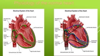
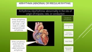
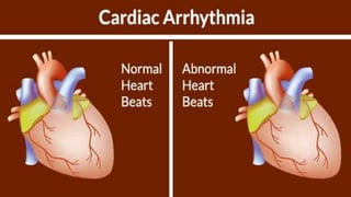
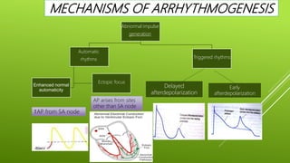
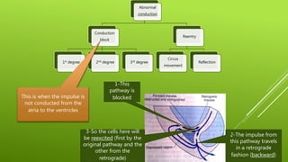
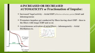
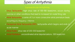
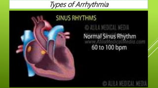
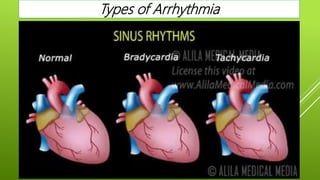
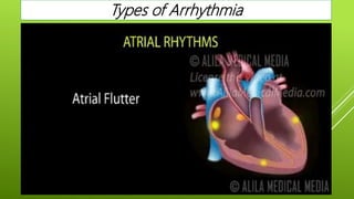
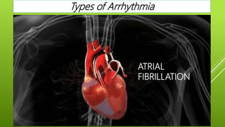
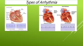
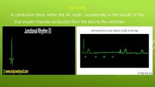
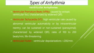
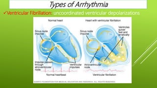
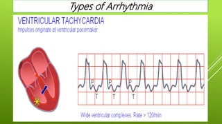
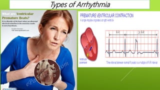
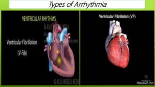
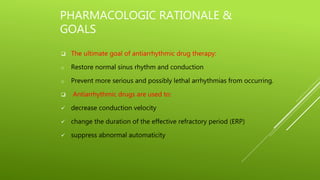
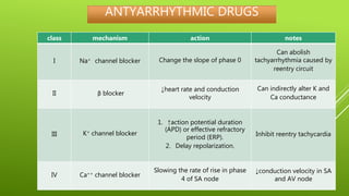
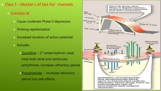
ANTIARRTHYMIC AGENTS
Mr. Sujit Karpe
Principal,
Sojar college of Pharmacy, Khandvi
Normal conduction pathway:
1- SA node generates
action potential and
delivers it to the atria
and the AV node
2- The AV node
delivers the impulse
to purkinje fibers
3- purkinje fibers
conduct the impulse
to the ventricles
Other types of
conduction that
occurs between
myocardial cells:
When a cell is
depolarized 
adjacent cell
depolarizes along
ARRHYTHMIA (ABNORMAL OR IRREGULAR RHYTHM)
If the arrhythmia
arises from the
ventricles it is
called ventricular
arrhythmia
If the arrhythmia
arises from atria,
SA node, or AV
node it is called
supraventricular
arrhythmia
Causes of
arrhythmia
arteriosclerosi
s
Coronary
artery spasm
Heart block
Myocardial
ischemia
MECHANISMS OF ARRHYTHMOGENESIS
Abnormal impulse
generation
Automatic
rhythms
Ectopic focus
Enhanced normal
automaticity
Triggered rhythms
Delayed
afterdepolarization
Early
afterdepolarization
↑AP from SA node
AP arises from sites
other than SA node
Abnormal
conduction
Conduction
block
1st degree 2nd degree 3rd degree
Reentry
Circus
movement
Reflection
This is when the impulse is
not conducted from the
atria to the ventricles
1-This
pathway is
blocked
2-The impulse from
this pathway travels
in a retrograde
fashion (backward)
3-So the cells here will
be reexcited (first by the
original pathway and the
other from the
retrograde)
Supraventricular Arrhythmias
 Sinus Tachycardia: high sinus rate of 100-180 beats/min, occurs during
exercise or other conditions that lead to increased SA nodal firing rate
 Atrial Tachycardia: a series of 3 or more consecutive atrial premature beats
occurring at a frequency >100/min
 Paroxysmal Atrial Tachycardia (PAT): tachycardia which begins and ends in
acute manner
 Atrial Flutter: sinus rate of 250-350 beats/min.
 Atrial Fibrillation: uncoordinated atrial depolarizations. 500 beats/min
Types of Arrhythmia
Types of Arrhythmia
Types of Arrhythmia
Types of Arrhythmia
Types of Arrhythmia
ATRIAL
FIBRILLATION
Types of Arrhythmia
AV blocks
A conduction block within the AV node , occasionally in the bundle of His,
that impairs impulse conduction from the atria to the ventricles.
 Ventricular Premature Beats (VPBs): caused by ectopic
ventricular foci; characterized by widened QRS.
 Ventricular Tachycardia (VT): high ventricular rate caused by
abnormal ventricular automaticity or by intraventricular
reentry; can be sustained or non-sustained (paroxysmal);
characterized by widened QRS; rates of 100 to 200
beats/min; life-threatening.
 Ventricular Flutter: ventricular depolarizations >200/min.
ventricular Arrhythmias
Types of Arrhythmia
Ventricular Fibrillation: uncoordinated ventricular depolarizations
Types of Arrhythmia
Types of Arrhythmia
Types of Arrhythmia
Types of Arrhythmia
PHARMACOLOGIC RATIONALE &
GOALS
 The ultimate goal of antiarrhythmic drug therapy:
o Restore normal sinus rhythm and conduction
o Prevent more serious and possibly lethal arrhythmias from occurring.
 Antiarrhythmic drugs are used to:
 decrease conduction velocity
 change the duration of the effective refractory period (ERP)
 suppress abnormal automaticity
ANTYARRHYTHMIC DRUGS
•Most antiarrhythmic drugs are pro-arrhythmic (promote
arrhythmia)
•They are classified according to Vaughan William into
four classes according to their effects on the cardiac
action potential
ANTYARRHYTHMIC DRUGS
class mechanism action notes
I Na+ channel blocker Change the slope of phase 0
Can abolish
tachyarrhythmia caused by
reentry circuit
II β blocker
↓heart rate and conduction
velocity
Can indirectly alter K and
Ca conductance
III K+ channel blocker
1. ↑action potential duration
(APD) or effective refractory
period (ERP).
2. Delay repolarization.
Inhibit reentry tachycardia
IV Ca++ channel blocker
Slowing the rate of rise in phase
4 of SA node
↓conduction velocity in SA
and AV node
 Class I – blocker’s of fast Na+ channels
 Subclass IA
 Cause moderate Phase 0 depression
 Prolong repolarization
 Increased duration of action potential
 Includes
Quinidine – 1st antiarrhythmic used,
treat both atrial and ventricular
arrhythmias, increases refractory period
Procainamide - increases refractory
period but side effects
 QUINIDINE
 Isomer of anti malarial drug Quinine
Pharmacological Actions
 Automaticity: depresses re entry of sodium into cell during depolarization, and there by
depresses diastolic depolarization – decreases automaticity.
 Excitability: depresses excitability and thus make ectopic impulse ineffective.
 Conduction Velocity: reduces conduction velocity, increases refractory period and reduces
excitability restore rhythm to normal.
 Refractory period: depresses potassium efflux during repolarization and thus prolong
refractory period.
 A. V. node conduction: depresses conduction between atria and purkinje system.
 Contractility: depresses entry of calcium ions – reduces contractility.
 QUINIDINE
Pharmacokinetic parameter
 Completely absorb from gut.
 Excreted through kidney.
Side effect
 Cinchonism characterized by, tinnitus (Ringing in the ear), vertigo, blurring of vision.
 Embolism: sudden restoration of normal rhythm, may dislodge the mural thrombi attached
to auricular appendages. This may occlude the blood vessels causing embolism (obstruction
of an artery, typically by a clot of blood or an air bubble).
 QUINIDINE
Therapeutic uses
 Atrial fibrillation
 Atrial Flutter
 Paroxysmal supraventricular tachycardia
 Ventricular arrhythmias.
Side effect
 Quinidine sulphate – 200 mg t. i. d. oral
 Quinidine gluconate – 200 mg oral
 PROCAINAMIDE
 Definite depressant effect on S. A. Node.
 Reduces Excitability.
 Reduces conduction velocity.
 Prolongs refractory period.
 Less potent than quinidine.
 Rapidly and completely absorbed from gut.
 Nausea, bitter taste, anorexia are common side effect.
 Used in ventricular arrhythmia
 Subclass IB
 Weak Phase 0 depression
 Shortened depolarization
 Decreased action potential duration
 Includes
Lidocane (also acts as local anesthetic)
– blocks Na+ channels mostly in
ventricular cells, also good for
digitalis-associated arrhythmias
Phenytoin – anticonvulsant that also
works as antiarrhythmic similar to
lidocane
 LIGNOCAINE
 Local anesthetic.
 Decreases duration of action potential.
 Enhances conduction velocity.
 Increases membrane responsiveness.
 Shortens duration of action potential.
 Reduces re-entry type of circuitous transmitters.
 Always administered I/V
 Excreted unchanged in urine.
 Used to treat ventricular arrhythmias.
 Subclass IC
 Strong Phase 0 depression
 No effect of depolarization
 No effect on action potential duration
 Includes
Flecainide (initially developed as a local
anesthetic)
Slows conduction in all parts of heart,
Also inhibits abnormal automaticity
Propafenone
Also slows conduction
Weak β – blocker
Also some Ca2+ channel blockade
 Class II – β–adrenergic blockers
 Based on two major actions
1) blockade of myocardial β–adrenergic receptors
2) Direct membrane-stabilizing effects related to Na+ channel blockade
 Propranolol
causes both myocardial β–adrenergic blockade and membrane-stabilizing effects
Slows SA node and ectopic pacemaking
Can block arrhythmias induced by exercise or apprehension
Prolongs nodal refractory period.
Completely absorbed through gut, metabolized by liver and excreted in urine.
Class III – K+ channel blockers
 Developed because some patients negatively
sensitive to Na channel blockers (they died!)
 Cause delay in repolarization and prolonged
refractory period
 Amiodarone – prolongs action potential by
delaying K+ efflux but many other effects
characteristic of other classes
 Bretylium – first developed to treat
hypertension but found to also suppress
ventricular fibrillation associated with myocardial
infarction
 Class IV – Ca2+ channel blockers
 Inhibit influx of Calcium through
cardiac cell membrane into cardiac
cell.
 slow rate of AV-conduction in
patients with atrial fibrillation
 Also called peripheral vasodilators.
 Includes
 Verapamil – blocks Na+ channels
in addition to Ca2+; also slows SA
node in tachycardia
 Diltiazem

Anti arrhythmic agents

  • 1.
    ANTIARRTHYMIC AGENTS Mr. SujitKarpe Principal, Sojar college of Pharmacy, Khandvi
  • 2.
    Normal conduction pathway: 1-SA node generates action potential and delivers it to the atria and the AV node 2- The AV node delivers the impulse to purkinje fibers 3- purkinje fibers conduct the impulse to the ventricles Other types of conduction that occurs between myocardial cells: When a cell is depolarized  adjacent cell depolarizes along
  • 4.
    ARRHYTHMIA (ABNORMAL ORIRREGULAR RHYTHM) If the arrhythmia arises from the ventricles it is called ventricular arrhythmia If the arrhythmia arises from atria, SA node, or AV node it is called supraventricular arrhythmia Causes of arrhythmia arteriosclerosi s Coronary artery spasm Heart block Myocardial ischemia
  • 6.
    MECHANISMS OF ARRHYTHMOGENESIS Abnormalimpulse generation Automatic rhythms Ectopic focus Enhanced normal automaticity Triggered rhythms Delayed afterdepolarization Early afterdepolarization ↑AP from SA node AP arises from sites other than SA node
  • 7.
    Abnormal conduction Conduction block 1st degree 2nddegree 3rd degree Reentry Circus movement Reflection This is when the impulse is not conducted from the atria to the ventricles 1-This pathway is blocked 2-The impulse from this pathway travels in a retrograde fashion (backward) 3-So the cells here will be reexcited (first by the original pathway and the other from the retrograde)
  • 9.
    Supraventricular Arrhythmias  SinusTachycardia: high sinus rate of 100-180 beats/min, occurs during exercise or other conditions that lead to increased SA nodal firing rate  Atrial Tachycardia: a series of 3 or more consecutive atrial premature beats occurring at a frequency >100/min  Paroxysmal Atrial Tachycardia (PAT): tachycardia which begins and ends in acute manner  Atrial Flutter: sinus rate of 250-350 beats/min.  Atrial Fibrillation: uncoordinated atrial depolarizations. 500 beats/min Types of Arrhythmia
  • 10.
  • 11.
  • 12.
  • 13.
  • 14.
  • 15.
    AV blocks A conductionblock within the AV node , occasionally in the bundle of His, that impairs impulse conduction from the atria to the ventricles.
  • 16.
     Ventricular PrematureBeats (VPBs): caused by ectopic ventricular foci; characterized by widened QRS.  Ventricular Tachycardia (VT): high ventricular rate caused by abnormal ventricular automaticity or by intraventricular reentry; can be sustained or non-sustained (paroxysmal); characterized by widened QRS; rates of 100 to 200 beats/min; life-threatening.  Ventricular Flutter: ventricular depolarizations >200/min. ventricular Arrhythmias Types of Arrhythmia
  • 17.
    Ventricular Fibrillation: uncoordinatedventricular depolarizations Types of Arrhythmia
  • 18.
  • 19.
  • 20.
  • 21.
    PHARMACOLOGIC RATIONALE & GOALS The ultimate goal of antiarrhythmic drug therapy: o Restore normal sinus rhythm and conduction o Prevent more serious and possibly lethal arrhythmias from occurring.  Antiarrhythmic drugs are used to:  decrease conduction velocity  change the duration of the effective refractory period (ERP)  suppress abnormal automaticity
  • 22.
    ANTYARRHYTHMIC DRUGS •Most antiarrhythmicdrugs are pro-arrhythmic (promote arrhythmia) •They are classified according to Vaughan William into four classes according to their effects on the cardiac action potential
  • 23.
    ANTYARRHYTHMIC DRUGS class mechanismaction notes I Na+ channel blocker Change the slope of phase 0 Can abolish tachyarrhythmia caused by reentry circuit II β blocker ↓heart rate and conduction velocity Can indirectly alter K and Ca conductance III K+ channel blocker 1. ↑action potential duration (APD) or effective refractory period (ERP). 2. Delay repolarization. Inhibit reentry tachycardia IV Ca++ channel blocker Slowing the rate of rise in phase 4 of SA node ↓conduction velocity in SA and AV node
  • 24.
     Class I– blocker’s of fast Na+ channels  Subclass IA  Cause moderate Phase 0 depression  Prolong repolarization  Increased duration of action potential  Includes Quinidine – 1st antiarrhythmic used, treat both atrial and ventricular arrhythmias, increases refractory period Procainamide - increases refractory period but side effects
  • 25.
     QUINIDINE  Isomerof anti malarial drug Quinine Pharmacological Actions  Automaticity: depresses re entry of sodium into cell during depolarization, and there by depresses diastolic depolarization – decreases automaticity.  Excitability: depresses excitability and thus make ectopic impulse ineffective.  Conduction Velocity: reduces conduction velocity, increases refractory period and reduces excitability restore rhythm to normal.  Refractory period: depresses potassium efflux during repolarization and thus prolong refractory period.  A. V. node conduction: depresses conduction between atria and purkinje system.  Contractility: depresses entry of calcium ions – reduces contractility.
  • 26.
     QUINIDINE Pharmacokinetic parameter Completely absorb from gut.  Excreted through kidney. Side effect  Cinchonism characterized by, tinnitus (Ringing in the ear), vertigo, blurring of vision.  Embolism: sudden restoration of normal rhythm, may dislodge the mural thrombi attached to auricular appendages. This may occlude the blood vessels causing embolism (obstruction of an artery, typically by a clot of blood or an air bubble).
  • 27.
     QUINIDINE Therapeutic uses Atrial fibrillation  Atrial Flutter  Paroxysmal supraventricular tachycardia  Ventricular arrhythmias. Side effect  Quinidine sulphate – 200 mg t. i. d. oral  Quinidine gluconate – 200 mg oral
  • 28.
     PROCAINAMIDE  Definitedepressant effect on S. A. Node.  Reduces Excitability.  Reduces conduction velocity.  Prolongs refractory period.  Less potent than quinidine.  Rapidly and completely absorbed from gut.  Nausea, bitter taste, anorexia are common side effect.  Used in ventricular arrhythmia
  • 29.
     Subclass IB Weak Phase 0 depression  Shortened depolarization  Decreased action potential duration  Includes Lidocane (also acts as local anesthetic) – blocks Na+ channels mostly in ventricular cells, also good for digitalis-associated arrhythmias Phenytoin – anticonvulsant that also works as antiarrhythmic similar to lidocane
  • 30.
     LIGNOCAINE  Localanesthetic.  Decreases duration of action potential.  Enhances conduction velocity.  Increases membrane responsiveness.  Shortens duration of action potential.  Reduces re-entry type of circuitous transmitters.  Always administered I/V  Excreted unchanged in urine.  Used to treat ventricular arrhythmias.
  • 31.
     Subclass IC Strong Phase 0 depression  No effect of depolarization  No effect on action potential duration  Includes Flecainide (initially developed as a local anesthetic) Slows conduction in all parts of heart, Also inhibits abnormal automaticity Propafenone Also slows conduction Weak β – blocker Also some Ca2+ channel blockade
  • 32.
     Class II– β–adrenergic blockers  Based on two major actions 1) blockade of myocardial β–adrenergic receptors 2) Direct membrane-stabilizing effects related to Na+ channel blockade  Propranolol causes both myocardial β–adrenergic blockade and membrane-stabilizing effects Slows SA node and ectopic pacemaking Can block arrhythmias induced by exercise or apprehension Prolongs nodal refractory period. Completely absorbed through gut, metabolized by liver and excreted in urine.
  • 33.
    Class III –K+ channel blockers  Developed because some patients negatively sensitive to Na channel blockers (they died!)  Cause delay in repolarization and prolonged refractory period  Amiodarone – prolongs action potential by delaying K+ efflux but many other effects characteristic of other classes  Bretylium – first developed to treat hypertension but found to also suppress ventricular fibrillation associated with myocardial infarction
  • 34.
     Class IV– Ca2+ channel blockers  Inhibit influx of Calcium through cardiac cell membrane into cardiac cell.  slow rate of AV-conduction in patients with atrial fibrillation  Also called peripheral vasodilators.  Includes  Verapamil – blocks Na+ channels in addition to Ca2+; also slows SA node in tachycardia  Diltiazem