Antiarrhythmic Drugs
By: Muhammad Aurangzeb
Lecturer-INS/KMU
By the end of this unit learners, will be able to:
• Discuss pharmacological management of
arrhythmias.
• Discuss nursing implication in relation to
antiarrhythmic drugs.
• Calculate the drug dosage accurately while
administering oral and parenteral medication
Objectives
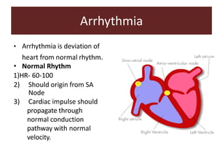
Arrhythmia
• Arrhythmia is deviation of
heart from normal rhythm.
• Normal Rhythm
1)HR- 60-100
2) Should origin from SA
Node
3) Cardiac impulse should
propagate through
normal conduction
pathway with normal
velocity.
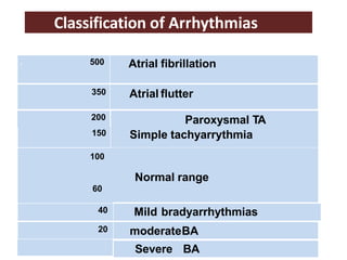
100
60
Normal range
150 Simple tachyarrythmia
200 Paroxysmal TA
. 500 Atrial fibrillation
350 Atrial flutter
40 Mild bradyarrhythmias
20 moderateBA
Severe BA
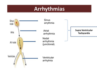
Classification of Arrhythmias
Arrhythmias
Sinus
arrythmia
Atrial
arrhythmia
Nodal
arrhythmia
(junctional)
Ventricular
arrhytmia
Supra Ventricular
Tachycardia
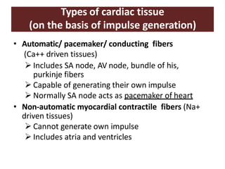
Types of cardiac tissue
(on the basis of impulse generation)
• Automatic/ pacemaker/ conducting fibers
(Ca++ driven tissues)
 Includes SA node, AV node, bundle of his,
purkinje fibers
 Capable of generating their own impulse
 Normally SA node acts as pacemaker of heart
• Non-automatic myocardial contractile fibers (Na+
driven tissues)
 Cannot generate own impulse
 Includes atria and ventricles
Electrophysiology of cardiac tissue
• Impulse generation and transmission
• Myocardial action potential
• Depolarization and repolarization waves as seen
in ECG
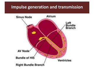
Impulse generation and transmission
Myocardial action potential
In automatic tissues In non-automatic tissues
(See video for it)
https://www.youtube.com/watch?v=9xSqezC
MHnw
Arrhythmia
• Definition:
– Disturbances in the heart rate, rhythm, impulse
generation or conduction of electrical impulses
responsible for membrane depolarization
– These disturbances can lead to alterations in
overall cardiac function that can be life
threatening.
• Antiarrhythmic drugs:
– Compounds used to prevent or treat cardiac
arrhythmias
Mechanism of arrhythmias
• Disturbances in impulse generation may be due to
– Abnormal automaticity
– Delayed after depolarization
• Disturbances of impulse conduction
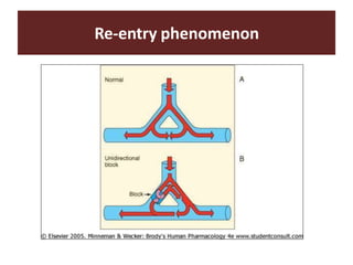
– The impulse may recirculate in heart causing
repeated activation (re-entry)
– Conduction blocks
Re-entry phenomenon
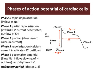
Phases of action potential of cardiac cells
Phase 0 rapid depolarisation
(inflow of Na+)
Phase 1 partial repolarisation
(inward Na+ current deactivated,
outflow of K+)
Phase 2 plateau (slow inward
calcium current)
Phase 3 repolarisation (calcium
current inactivates, K+ outflow)
Phase 4 pacemaker potential
(Slow Na+ inflow, slowing of K+
outflow) ‘autorhythmicity’
Refractory period (phases 1-3)
Phase 4
II
Phase
0
Phase 1
Phase 3
0
mV
-80mV
I
III
IV
Phase 2
Cont…
• Phase 0: Fast sodium channels are responsible for
initial rapid depolarization
• Phase 1: Early fast repolarization (K+ efflux)
• Phase 2: Prolonged depolarization “plateau” due to
slow calcium influx
• Phase 3: Repolarization due to closing of the calcium
channels and potassium efflux
• Phase 4: Resting membrane potential is restored
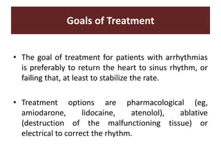
Goals of Treatment
• The goal of treatment for patients with arrhythmias
is preferably to return the heart to sinus rhythm, or
failing that, at least to stabilize the rate.
• Treatment options are pharmacological (eg,
amiodarone, lidocaine, atenolol), ablative
(destruction of the malfunctioning tissue) or
electrical to correct the rhythm.
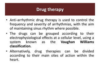
Drug therapy
• Anti-arrhythmic drug therapy is used to control the
frequency and severity of arrhythmias, with the aim
of maintaining sinus rhythm where possible.
• The drugs can be grouped according to their
electrophysiological effects at a cellular level, using a
system known as the Vaughan Williams
classification.
• Alternatively, drug therapies can be divided
according to their main sites of action within the
heart.
Vaughan Williams classification
• All anti-arrhythmic drugs act by altering the
movement of electrolytes within the electrical
conduction pathways of the myocardium.
• The Vaughan Williams classification system groups
drugs according to their ability to block the
movement of one or more of these ions across the
myocardial cell membrane.
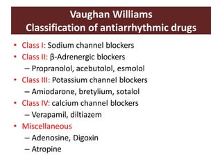
Vaughan Williams
Classification of antiarrhythmic drugs
• Class I: Sodium channel blockers
• Class II: β-Adrenergic blockers
– Propranolol, acebutolol, esmolol
• Class III: Potassium channel blockers
– Amiodarone, bretylium, sotalol
• Class IV: calcium channel blockers
– Verapamil, diltiazem
• Miscellaneous
– Adenosine, Digoxin
– Atropine
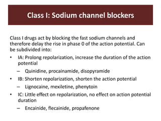
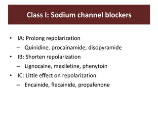
Class I: Sodium channel blockers
Class I drugs act by blocking the fast sodium channels and
therefore delay the rise in phase 0 of the action potential. Can
be subdivided into:
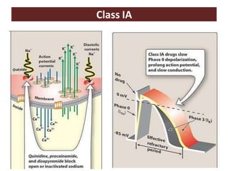
• IA: Prolong repolarization, increase the duration of the action
potential
– Quinidine, procainamide, disopyramide
• IB: Shorten repolarization, shorten the action potential
– Lignocaine, mexiletine, phenytoin
• IC: Little effect on repolarization, no effect on action potential
duration
– Encainide, flecainide, propafenone
Class I: Sodium channel blockers
• IA: Prolong repolarization
– Quinidine, procainamide, disopyramide
• IB: Shorten repolarization
– Lignocaine, mexiletine, phenytoin
• IC: Little effect on repolarization
– Encainide, flecainide, propafenone
Class IA
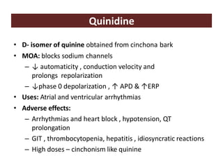
Quinidine
• D- isomer of quinine obtained from cinchona bark
• MOA: blocks sodium channels
– ↓ automaticity , conduction velocity and
prolongs repolarization
– ↓phase 0 depolarization , ↑ APD & ↑ERP
• Uses: Atrial and ventricular arrhythmias
• Adverse effects:
– Arrhythmias and heart block , hypotension, QT
prolongation
– GIT , thrombocytopenia, hepatitis , idiosyncratic reactions
– High doses – cinchonism like quinine
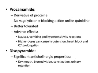
• Procainamide:
– Derivative of procaine
– No vagolytic or α-blocking action unlike quinidine
– Better tolerated
– Adverse effects:
• Nausea, vomiting and hypersensitivity reactions
• Higher doses can cause hypotension, heart block and
QT prolongation
• Disopyramide:
– Significant anticholinergic properties:
• Dry mouth, blurred vision, constipation, urinary
retention
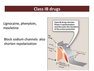
Class IB drugs
Lignocaine, phenytoin,
mexiletine
Block sodium channels also
shorten repolarization
Lignocaine
• Local anesthetic
• Raises threshold for action potential,
• ↓automaticity
• Suppress electrical activity of arrhythmogenic
tissues, normal tissues less effected
• High first pass metabolism so given
parenterally
• Use: ventricular arrhythmias
• Adverse effects:
– Drowsiness, hypotension, blurred vision,
confusion and convulsions
• Phenytoin:
– Antiepileptic also useful in ventricular arrhythmias
(not preferred) and digitalis induced arrhythmias
• Mexiletine:
– Can be used orally causes dose related
neurological adverse events like tremors and
blurred vision
– Nausea is common
– Used as alternative to lignocaine in ventricular
arrhythmias
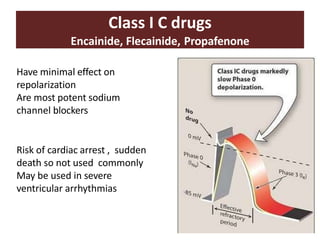
Class I C drugs
Encainide, Flecainide, Propafenone
Have minimal effect on
repolarization
Are most potent sodium
channel blockers
Risk of cardiac arrest , sudden
death so not used commonly
May be used in severe
ventricular arrhythmias
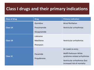
Class I drugs and their primary indications
Class of drug Drug Primary indication
Class 1A
Quinidine
Procainamide
Disopyramide
Atrial fibrillation
Ventricular arrhythmias
Class 1B
Lidocaine
Mexilitene
Phenytoin
Ventricular arrhythmias
Class 1C
Flecainide
Propafenone
AV nodal re-entry
Wolff-Parkinson White
syndrome-related arrhythmias
Ventricular arrhythmias (but
increased risk of mortality
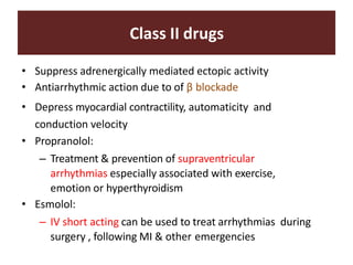
Class II drugs
• Suppress adrenergically mediated ectopic activity
• Antiarrhythmic action due to of β blockade
• Depress myocardial contractility, automaticity and
conduction velocity
• Propranolol:
– Treatment & prevention of supraventricular
arrhythmias especially associated with exercise,
emotion or hyperthyroidism
• Esmolol:
– IV short acting can be used to treat arrhythmias during
surgery , following MI & other emergencies
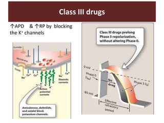
↑APD & ↑RP by blocking
the K+ channels
Class III drugs
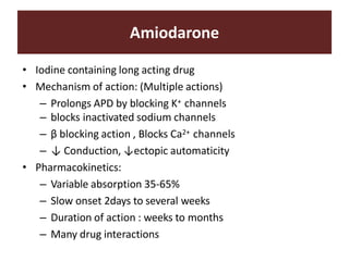
Amiodarone
• Iodine containing long acting drug
• Mechanism of action: (Multiple actions)
– Prolongs APD by blocking K+ channels
– blocks inactivated sodium channels
– β blocking action , Blocks Ca2+ channels
– ↓ Conduction, ↓ectopic automaticity
• Pharmacokinetics:
– Variable absorption 35-65%
– Slow onset 2days to several weeks
– Duration of action : weeks to months
– Many drug interactions
Amiodarone
• Uses:
– Can be used for both supraventricular and ventricular
tachycardia
• Adverse effects:
– Cardiac: heart block , QT prolongation, bradycardia,
cardiac failure, hypotension
– Pulmonary: pneumonitis leading to pulmonary fibrosis
– Bluish discoloration of skin
– GIT disturbances, hepatotoxicity
– Blocks peripheral conversion of T4 to T3 can cause
hypothyroidism or hyperthyroidism
Other Class III Drugs
• Bretylium:
– Adrenergic neuron blocker used in resistant
ventricular arrhythmias
• Sotalol:
– Beta blocker
• Dofetilide:
– Selective K+ channel blocker, less adverseevents
– Oral use in AF to convert or maintain sinus rhythm
• Ibutilide:
– K+ channel blocker used as IV infusion in AF or flutter
can cause QT prolongation
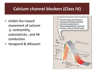
Calcium channel blockers (Class IV)
• Inhibit the inward
movement of calcium
↓ contractility,
automaticity , and AV
conduction.
• Verapamil & diltiazem
Verapamil
• Uses:
– Terminate PSVT
– control ventricular rate in atrial flutter or
fibrillation
• Drug interactions:
– Displaces digoxin from binding sites
– ↓ renal clearance of digoxin
Other antiarrhythmics
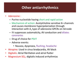
• Adenosine :
– Purine nucleotide having short and rapid action
– Mechanism of action: Acetylcholine sensitive K+ channels
and causes membrane hyperpolarization through
interaction with A1 type of adenosine GPCRs on SAnode
– IV suppresses automaticity, AV conduction and dilates
coronaries
– Drug of choice for PSVT
– Adverse events:
• Nausea, dyspnoea, flushing, headache
• Atropine: Used in sinus bradycardia, AV block
• Digitalis: Atrial fibrillation and atrial flutter
• Magnesium SO4: digitalis induced arrhythmias
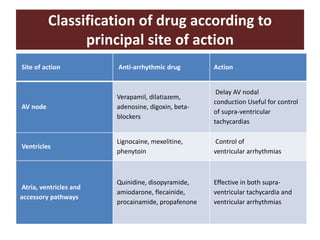
Classification of drug according to
principal site of action
Site of action Anti-arrhythmic drug Action
AV node
Verapamil, dilatiazem,
adenosine, digoxin, beta-
blockers
Delay AV nodal
conduction Useful for control
of supra-ventricular
tachycardias
Ventricles
Lignocaine, mexelitine,
phenytoin
Control of
ventricular arrhythmias
Atria, ventricles and
accessory pathways
Quinidine, disopyramide,
amiodarone, flecainide,
procainamide, propafenone
Effective in both supra-
ventricular tachycardia and
ventricular arrhythmias
Nursing Considerations for Antiarrhythmic
drugs
• Assess for the mentioned contraindications to this drug (e.g.
renal dysfunction, heart blocks, hypersensitivity, etc.) to
prevent potential adverse effects.
• Conduct thorough physical assessment before beginning drug
therapy to establish baseline status, and to evaluate potential
adverse effects.
• Assess cardiac status closely (e.g. blood pressure, heart rate
and rhythm, heart sounds, ec.) to determine whether change
in drug dose is imperative.
• Monitor laboratory test results including complete blood
count, renal and liver function tests to determine the need for
possible change in dose and identify toxic effects.
Nursing Intervention
These are vital nursing interventions done in patients who are
taking antiarrhythmic drugs:
• Titrate the dose to the smallest amount enough to manage
arrhythmia to decrease the risk of drug toxicity.
• Monitor cardiac rhythm closely to detect potentially serious
adverse effects and to evaluate drug effectiveness.
• Ensure maintenance of emergency drugs and equipment at
bedside to promote prompt treatment in cases of severe
toxicity.
• Educate patient on drug therapy including drug name, its
indication, and adverse effects to watch out for to enhance
patient understanding on drug therapy and thereby promote
adherence to drug regimen.
References
• Karch, A. M., & Karch. (2011). Focus on nursing pharmacology.
Wolters Kluwer Health/Lippincott Williams & Wilkins. [Link]
• Katzung, B. G. (2017). Basic and clinical pharmacology.
McGraw-Hill Education.
• Lehne, R. A., Moore, L. A., Crosby, L. J., & Hamilton, D. B.
(2004). Pharmacology for nursing care.
• Smeltzer, S. C., & Bare, B. G. (1992). Brunner & Suddarth’s
textbook of medical-surgical nursing. Philadelphia: JB
Lippincott