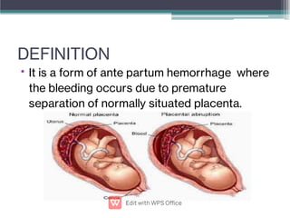
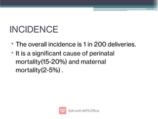
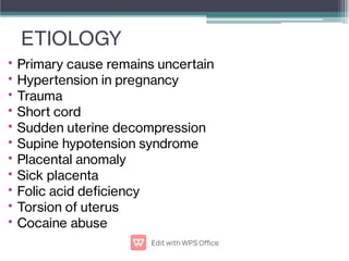
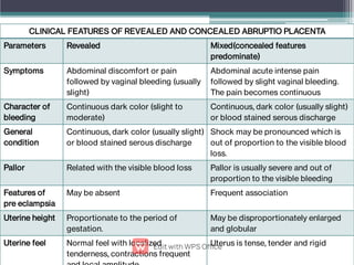
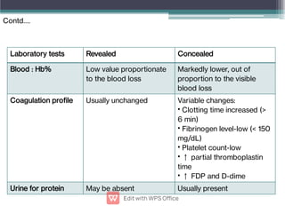
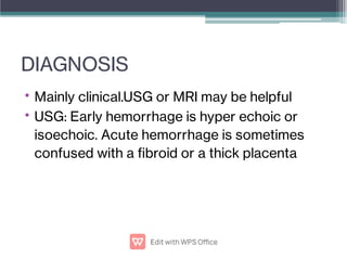
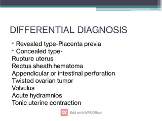
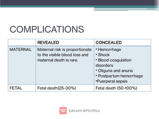
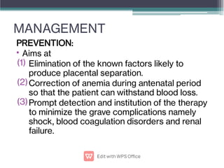
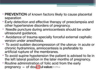
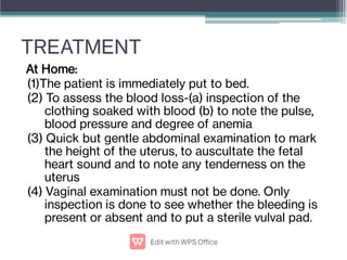
ABRUPTIO PLACENTA
DEFINITION
• It is a form of ante partum hemorrhage where
the bleeding occurs due to premature
separation of normally situated placenta.
INCIDENCE
•
•
The overall incidence is 1 in 200 deliveries.
It is a signicant cause of perinatal
mortality(15-20%) and maternal
mortality(2-5%) .
ETIOLOGY
•
•
•
•
•
•
•
•
•
•
•
Primary cause remains uncertain
Hypertension in pregnancy
Trauma
Short cord
Sudden uterine decompression
Supine hypotension syndrome
Placental anomaly
Sick placenta
Folic acid deciency
Torsion of uterus
Cocaine abuse
•
•
•
Thrombophilias
Prior abruption
Maternal risk factor
High birth order pregnancies with gravida 5 &above
Advancing age of mother
Poor socio-economic condition
Malnutrition
Smoking(vasospasm)
PATHOGENESIS
Placental abruption initiated by hemorrhage into
decidua basalis
Decidual hematoma
Retroplacental hematoma Accumulated blood
seeps into myometrium
Couvelaire uterus
(uteroplacental apoplexy)
•
•
Features:
Depression found on
maternal surface of the
placenta with a clot which
may be found rmly
attached to the area.
Areas of infarction with
varying degree of
organization.
COUVELAIRE UTERUS
•
•
•
•
•
•
Massive intravasation of blood into
uterine musculature up to the serous
coat.
Can be diagnosed only on laparotomy.
Features:
Uterus is of dark port wine colour
(patchy/diffuse)
Sub peritoneal petechial hemorrhages
are found under the uterine peritoneum
and may extend into the broad ligament.
There may be free blood in the
peritoneal cavity or broad ligament.
It rarely interferes with uterine
TYPES
•
•
•
Revealed-Most common type
Following separation of placenta ,the blood insinuates
downwards between the membranes and the decidua to be
visible externally
Concealed-Rare type
The blood collects behind the separated placenta or
collected in between the membranes and decidua. Collected
blood is prevented from coming out of the cervix by the
presenting part which presses on the lower segment.
Mixed-In this type ,some part of blood collects inside
(concealed ) and a part is expelled out(revealed).Usually one
type predominates over other.
Revealed Concealed
CLINICAL CLASSIFICATION
•
•
•
•
Grade 0-Clinical features absent. Diagnosed after
inspection of placenta following delivery.
Grade 1-(i) vaginal bleeding is slight (ii) uterus: irritable,
tenderness may be minimal or absent (iii) maternal BP and
brinogen levels unaffected (iv) FHS is good
Grade 2-(i) vaginal bleeding : mild to moderate (ii) uterine
tenderness is always present (iii) maternal pulse ↑, BP is
maintained (iv) brinogen level may be decreased (v)
shock is absent (vi) fetal distress or even fetal death
occurs
Grade 3- (i) Bleeding is moderate to severe or may be
concealed (ii) uterine tenderness is marked (iii) shock is
pronounced (iv) fetal death is the rule (v) associated
coagulation defect or anuria may complicate.
CLINICAL FEATURES
• Depend on:
a)Degree of separation of placenta
b)Speed at which separation occurs
c)Amount of blood concealed inside the uterine
cavity
CLINICAL FEATURES OF REVEALED AND CONCEALED ABRUPTIO PLACENTA
Parameters Revealed Mixed(concealed features
predominate)
Symptoms Abdominal discomfort or pain
followed by vaginal bleeding (usually
slight)
Abdominal acute intense pain
followed by slight vaginal bleeding.
The pain becomes continuous
Character of
bleeding
Continuous dark color (slight to
moderate)
Continuous, dark color (usually slight)
or blood stained serous discharge
General
condition
Continuous, dark color (usually slight)
or blood stained serous discharge
Shock may be pronounced which is
out of proportion to the visible blood
loss.
Pallor Related with the visible blood loss Pallor is usually severe and out of
proportion to the visible bleeding
Features of
pre eclampsia
May be absent Frequent association
Uterine height Proportionate to the period of
gestation.
May be disproportionately enlarged
and globular
Uterine feel Normal feel with localized
tenderness, contractions frequent
Uterus is tense, tender and rigid
Laboratory tests Revealed Concealed
Blood : Hb% Low value proportionate
to the blood loss
Markedly lower, out of
proportion to the visible
blood loss
Coagulation prole Usually unchanged Variable changes:
Clotting time increased (>
6 min)
Fibrinogen level-low (< 150
mg/dL)
Platelet count-low
↑ partial thromboplastin
time
↑ FDP and D-dime
•
•
•
•
•
Urine for protein May be absent Usually present
Contd….
DIAGNOSIS
•
•
Mainly clinical.USG or MRI may be helpful
USG: Early hemorrhage is hyper echoic or
isoechoic. Acute hemorrhage is sometimes
confused with a broid or a thick placenta
DIFFERENTIAL DIAGNOSIS
•
•
Revealed type-Placenta previa
Concealed type-
Rupture uterus
Rectus sheath hematoma
Appendicular or intestinal perforation
Twisted ovarian tumor
Volvulus
Acute hydramnios
Tonic uterine contraction
COMPLICATIONS
REVEALED CONCEALED
MATERNAL Maternal risk is proportionate
to the visible blood loss and
maternal death is rare.
Hemorrhage
Shock
Blood coagulation
disorders
Oliguria and anuria
Postpartum hemorrhage
Puerperal sepsis
•
•
•
•
•
•
FETAL Fetal death(25-30%) Fetal death (50-100%)
MANAGEMENT
•
(1)
(2)
(3)
PREVENTION:
Aims at
Elimination of the known factors likely to
produce placental separation.
Correction of anemia during antenatal period
so that the patient can withstand blood loss.
Prompt detection and institution of the therapy
to minimize the grave complications namely
shock, blood coagulation disorders and renal
failure.
•
•
•
•
•
•
•
PREVENTION of known factors likely to cause placental
separation
Early detection and effective therapy of preeclampsia and
other hypertensive disorders of pregnancy.
Needle puncture during amniocentesis should be under
ultrasound guidance.
Avoidance of trauma-specially forceful external cephalic
version under anesthesia.
To avoid sudden decompression of the uterus- in acute or
chronic hydramnios, amniocentesis is preferable to
articial rupture of the membranes.
To avoid supine hypotension the patient is advised to lie in
the left lateral position in the later months of pregnancy.
Routine administration of folic acid from the early
pregnancy — of doubtful value.
TREATMENT
At Home:
(1)The patient is immediately put to bed.
(2) To assess the blood loss-(a) inspection of the
clothing soaked with blood (b) to note the pulse,
blood pressure and degree of anemia
(3) Quick but gentle abdominal examination to mark
the height of the uterus, to auscultate the fetal
heart sound and to note any tenderness on the
uterus
(4) Vaginal examination must not be done. Only
inspection is done to see whether the bleeding is
present or absent and to put a sterile vulval pad.
In the Hospital
Assess:
1)Amount of blood loss
2)Maturity of the fetus
3) Whether the patient is in labor or not (usually
labor starts)
4)Presence of any complication
5) Type and grade of placental abruption
Contd….
EMERGENCY MEASURES:
1)Blood is sent for hemoglobin and hematocrit
estimation, coagulation prole (brinogen level,
FDP, prothrombin time, activated partial
thromboplastin time and platelets), ABO and Rh
grouping and urine for detection of protein
2) Ringer’s solution drip is started with a wide bore
cannula and arrangement for blood transfusion is
made for resuscitation. Close monitoring of
maternal and fetal condition is done
Contd….
Management options are:
a)Immediate delivery
b)Management of complications if there is any
c)Expectant management
Contd….
Abruptio
Placenta
Resuscitation
Revealed
Patient in labour
ARM±Oxytocin
Vaginal
delivery
Patient not in
labour
Delivery
ARM±Oxyto
cin
Vaginal
delivery
Cesarean
delivery(indica
tions)
Concealed
Delivery
ARM±Oxytocin
Vaginal
delivery(selected
cases)
Cesarean
delivery(indications)
SCHEME OF MANAGEMENT OF ABRUPTIO PLACENTA
Oxytocics to be continued to improve uterine tone along with blood transfusion
Contd….
THANK YOU
THANK YOU

Abruptio Plcenta.pptx.pdf

  • 1.
  • 2.
    DEFINITION • It isa form of ante partum hemorrhage where the bleeding occurs due to premature separation of normally situated placenta.
  • 3.
    INCIDENCE • • The overall incidenceis 1 in 200 deliveries. It is a signicant cause of perinatal mortality(15-20%) and maternal mortality(2-5%) .
  • 4.
    ETIOLOGY • • • • • • • • • • • Primary cause remainsuncertain Hypertension in pregnancy Trauma Short cord Sudden uterine decompression Supine hypotension syndrome Placental anomaly Sick placenta Folic acid deciency Torsion of uterus Cocaine abuse
  • 5.
    • • • Thrombophilias Prior abruption Maternal riskfactor High birth order pregnancies with gravida 5 &above Advancing age of mother Poor socio-economic condition Malnutrition Smoking(vasospasm)
  • 6.
    PATHOGENESIS Placental abruption initiatedby hemorrhage into decidua basalis Decidual hematoma Retroplacental hematoma Accumulated blood seeps into myometrium Couvelaire uterus (uteroplacental apoplexy)
  • 7.
    • • Features: Depression found on maternalsurface of the placenta with a clot which may be found rmly attached to the area. Areas of infarction with varying degree of organization.
  • 8.
    COUVELAIRE UTERUS • • • • • • Massive intravasationof blood into uterine musculature up to the serous coat. Can be diagnosed only on laparotomy. Features: Uterus is of dark port wine colour (patchy/diffuse) Sub peritoneal petechial hemorrhages are found under the uterine peritoneum and may extend into the broad ligament. There may be free blood in the peritoneal cavity or broad ligament. It rarely interferes with uterine
  • 9.
    TYPES • • • Revealed-Most common type Followingseparation of placenta ,the blood insinuates downwards between the membranes and the decidua to be visible externally Concealed-Rare type The blood collects behind the separated placenta or collected in between the membranes and decidua. Collected blood is prevented from coming out of the cervix by the presenting part which presses on the lower segment. Mixed-In this type ,some part of blood collects inside (concealed ) and a part is expelled out(revealed).Usually one type predominates over other.
  • 10.
  • 11.
    CLINICAL CLASSIFICATION • • • • Grade 0-Clinicalfeatures absent. Diagnosed after inspection of placenta following delivery. Grade 1-(i) vaginal bleeding is slight (ii) uterus: irritable, tenderness may be minimal or absent (iii) maternal BP and brinogen levels unaffected (iv) FHS is good Grade 2-(i) vaginal bleeding : mild to moderate (ii) uterine tenderness is always present (iii) maternal pulse ↑, BP is maintained (iv) brinogen level may be decreased (v) shock is absent (vi) fetal distress or even fetal death occurs Grade 3- (i) Bleeding is moderate to severe or may be concealed (ii) uterine tenderness is marked (iii) shock is pronounced (iv) fetal death is the rule (v) associated coagulation defect or anuria may complicate.
  • 12.
    CLINICAL FEATURES • Dependon: a)Degree of separation of placenta b)Speed at which separation occurs c)Amount of blood concealed inside the uterine cavity
  • 13.
    CLINICAL FEATURES OFREVEALED AND CONCEALED ABRUPTIO PLACENTA Parameters Revealed Mixed(concealed features predominate) Symptoms Abdominal discomfort or pain followed by vaginal bleeding (usually slight) Abdominal acute intense pain followed by slight vaginal bleeding. The pain becomes continuous Character of bleeding Continuous dark color (slight to moderate) Continuous, dark color (usually slight) or blood stained serous discharge General condition Continuous, dark color (usually slight) or blood stained serous discharge Shock may be pronounced which is out of proportion to the visible blood loss. Pallor Related with the visible blood loss Pallor is usually severe and out of proportion to the visible bleeding Features of pre eclampsia May be absent Frequent association Uterine height Proportionate to the period of gestation. May be disproportionately enlarged and globular Uterine feel Normal feel with localized tenderness, contractions frequent Uterus is tense, tender and rigid
  • 14.
    Laboratory tests RevealedConcealed Blood : Hb% Low value proportionate to the blood loss Markedly lower, out of proportion to the visible blood loss Coagulation prole Usually unchanged Variable changes: Clotting time increased (> 6 min) Fibrinogen level-low (< 150 mg/dL) Platelet count-low ↑ partial thromboplastin time ↑ FDP and D-dime • • • • • Urine for protein May be absent Usually present Contd….
  • 15.
    DIAGNOSIS • • Mainly clinical.USG orMRI may be helpful USG: Early hemorrhage is hyper echoic or isoechoic. Acute hemorrhage is sometimes confused with a broid or a thick placenta
  • 16.
    DIFFERENTIAL DIAGNOSIS • • Revealed type-Placentaprevia Concealed type- Rupture uterus Rectus sheath hematoma Appendicular or intestinal perforation Twisted ovarian tumor Volvulus Acute hydramnios Tonic uterine contraction
  • 17.
    COMPLICATIONS REVEALED CONCEALED MATERNAL Maternalrisk is proportionate to the visible blood loss and maternal death is rare. Hemorrhage Shock Blood coagulation disorders Oliguria and anuria Postpartum hemorrhage Puerperal sepsis • • • • • • FETAL Fetal death(25-30%) Fetal death (50-100%)
  • 18.
    MANAGEMENT • (1) (2) (3) PREVENTION: Aims at Elimination ofthe known factors likely to produce placental separation. Correction of anemia during antenatal period so that the patient can withstand blood loss. Prompt detection and institution of the therapy to minimize the grave complications namely shock, blood coagulation disorders and renal failure.
  • 19.
    • • • • • • • PREVENTION of knownfactors likely to cause placental separation Early detection and effective therapy of preeclampsia and other hypertensive disorders of pregnancy. Needle puncture during amniocentesis should be under ultrasound guidance. Avoidance of trauma-specially forceful external cephalic version under anesthesia. To avoid sudden decompression of the uterus- in acute or chronic hydramnios, amniocentesis is preferable to articial rupture of the membranes. To avoid supine hypotension the patient is advised to lie in the left lateral position in the later months of pregnancy. Routine administration of folic acid from the early pregnancy — of doubtful value.
  • 20.
    TREATMENT At Home: (1)The patientis immediately put to bed. (2) To assess the blood loss-(a) inspection of the clothing soaked with blood (b) to note the pulse, blood pressure and degree of anemia (3) Quick but gentle abdominal examination to mark the height of the uterus, to auscultate the fetal heart sound and to note any tenderness on the uterus (4) Vaginal examination must not be done. Only inspection is done to see whether the bleeding is present or absent and to put a sterile vulval pad.
  • 21.
    In the Hospital Assess: 1)Amountof blood loss 2)Maturity of the fetus 3) Whether the patient is in labor or not (usually labor starts) 4)Presence of any complication 5) Type and grade of placental abruption Contd….
  • 22.
    EMERGENCY MEASURES: 1)Blood issent for hemoglobin and hematocrit estimation, coagulation prole (brinogen level, FDP, prothrombin time, activated partial thromboplastin time and platelets), ABO and Rh grouping and urine for detection of protein 2) Ringer’s solution drip is started with a wide bore cannula and arrangement for blood transfusion is made for resuscitation. Close monitoring of maternal and fetal condition is done Contd….
  • 23.
    Management options are: a)Immediatedelivery b)Management of complications if there is any c)Expectant management Contd….
  • 24.
    Abruptio Placenta Resuscitation Revealed Patient in labour ARM±Oxytocin Vaginal delivery Patientnot in labour Delivery ARM±Oxyto cin Vaginal delivery Cesarean delivery(indica tions) Concealed Delivery ARM±Oxytocin Vaginal delivery(selected cases) Cesarean delivery(indications) SCHEME OF MANAGEMENT OF ABRUPTIO PLACENTA Oxytocics to be continued to improve uterine tone along with blood transfusion Contd….
  • 25.