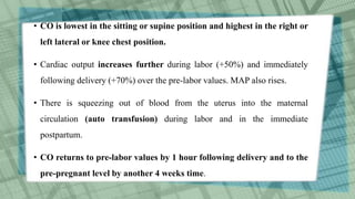
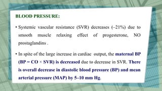
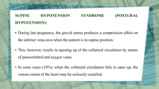
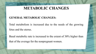
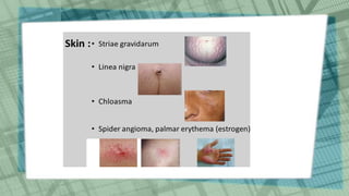
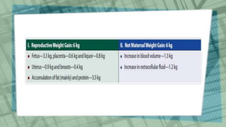
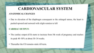
This document summarizes the physiological changes that occur during pregnancy across multiple body systems. Key changes include increased blood volume and cardiac output to support the growing fetus. The uterus enlarges enormously and the cervix softens to facilitate birth. Breast size increases in preparation for lactation. Skin pigmentation and stretch marks commonly occur. Weight gain supplies nutrients for fetal growth. Hematological changes include anemia due to hemodilution. The cardiovascular system adapts to support increased demands through higher cardiac output and lower blood pressure.
Physiological changes in pregnancy. It includes changes in the genital organs, uterus, cardiovascular changes, respiratory, metabolic, alimentary, skin, skeleton, psychological changes, urinary changes and weight gain in pregnancy.
It explains the mechanism of normal labour to medical and para-medical staff.It also puts light on principle movements underlying mechanism of normal labour with pictures.Thank You Like an share it to the maximum.
This topic contains detailed description regarding Normal puerperium, it's definition, duration, phases, involution of uterus and other pelvic organs, lochia, general physiological changes of puerperium, lactation, management of normal puerperium, management of ailments and postnatal care.
This topic includes difference between female and male pelvis, various pelvis types, general description of pelvis bones, division of pelvis, landmarks of pelvis, plane, axis, sacral angle, diameters of inlet, cavity and outlet.
introduction
anatomy and physiologic changes-UTERUS: At the end of third stage of labour, the uterus is in the midline , about 2cm below the level of umbilicus and weight 1000g
Puerperium is the period following childbirth during which the body tissues, specially the pelvic organs revert back approximately to the pre-pregnant state both anatomically and physiologically. puerperium begins as soon as the placenta is expelled and lasts for approximately 6 weeks when the uterus becomes regressed almost to the non-pregnant size.
NORMAL LABOR.. (EUTOCIA) ABNORMAL LABOR ALSO EXPLAINED. Series of events that take place in the genital organs in an effort to expel the viable products of conception out of the womb through the vagina into the outer world is called LABOR.
USMLE GENERAL EMBRYOLOGY 019 Anatomical changes during pregnancy.pdfAHMED ASHOUR
Throughout the antenatal period, cervical examinations were complemented by assessments of cervical consistency, effacement, and fetal station to provide a comprehensive evaluation of cervical readiness for labor and delivery.
At 39 weeks gestation, the patient spontaneously entered labor, and cervical examination revealed complete effacement and dilation to 4 centimeters, consistent with active labor. The patient progressed through the stages of labor and delivered a healthy infant via uncomplicated vaginal delivery.
It is a chapter in obstetrics. it is important to know what happens after pregnancy. it includes definition, involution of the uterus,lochia, general physiological changes , lactation, physiology of lactation etc. it is very knowledgeable ppt. please read this vey carefully.
This power point presentation explains the female reproductive system briefly. It explains about different stages of female reproduction i.e. puberty, menarche, menstruation, pregnancy, menopause etc.
Navigating Challenges: Mental Health, Legislation, and the Prison System in B...Guillermo Rivera
This conference will delve into the intricate intersections between mental health, legal frameworks, and the prison system in Bolivia. It aims to provide a comprehensive overview of the current challenges faced by mental health professionals working within the legislative and correctional landscapes. Topics of discussion will include the prevalence and impact of mental health issues among the incarcerated population, the effectiveness of existing mental health policies and legislation, and potential reforms to enhance the mental health support system within prisons.
R3 Stem Cells and Kidney Repair A New Horizon in Nephrology.pptxR3 Stem Cell
R3 Stem Cells and Kidney Repair: A New Horizon in Nephrology" explores groundbreaking advancements in the use of R3 stem cells for kidney disease treatment. This insightful piece delves into the potential of these cells to regenerate damaged kidney tissue, offering new hope for patients and reshaping the future of nephrology.
CHAPTER 1 SEMESTER V PREVENTIVE-PEDIATRICS.pdfSachin Sharma
This content provides an overview of preventive pediatrics. It defines preventive pediatrics as preventing disease and promoting children's physical, mental, and social well-being to achieve positive health. It discusses antenatal, postnatal, and social preventive pediatrics. It also covers various child health programs like immunization, breastfeeding, ICDS, and the roles of organizations like WHO, UNICEF, and nurses in preventive pediatrics.
We understand the unique challenges pickleball players face and are committed to helping you stay healthy and active. In this presentation, we’ll explore the three most common pickleball injuries and provide strategies for prevention and treatment.
Welcome to Secret Tantric, London’s finest VIP Massage agency. Since we first opened our doors, we have provided the ultimate erotic massage experience to innumerable clients, each one searching for the very best sensual massage in London. We come by this reputation honestly with a dynamic team of the city’s most beautiful masseuses.
The dimensions of healthcare quality refer to various attributes or aspects that define the standard of healthcare services. These dimensions are used to evaluate, measure, and improve the quality of care provided to patients. A comprehensive understanding of these dimensions ensures that healthcare systems can address various aspects of patient care effectively and holistically. Dimensions of Healthcare Quality and Performance of care include the following; Appropriateness, Availability, Competence, Continuity, Effectiveness, Efficiency, Efficacy, Prevention, Respect and Care, Safety as well as Timeliness.
Struggling with intense fears that disrupt your life? At Renew Life Hypnosis, we offer specialized hypnosis to overcome fear. Phobias are exaggerated fears, often stemming from past traumas or learned behaviors. Hypnotherapy addresses these deep-seated fears by accessing the subconscious mind, helping you change your reactions to phobic triggers. Our expert therapists guide you into a state of deep relaxation, allowing you to transform your responses and reduce anxiety. Experience increased confidence and freedom from phobias with our personalized approach. Ready to live a fear-free life? Visit us at Renew Life Hypnosis..
QA Paediatric dentistry department, Hospital Melaka 2020Azreen Aj
QA study - To improve the 6th monthly recall rate post-comprehensive dental treatment under general anaesthesia in paediatric dentistry department, Hospital Melaka
2. • During pregnancy there is progressive anatomical, physiological
and biochemical change not only confined to the genital organs
but also to all systems of the body.
• This is principally a phenomenon of maternal adaptation to the
increasing demands of the growing fetus.
• Unless well understood, these physiological adaptations of
normal pregnancy can be misinterpreted as pathological.
4. • VULVA: Vulva becomes edematous and more vascular;
superficial varicosities may appear especially in multipara. Labia
minora are pigmented and hypertrophied.
• VAGINA: Vaginal walls become hypertrophied, edematous and
more vascular. Increased blood supply of the venous plexus
surrounding the walls gives the bluish coloration of the mucosa
(Jacquemier’s sign). The length of the anterior vaginal wall is
increased.
5. • Secretion: The secretion becomes copious, thin and curdy white
due to marked exfoliated cells and bacteria.
7. • There is enormous growth of the uterus during pregnancy.
• The uterus which in non-pregnant state weighs about 60 g, with a
cavity of 5–10 mL and measures about 7.5 cm in length, at term,
weighs 900–1,000 g and measures 35 cm in length.
• The capacity is increased by 500–1,000 times.
• Changes occur in all the parts of the uterus—body, isthmus and
cervix.
8. BODY OF THE UTERUS
There is increase in growth and enlargement of the body of the uterus.
Enlargement: The enlargement of the uterus is affected by the following
factors: Changes in the muscles—
(1) Hypertrophy and hyperplasia: Not only the individual muscle fiber
increases in length and breadth but also there is limited addition of
new muscle fibers. These occur under the influence of the
hormones—estrogen and progesterone—limited to the first half of
pregnancy but pronounced up to 12 weeks.
9. 2) Stretching: The muscle fibers further elongate beyond 20
weeks due to distension by the growing fetus. The wall
becomes thinner and, at term, measures about 1.5 cm or less.
The uterus feels soft and elastic in contrast to firm feel of the
nongravid uterus.
10. • There is simultaneous increase in number and size of the
supporting fibrous and elastic tissues.
• Vascular system—Whereas in the non-pregnant state, the blood
supply to the uterus is mainly through the uterine and least
through the ovarian but, in the pregnant state, the latter carries
as much the blood as the former. There is marked spiraling of
the arteries, reaching the maximum at 20 weeks; thereafter, they
straighten out.
11. • The uterine enlargement is not a symmetrical one. The fundus
enlarges more than the body.
• Weight: The increase in weight is due to the increased growth of the
uterine muscles, connective tissues and vascular channels.
• Shape—Non-pregnant pyriform shape is maintained in early months.
It becomes globular at 12 weeks. As the uterus enlarges, the shape
once more becomes pyriform or ovoid by 28 weeks and changes to
spherical beyond 36th week
12. Position
• Normal anteverted position is exaggerated up to 8 weeks. Thus,
the enlarged uterus may lie on the bladder rendering it incapable
of filling, clinically evident by frequency of micturition.
• Afterwards, it becomes erect, the long axis of the uterus
conforms more or less to the axis of the inlet.
13. • As the term approaches, especially in multipara with lax
abdominal wall, there is a tendency of anteversion.
• But in primigravida with good tone of the abdominal muscles, it
is held firmly against the maternal spine.
14. Contractions (Braxton-Hicks)
• Uterine contraction in pregnancy has been named after Braxton-
Hicks who first described its entity during pregnancy.
• From the very early weeks of pregnancy, the uterus undergoes
spontaneous contraction.
• This can be felt during bimanual palpation in early weeks or during
abdominal palpation when the uterus feels firmer at one moment and
softer at another.
15. • Although spontaneous, the contractions may be excited by
rubbing the uterus.
• The contractions are irregular, infrequent, spasmodic and
painless without any effect on dilatation of the cervix. The
patient is not conscious about the contractions.
16. Endometrium: The changes of the endometrium of the non-
pregnant uterus into decidua of pregnancy
17. ISTHMUS
• During the first trimester, isthmus hypertrophies and elongates
to about 3 times its original length. It becomes softer. With
advancing pregnancy beyond 12 weeks, it progressively unfolds
from above, downward until it is incorporated into the uterine
cavity. The circularly arranged muscle fibers in the region.
18. • function as a sphincter in early pregnancy and thus help to retain
the fetus within the uterus. Incompetency of the sphincteric
action leads to mid-trimester abortion and the encirclage
operation done to rectify the defect is based on the principle of
restoration of the retentive function of the isthmus
19. CERVIX
Stroma:
• There are hypertrophy and hyperplasia of the elastic and
connective tissues. Fluids accumulate inside and in between the
fibers.
• Vascularity is increased especially beneath the squamous
epithelium of the portio vaginalis which is responsible for its
bluish coloration.
20. • There are marked hypertrophy and hyperplasia of the glands
which occupy about half the bulk of the cervix.
• All these lead to marked softening of the cervix (Goodell’s
sign) It not only provides diagnostic aid in pregnancy but also
the changes in the cervix facilitate its dilatation during labor.
21. Epithelium: There is marked proliferation of the endocervical
mucosa with downward extension beyond the squamocolumnar
junction.
22. • Secretion: The secretion is copious and tenacious-physiological
leucorrhea of pregnancy.
• This is due to the effect of progesterone.
• There is no alteration in the relation of the cervix. There is unfolding
of the isthmus; beginning 12 weeks onwards and takes part in the
formation of the lower uterine segment.
• Variable amount of effacement is noticed near term in primigravidae.
In multiparae, the canal is slightly dilated.
23. FALLOPIAN TUBE
• As the uterine end rises up and the fimbrial end is held up by the
infundibulopelvic ligament, it is placed almost vertical by the
side of the uterus.
• The total length is somewhat increased. The tube becomes
congested. Muscles undergo hypertrophy. Epithelium becomes
flattened, and patches of decidual reaction are observed.
24. OVARY
• The growth and function of the corpus luteum reaches its maximum
at 8th week when it measures about 2.5 cm and becomes cystic. It
looks bright orange, later on becomes yellow and, finally pale.
Regression occurs following decline in the secretion of human
chorionic gonadotropin (hCG) from the placenta.
• Hormones—estrogen and progesterone—secreted by the corpus
luteum maintain the environment for the growing ovum before the
action is taken over by the placenta.
25. • These hormones not only control the formation and
maintenance of decidua of pregnancy but also inhibit ripening
of the follicles. Thus both the ovarian and uterine cycles of the
normal menstruation remain suspended.
• Luteoma of pregnancy results from exaggerated luteinization
reaction of the ovary.
26. BREAST
• The changes in the breasts are best evident in a primigravida. In multipara,
who has once lactated, the changes are not clearly defined.
• SIZE: Increased size of the breasts becomes evident even in early weeks.
This is due to marked hypertrophy and proliferation of the ducts (estrogen)
and the alveoli (estrogen and progesterone) which are marked in the
peripheral lobules. There is also hypertrophy of the connective tissue
stroma. Quite often, the “axillary tail” becomes enlarged and painful.
27. • NIPPLES AND AREOLA: The nipples become larger, erectile and
deeply pigmented. Variable number of sebaceous glands (5–15)
which remain invisible in the non-pregnant state in the areola,
become hypertrophied and are called Montgomery’s tubercles.
Those are placed surrounding the nipples. Their secretion keeps the
nipple and the areola moist and healthy. An outer zone of less marked
and irregular pigmented area appears in second trimester and is
called secondary areola.
28. • SECRETION: Secretion (colostrum) can be squeezed out of
the breast at about 12th week which at first becomes sticky.
Later on, by 16th week, it becomes thick and yellowish. The
demonstration of secretion from the breast of a woman who has
never lactated is an important sign of pregnancy. In latter
months, colostrum may be expressed from the nipples.
29. CUTANEOUS CHANGES
1. Face (chloasma gravidarum or pregnancy mask): It is an extreme
form of pigmentation around the cheek, forehead and around the eyes.
It may be patchy or diffuse; disappears spontaneously after delivery.
2. Breast:
3. Abdomen:
• Linea nigra: It is a brownish black pigmented area in the midline
stretching from the xiphisternum to the symphysis pubis. The
pigmentation disappears after delivery.
30. Striae gravidarum:
• These are slightly depressed linear marks with varying length
and breadth found in pregnancy. They are predominantly found
in the abdominal wall below the umbilicus, sometimes over the
thighs and breasts. These stretch marks represent the scar tissues
in the deeper layer of the cutis.
31. • Apart from the mechanical stretching of the skin, increase in
aldosterone production during pregnancy are the responsible
factors.
• Controlled weight gain during pregnancy and massaging the
abdominal wall by lubricants like olive oil may be helpful in
reducing their formation.
32. OTHER CUTANEOUS CHANGES:
• These include vascular spider and palmar erythema which are
due to high estrogen level. Mild degrees of hirsutism may be
observed and in puerperium the excess hair is lost.
33.
34. WEIGHT GAIN
• The total weight gain during the course of a singleton pregnancy
for a healthy woman averages 11 kg (24 lb).
• This has been distributed to 1 kg in first trimester and 5 kg each
in second and third trimester.
• The total weight gain at term is distributed approximately as
follows:
35.
36. • Ideally weight gain should depend on pre-pregnancy body mass
index (BMI) level. Weight gain for a woman with normal BMI (20–
26) is 11–16 kg. An obese woman (BMI > 30) should not gain more
than 7 kg, whereas an underweight woman (BMI < 19) may be
allowed to gain up to 18 kg.
• Maternal nutrition and weight gain during pregnancy are directly
related to the newborn weight. However, it may not be a specific
indicator as there are other factors for low birth weight infant.
37. BODY WATER METABOLISM
• During pregnancy, the amount of water retained at term is about 6.5 liters.
The water content of the fetus, placenta and amniotic fluid is about 3.5
liters.
• Pregnancy is a state of hypervolemia. There is active retention of sodium
(900 mEq), potassium (300 mEq) and water.
• The important causes of sodium retention and volume overload are:
(i) changes in maternal osmoregulation,
38. (ii) increased estrogen and progesterone,
(iii) increase in renin-angiotensin-aldosterone system (RAAS) activity,
(iv) increased aldosterone, deoxycorticosterone,
(v) control by arginine vasopressin (AVP) from posterior pituitary and
(vi) atrial natriuretic peptide.
39. • Increase in water intake due to lowered osmotic threshold for
thirst causes polyuria in early pregnancy.
40. HEMATOLOGICAL CHANGES
BLOOD VOLUME:
• During pregnancy, there is increased vascularity of the enlarging
uterus with the interposition of uteroplacental circulation.
• The activities of all the systems are increased.
• Blood volume is markedly raised during pregnancy.
41. • The rise is progressive and inconsistent. All the constituents of
blood are affected with increased blood volume.
• The blood volume starts to increase from about 6th week,
expands rapidly thereafter to maximum 40–50% above the
nonpregnant level at 30–34 weeks. The level remains almost
static till delivery
42. PLASMA VOLUME:
• It starts to increase by 6 weeks and it plateaus at 30 weeks of
gestation.
• The rate of increase almost parallels to that of blood volume but the
maximum is reached to the extent of 50%.
• Total plasma volume increases to the extent of 1.25 liters.
• The increase is greater in multigravida, in multiple pregnancy and
with large baby.
43. RBC AND HEMOGLOBIN:
• The RBC mass is increased to the extent of 20–30%.
• The total increase in volume is about 350 mL.
• This increase is regulated by the increased demand of oxygen transport during
pregnancy.
• RBC mass begins to increase at about 10 weeks and continues till term without
plateauing.
• Iron supplementation increases the RBC mass by 30%. Reticulocyte count
increases by 2%. Erythropoietin level is raised.
44.
45. • The disproportionate increase in plasma and RBC volume produces a state of
hemodilution (fall in hematocrit) during pregnancy.
• Thus, even though the total hemoglobin mass increases during pregnancy to the
extent of 18–20%, there is apparent fall in hemoglobin concentration. At term, the
fall is about 2 g% from the non-pregnant value.
• There is simultaneous fall in number of red cells by 15–20% and the hematocrit
level.
• The excess of circulating hemoglobin left behind after delivery yields iron for
storage.
46. LEUKOCYTES and IMMUNE SYSTEM:
• Neutrophilic leukocytosis occurs to the extent of 8,000/mm3
and even to 20,000/mm3 in labor. The increase may be due to
rise in the levels of estrogen and cortisol.
• The major change in the immune system is the modulation
away from cell-mediated cytotoxic immune response toward
increased humoral innate immune responses
49. CARDIOVASCULAR SYSTEM
ANATOMICAL CHANGES
• Due to elevation of the diaphragm consequent to the enlarged uterus, the heart is
pushed upward and outward with slight rotation to left
CARDIAC OUTPUT:
• The cardiac output (CO) starts to increase from 5th week of pregnancy and reaches
its peak 40–50% at about 30–34 weeks.
• Thereafter the CO remains static till term.
50. • CO is lowest in the sitting or supine position and highest in the right or
left lateral or knee chest position.
• Cardiac output increases further during labor (+50%) and immediately
following delivery (+70%) over the pre-labor values. MAP also rises.
• There is squeezing out of blood from the uterus into the maternal
circulation (auto transfusion) during labor and in the immediate
postpartum.
• CO returns to pre-labor values by 1 hour following delivery and to the
pre-pregnant level by another 4 weeks time.
51. BLOOD PRESSURE:
• Systemic vascular resistance (SVR) decreases (–21%) due to
smooth muscle relaxing effect of progesterone, NO
prostaglandins .
• In spite of the large increase in cardiac output, the maternal BP
(BP = CO × SVR) is decreased due to decrease in SVR. There
is overall decrease in diastolic blood pressure (BP) and mean
arterial pressure (MAP) by 5–10 mm Hg.
52. SUPINE HYPOTENSION SYNDROME (POSTURAL
HYPOTENSION):
• During late pregnancy, the gravid uterus produces a compression effect on
the inferior vena cava when the patient is in supine position.
• This, however, results in opening up of the collateral circulation by means
of paravertebral and azygos veins.
• In some cases (10%), when the collateral circulation fails to open up, the
venous return of the heart may be seriously curtailed.
53. • This results in production of hypotension, tachycardia and
syncope.
• The normal blood pressure is quickly restored by turning
the patient to lateral position.
• The augmentation of the venous return during uterine
contraction prevents the manifestation from developing during
labor.
54. METABOLIC CHANGES
GENERAL METABOLIC CHANGES:
Total metabolism is increased due to the needs of the growing
fetus and the uterus.
Basal metabolic rate is increased to the extent of 30% higher than
that of the average for the nonpregnant women.
55. SYSTEMIC CHANGES
RESPIRATORY SYSTEM:
• With the enlargement of the uterus, especially in the later
months, there is elevation of the diaphragm by 4 cm.
• Total lung capacity is reduced by 5% due to this elevation.
However, diaphragmatic excursion is increased by 1–2 cm and
breathing becomes diaphragmatic.
56. • Total pulmonary resistance is reduced due to progesterone
effect.
• The mucosa of the nasopharynx becomes hyperemic and
edematous. This may cause nasal stuffiness and rarely epistaxis.
• The woman feels shortness of breath.
57. URINARY SYSTEM:
Kidney—
• There is dilatation of the ureters, renal pelvis and the calyces. The
kidneys enlarge in length by 1 cm.
• Renal plasma flow is increased by 50–75%, maximum by the 16
weeks and is maintained until 34 weeks. Thereafter it falls by 25%.
• Glomerular filtration rate (GFR) is increased by 50% all throughout
pregnancy.
58. Ureter:
• Ureters become atonic due to high progesterone level.
• Dilatation of the ureter above the pelvic brim with stasis is marked on the
right side especially in primigravidae.
• There is marked hypertrophy of the muscle and the sheath of the ureter
especially the pelvic part probably due to estrogen.
• There is elongation, kinking and outward displacement of the ureters.
The dilatation effect resolves by 6 weeks postpartum.
59. Bladder:
• There is marked congestion with hypertrophy of the muscles
and elastic tissues of the wall.
• Increased frequency of micturition is noticed at 6–8 weeks of
pregnancy which subsides after 12 weeks.
• It may be due to resetting of osmoregulation causing
increased water intake and polyuria.
• .
60. • In late pregnancy, frequency of micturition once more
reappears due to pressure on the bladder as the presenting part
descends down the pelvis.
• Stress incontinence may be observed in late pregnancy due to
urethral sphincter weakness
61. ALIMENTARY SYSTEM:
• The gums become congested and spongy and may bleed to touch.
• Muscle tone and motility of the entire gastrointestinal tract are
diminished due to high progesterone level.
• Cardiac sphincter is relaxed and regurgitation of acid gastric content
into the esophagus may produce chemical esophagitis and heart burn.
62. • Dyspepsia is common. There is diminished gastric secretion and
delayed emptying time of the stomach.
• Risk of peptic ulcer disease is reduced.
• Atonicity of the gut leads to constipation, while diminished
peristalsis facilitates more absorption of food materials.
63. LIVER AND GALLBLADDER:
Although there is no histological change in the liver cells, but the
functions are depressed.
With the exception of raised alkaline phosphatase levels, other
liver function tests (serum levels of bilirubin, AST, ALT, CPK,
LDH) are unchanged.
64. • There is mild cholestasis (estrogen effect).
• There is marked atonicity of the gallbladder (progesterone
effect). This, together with high blood cholesterol level during
pregnancy, favors stone formation.
65. NERVOUS SYSTEM:
• Some sorts of temperamental changes are found during
pregnancy and in the puerperium.
• Nausea, vomiting, mental irritability and sleep disorders are
probably due to some psychological background.
• Postpartum blues, depression or psychosis may develop in a
susceptible Individual.
66. • Compression of the median nerve underneath the flexor
retinaculum over the wrist joint leading to pain and paresthesia
in the hands and arm (Carpal tunnel syndrome) may appear in
the later months of pregnancy.
• Similarly, paresthesia and sensory loss over the anterolateral
aspect of the thigh may occur. It is due to compression of the
lateral cutaneous nerve of the thigh.