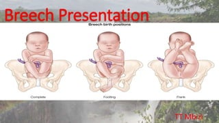
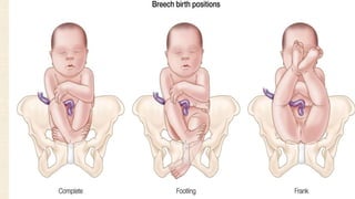
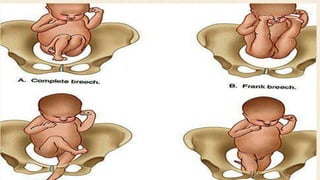
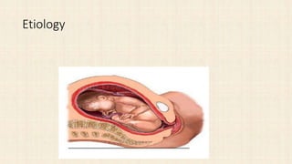
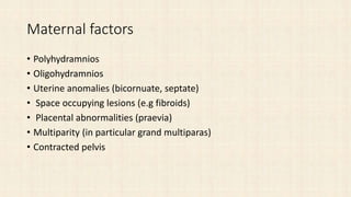
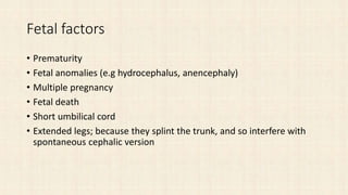
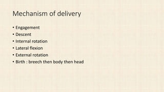
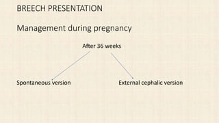
Breech Presentation
TT Mbizi
Etiology
Maternal factors
• Polyhydramnios
• Oligohydramnios
• Uterine anomalies (bicornuate, septate)
• Space occupying lesions (e.g fibroids)
• Placental abnormalities (praevia)
• Multiparity (in particular grand multiparas)
• Contracted pelvis
Fetal factors
• Prematurity
• Fetal anomalies (e.g hydrocephalus, anencephaly)
• Multiple pregnancy
• Fetal death
• Short umbilical cord
• Extended legs; because they splint the trunk, and so interfere with
spontaneous cephalic version
Mechanism of delivery
• Engagement
• Descent
• Internal rotation
• Lateral flexion
• External rotation
• Birth : breech then body then head
Diagnosis of Breech
• Clinical
• Hx exaggerated subcoastal discomfort or lump under the ribs
• Fetal mvts felt predominantly in lower part of uterus
• Examination
• Ballotable round head at fundus and soft presenting part at lower uterine pole
• Fetal heart sounds above umblicus
• Meconium seen in labour
• Ultrasound
• Xray
Clinical Diagnosis
• Abdominal examination
Palpation
1. Fundal grips; the head is felt with its characters
2. Pelvic grip; the breech is felt, with its characters.
3. Auscultation
The fetal heart sounds are heard just at, or above the level of the umbilicus
Vaginal examination
• 1. Slow dilatation of cervix, sausage-chapel bag of fore-waters, and
liability to premature rupture of the membrane and prolapse of the cord.
• 2. After rupture of the membranes, the presenting part is felt, that is ,
the two buttocks with the anus in between , the genitalia on one side and
the sacral spines on the opposite side.
• 3. In case of complete breech, the feet are felt on the same level as the
buttocks.
• 4. In case of breech with extended legs, the buttocks only are felt. In
case of footling presentation, the feet are at a lower level than the
buttocks. In case of knee presentation, the knees are a lower level than the
buttocks.
BREECH PRESENTATION
Management during pregnancy
After 36 weeks
Spontaneous version External cephalic version
Version
• External cephalic version
• Internal podalic version
External Cephalic Version
In delivery room
NPO and ready for c/s
CTG & USS
Tocolytic
Head down position
Dislodge breech then
gently turn around
US and CTG after procedure.
Internal podalic version
Risks of External Cephalic Version
• Placental abruption
• Premature rupture of the membranes
• Cord accident
• Transplacental haemorrhage(remember anti-D administration in
Rhesus-negative women)
• Fetal bradycardia
Contraindications of External Cephalic Version
• Previous scar on the uterus
• Placenta praevia
• Unexplained APH
• Pre-eclampsia
• Multiple pregnancy
• Rhesus isoimmunisation
• Elderly primigravida
• IUGR
• Oligohydramnios
• Polyhydramnios
Caesarean section
Vaginal delivery
• Spontaneous breech delivery
• Assisted breech delivery
• Total breech extraction
Indications of vaginal delivery
a) Frank or complete breech presentation
b) Gestational age > 36 weeks
c) Estimated foetal weight 2.5-3.5 kg
d) Foetal head must be flexed
e) Adequate maternal pelvis
f) No other obstetric complications.
Management during labour
• During labour:
If there is contracted pelvic, and fetus is living and good; do
caesarean section.
Rest in bed and avoid repeated vaginal examination to prevent PROM
PV is done after ROM to exclude cord prolapse.
• Pinard manoeuvre to deliver extended knees
• Second stage :Delivery of the aftercoming head
• Burns Marshall method
• Mauriceau-Smellie-veit maneuver
• Wigand Martin method
• Prague maneuver
• Piper forceps
Burns Marshall Method
Mauriceau-Smellie-Veit Maneuver
Prague maneuver
The back of the fetus fail to rotate to the anterior
Piper Forceps
Total breech extraction
Indication
• 1. Prolonged second stage of labor
• 2. Twins
• 3. Maternal disease
• 4. Prolapsed cord
• 5. Fetal distress
Total Breech Extraction
Cesarean section
• Indications:
• Large fetus
• Contraction or unfavorable shape of pelvis
• Hyperextended head(Star gazing)
• Uterine dysfunction
• Incomplete or footling presentation
• Primigravida
Indications of Cs in Breech
• Healthy preterm
• Severe fetal growth restriction
• Previous perinatal death or newborn
• complication of birth trauma
• Lack of an experienced operator
Complications of Breech Delivery
• Maternal complications
Risk of Operative intervention
Risk of infection due to Manipulations
Intrauterine maneuvers : Rupture of the
uterus +/- lacerations of Cx
• Extensions of the episiotomy
• Uterine atony
• Postpartum hemorrhage
Complications cont.
• Fetal complications Preterm delivery & low birth weight & IUGR Prolapse
cord Birth aphyxia Fetal Injuries
• Fx of humerous and clavicle
• Fx of femur
• Hematomas of sternocleidomastoid
• Separation of epiphyses of scapular,humerus or femur
• Brachial plexus
• Avulsion of upper C-spine
• Skull Fx , intracerebral injury
GRACIAS

Breech presentation [autosaved]

  • 1.
  • 4.
  • 5.
    Maternal factors • Polyhydramnios •Oligohydramnios • Uterine anomalies (bicornuate, septate) • Space occupying lesions (e.g fibroids) • Placental abnormalities (praevia) • Multiparity (in particular grand multiparas) • Contracted pelvis
  • 6.
    Fetal factors • Prematurity •Fetal anomalies (e.g hydrocephalus, anencephaly) • Multiple pregnancy • Fetal death • Short umbilical cord • Extended legs; because they splint the trunk, and so interfere with spontaneous cephalic version
  • 7.
    Mechanism of delivery •Engagement • Descent • Internal rotation • Lateral flexion • External rotation • Birth : breech then body then head
  • 8.
    Diagnosis of Breech •Clinical • Hx exaggerated subcoastal discomfort or lump under the ribs • Fetal mvts felt predominantly in lower part of uterus • Examination • Ballotable round head at fundus and soft presenting part at lower uterine pole • Fetal heart sounds above umblicus • Meconium seen in labour • Ultrasound • Xray
  • 9.
    Clinical Diagnosis • Abdominalexamination Palpation 1. Fundal grips; the head is felt with its characters 2. Pelvic grip; the breech is felt, with its characters. 3. Auscultation The fetal heart sounds are heard just at, or above the level of the umbilicus
  • 10.
    Vaginal examination • 1.Slow dilatation of cervix, sausage-chapel bag of fore-waters, and liability to premature rupture of the membrane and prolapse of the cord. • 2. After rupture of the membranes, the presenting part is felt, that is , the two buttocks with the anus in between , the genitalia on one side and the sacral spines on the opposite side. • 3. In case of complete breech, the feet are felt on the same level as the buttocks. • 4. In case of breech with extended legs, the buttocks only are felt. In case of footling presentation, the feet are at a lower level than the buttocks. In case of knee presentation, the knees are a lower level than the buttocks.
  • 11.
    BREECH PRESENTATION Management duringpregnancy After 36 weeks Spontaneous version External cephalic version
  • 12.
    Version • External cephalicversion • Internal podalic version
  • 13.
    External Cephalic Version Indelivery room NPO and ready for c/s CTG & USS Tocolytic Head down position Dislodge breech then gently turn around US and CTG after procedure.
  • 14.
  • 15.
    Risks of ExternalCephalic Version • Placental abruption • Premature rupture of the membranes • Cord accident • Transplacental haemorrhage(remember anti-D administration in Rhesus-negative women) • Fetal bradycardia
  • 16.
    Contraindications of ExternalCephalic Version • Previous scar on the uterus • Placenta praevia • Unexplained APH • Pre-eclampsia • Multiple pregnancy • Rhesus isoimmunisation • Elderly primigravida • IUGR • Oligohydramnios • Polyhydramnios
  • 17.
    Caesarean section Vaginal delivery •Spontaneous breech delivery • Assisted breech delivery • Total breech extraction
  • 18.
    Indications of vaginaldelivery a) Frank or complete breech presentation b) Gestational age > 36 weeks c) Estimated foetal weight 2.5-3.5 kg d) Foetal head must be flexed e) Adequate maternal pelvis f) No other obstetric complications.
  • 19.
    Management during labour •During labour: If there is contracted pelvic, and fetus is living and good; do caesarean section. Rest in bed and avoid repeated vaginal examination to prevent PROM PV is done after ROM to exclude cord prolapse.
  • 20.
    • Pinard manoeuvreto deliver extended knees
  • 21.
    • Second stage:Delivery of the aftercoming head • Burns Marshall method • Mauriceau-Smellie-veit maneuver • Wigand Martin method • Prague maneuver • Piper forceps
  • 22.
  • 23.
  • 24.
    Prague maneuver The backof the fetus fail to rotate to the anterior
  • 25.
  • 26.
    Total breech extraction Indication •1. Prolonged second stage of labor • 2. Twins • 3. Maternal disease • 4. Prolapsed cord • 5. Fetal distress
  • 27.
  • 28.
    Cesarean section • Indications: •Large fetus • Contraction or unfavorable shape of pelvis • Hyperextended head(Star gazing) • Uterine dysfunction • Incomplete or footling presentation • Primigravida
  • 29.
    Indications of Csin Breech • Healthy preterm • Severe fetal growth restriction • Previous perinatal death or newborn • complication of birth trauma • Lack of an experienced operator
  • 30.
    Complications of BreechDelivery • Maternal complications Risk of Operative intervention Risk of infection due to Manipulations Intrauterine maneuvers : Rupture of the uterus +/- lacerations of Cx • Extensions of the episiotomy • Uterine atony • Postpartum hemorrhage
  • 31.
    Complications cont. • Fetalcomplications Preterm delivery & low birth weight & IUGR Prolapse cord Birth aphyxia Fetal Injuries • Fx of humerous and clavicle • Fx of femur • Hematomas of sternocleidomastoid • Separation of epiphyses of scapular,humerus or femur • Brachial plexus • Avulsion of upper C-spine • Skull Fx , intracerebral injury
  • 32.