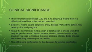
ANKLE-BRACHIAL INDEX
BY DR. HIMANSHU PATEL
DR. HIMANSHU PATEL (PT) 1
INTRODUCTION
• The ankle-brachial index, ankle-arm index, ankle-arm ratio, or the Winsor
Index (first described in 1950 by Winsor) is a quick, non-invasive,
inexpensive technique used widely to check the peripheral arterial
disease (PAD). It assesses the severity of arterial insufficiency of arterial
narrowing during walking.
• As the blood pressure of arms is an indicator for the general blood
pressure of the body, but when blood go farther in the body through the
arteries the pressure changes according to the condition of the patient’s
arteries and the general health also resting and exercising situations will
show different changes in reading.
DR. HIMANSHU PATEL (PT) 2
• The ankle-brachial index ABI is measured by dividing the ankle
systolic pressure by brachial systolic pressure.
• PAD ( Peripheral Arterial Disease ) is the reduction in the arterial
blood flow to the limbs during walking causing pain and may
predispose to heart attack and stroke, it affects over 13% of
populations who are above 50 years.
DR. HIMANSHU PATEL (PT) 3
INDICATIONS
• People presented with pain, cramps, or claudications in
the calf, hamstring, G.max during walking.
• Risk factors for PAD, such as:
• History of tobacco use
• Diabetes
• High blood pressure
• High cholesterol
• Restricted blood flow (atherosclerosis) in other parts of your body.
DR. HIMANSHU PATEL (PT) 4
CONTRAINDICATIONS
• Patients with confirmed or suspected DVT, because of the possibility
of breaking of the thrombus.
• Severe leg pain.
DR. HIMANSHU PATEL (PT) 5
PROCEDURES
• Tools
Hand-held Doppler, or hand-
held ultrasound device that's
pressed on your skin
Inflated cuff
DR. HIMANSHU PATEL (PT) 6
PREPARATION
• Rest for minutes before the measurements 10-30 minutes like normal
blood pressure taken during normal visits.
• Loose, comfortable clothes will be a better choice for easily wearing
the inflated cuff on the upper arm.
• Avoid nicotine/ smoking as it shows an increase in the peripheral
systolic pressure resulting in elevated ABI value
DR. HIMANSHU PATEL (PT) 7
CONT….
• Supported, comfortable position, and avoid limb movement during
measurements.
• The supine position is the position of choice as the sitting position
shows about 0.3 increase in the ABI
DR. HIMANSHU PATEL (PT) 8
TECHNIQUE
• The same procedures as in the measurement of normal blood
pressure, with the patient in rested position, it is better to measure the
brachial pressure before the ankle pressure:
• 1- Place the cuff around the patient's arm, use the hand-held
ultrasound device or hand doppler and press against the skin at the
level of the brachial artery in the arm in order to hear the waves of the
systolic pressure, repeat on the other arm and record the highest
measurement.
DR. HIMANSHU PATEL (PT) 9
CONT….
• 2- Place the cuff around the lower leg and use the hand-held
ultrasound device to hear the waves of the systolic pressure at two
levels :
• Dorsalis pedis, which is a branch from the anterior tibial artery, it is
located between the tibialis anterior and extensor hallucis longus
• Posterior tibial pulse, that is behind the lateral malleoli.
• 3- Record the highest pulse between two measurements of dorsalis
pedis and posterior tibial arteries.
DR. HIMANSHU PATEL (PT) 10
CONT….
• - The ankle-brachial index calculated by dividing the highest value of
ankle systolic pressure between the two arteries by the highest
brachial pressure between two sides.
• 5- Repeat the ankle measurement for the other leg in order to
calculate its ABI.
• 6- The normal ABI is between 0.90 and 1.30, below indicate PAD,
above may be a sign of arterial calcification.
DR. HIMANSHU PATEL (PT) 11
CLINICAL SIGNIFICANCE
• The normal range is between 0.90 and 1.30. below 0.8 means there is a
difficulty of blood flow to the foot and lower limb.
• Below 0.7 means severe peripheral artery disease PAD and the patient may
develop ulcer and gangrene.
• Above the normal level, 1.30 is a sign of calcification of arterial walls that
may happen in case of diabetic patients, chronic kidney disease, in this
situation you need to measure the ankle pressure at distal digital arteries
that is less likely to develop or be calcified
• This ABI is not a confirmation for the diagnosis for arteries calcification
DR. HIMANSHU PATEL (PT) 12
DR. HIMANSHU PATEL (PT) 13

Ankle brachial index

  • 1.
    ANKLE-BRACHIAL INDEX BY DR.HIMANSHU PATEL DR. HIMANSHU PATEL (PT) 1
  • 2.
    INTRODUCTION • The ankle-brachialindex, ankle-arm index, ankle-arm ratio, or the Winsor Index (first described in 1950 by Winsor) is a quick, non-invasive, inexpensive technique used widely to check the peripheral arterial disease (PAD). It assesses the severity of arterial insufficiency of arterial narrowing during walking. • As the blood pressure of arms is an indicator for the general blood pressure of the body, but when blood go farther in the body through the arteries the pressure changes according to the condition of the patient’s arteries and the general health also resting and exercising situations will show different changes in reading. DR. HIMANSHU PATEL (PT) 2
  • 3.
    • The ankle-brachialindex ABI is measured by dividing the ankle systolic pressure by brachial systolic pressure. • PAD ( Peripheral Arterial Disease ) is the reduction in the arterial blood flow to the limbs during walking causing pain and may predispose to heart attack and stroke, it affects over 13% of populations who are above 50 years. DR. HIMANSHU PATEL (PT) 3
  • 4.
    INDICATIONS • People presentedwith pain, cramps, or claudications in the calf, hamstring, G.max during walking. • Risk factors for PAD, such as: • History of tobacco use • Diabetes • High blood pressure • High cholesterol • Restricted blood flow (atherosclerosis) in other parts of your body. DR. HIMANSHU PATEL (PT) 4
  • 5.
    CONTRAINDICATIONS • Patients withconfirmed or suspected DVT, because of the possibility of breaking of the thrombus. • Severe leg pain. DR. HIMANSHU PATEL (PT) 5
  • 6.
    PROCEDURES • Tools Hand-held Doppler,or hand- held ultrasound device that's pressed on your skin Inflated cuff DR. HIMANSHU PATEL (PT) 6
  • 7.
    PREPARATION • Rest forminutes before the measurements 10-30 minutes like normal blood pressure taken during normal visits. • Loose, comfortable clothes will be a better choice for easily wearing the inflated cuff on the upper arm. • Avoid nicotine/ smoking as it shows an increase in the peripheral systolic pressure resulting in elevated ABI value DR. HIMANSHU PATEL (PT) 7
  • 8.
    CONT…. • Supported, comfortableposition, and avoid limb movement during measurements. • The supine position is the position of choice as the sitting position shows about 0.3 increase in the ABI DR. HIMANSHU PATEL (PT) 8
  • 9.
    TECHNIQUE • The sameprocedures as in the measurement of normal blood pressure, with the patient in rested position, it is better to measure the brachial pressure before the ankle pressure: • 1- Place the cuff around the patient's arm, use the hand-held ultrasound device or hand doppler and press against the skin at the level of the brachial artery in the arm in order to hear the waves of the systolic pressure, repeat on the other arm and record the highest measurement. DR. HIMANSHU PATEL (PT) 9
  • 10.
    CONT…. • 2- Placethe cuff around the lower leg and use the hand-held ultrasound device to hear the waves of the systolic pressure at two levels : • Dorsalis pedis, which is a branch from the anterior tibial artery, it is located between the tibialis anterior and extensor hallucis longus • Posterior tibial pulse, that is behind the lateral malleoli. • 3- Record the highest pulse between two measurements of dorsalis pedis and posterior tibial arteries. DR. HIMANSHU PATEL (PT) 10
  • 11.
    CONT…. • - Theankle-brachial index calculated by dividing the highest value of ankle systolic pressure between the two arteries by the highest brachial pressure between two sides. • 5- Repeat the ankle measurement for the other leg in order to calculate its ABI. • 6- The normal ABI is between 0.90 and 1.30, below indicate PAD, above may be a sign of arterial calcification. DR. HIMANSHU PATEL (PT) 11
  • 12.
    CLINICAL SIGNIFICANCE • Thenormal range is between 0.90 and 1.30. below 0.8 means there is a difficulty of blood flow to the foot and lower limb. • Below 0.7 means severe peripheral artery disease PAD and the patient may develop ulcer and gangrene. • Above the normal level, 1.30 is a sign of calcification of arterial walls that may happen in case of diabetic patients, chronic kidney disease, in this situation you need to measure the ankle pressure at distal digital arteries that is less likely to develop or be calcified • This ABI is not a confirmation for the diagnosis for arteries calcification DR. HIMANSHU PATEL (PT) 12
  • 13.