PRESENTER
ASADULLAH RIND
RN, DCHN,BSN, MSN
LECTURER- CON SIR C.J INSTITUTE
PSYCHIATRY HYDERABAD
1
Anatomy and Physiology-I
Topic: Tissue
2.
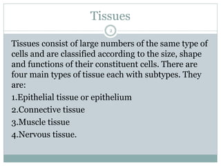
Tissues
2
Tissues consist oflarge numbers of the same type of
cells and are classified according to the size, shape
and functions of their constituent cells. There are
four main types of tissue each with subtypes. They
are:
1.Epithelial tissue or epithelium
2.Connective tissue
3.Muscle tissue
4.Nervous tissue.
3.
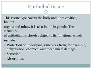
Epithelial tissue
3
This tissuetype covers the body and lines cavities,
hollow
organs and tubes. It is also found in glands. The
structure
of epithelium is closely related to its functions, which
include:
✓ Protection of underlying structures from, for example,
dehydration, chemical and mechanical damage
✓ Secretion
✓ Absorption.
4.
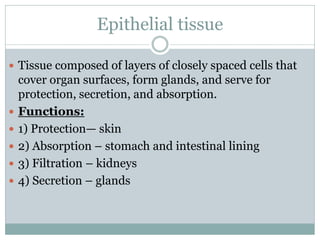
Epithelial tissue
Tissuecomposed of layers of closely spaced cells that
cover organ surfaces, form glands, and serve for
protection, secretion, and absorption.
Functions:
1) Protection— skin
2) Absorption – stomach and intestinal lining
3) Filtration – kidneys
4) Secretion – glands
5.
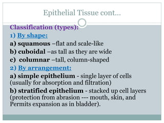
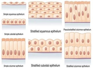
Epithelial Tissue cont…
Classification(types):
1) By shape:
a) squamous –flat and scale-like
b) cuboidal –as tall as they are wide
c) columnar –tall, column-shaped
2) By arrangement:
a) simple epithelium - single layer of cells
(usually for absorption and filtration)
b) stratified epithelium - stacked up cell layers
(protection from abrasion --- mouth, skin, and
Permits expansion as in bladder).
6.
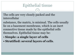
6
The cells arevery closely packed and the
intercellular
substance, the matrix, is minimal. The cells usually
lie on a basement membrane, which is an inert
connective tissue made by the epithelial cells
themselves. Epithelial tissue may be:
• Simple: a single layer of cells
• Stratified: several layers of cells.
Epithelial tissue
Simple epithelium
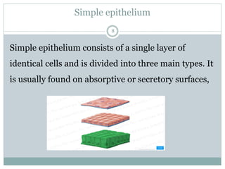
8
Simple epitheliumconsists of a single layer of
identical cells and is divided into three main types. It
is usually found on absorptive or secretory surfaces,
9.
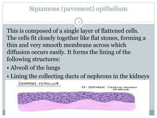
Squamous (pavement) epithelium
9
Thisis composed of a single layer of flattened cells.
The cells fit closely together like flat stones, forming a
thin and very smooth membrane across which
diffusion occurs easily. It forms the lining of the
following structures:
• Alveoli of the lungs
• Lining the collecting ducts of nephrons in the kidneys
10.
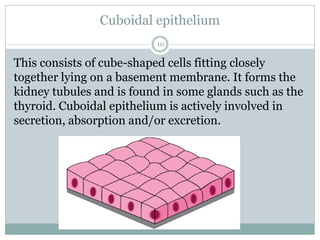
Cuboidal epithelium
10
This consistsof cube-shaped cells fitting closely
together lying on a basement membrane. It forms the
kidney tubules and is found in some glands such as the
thyroid. Cuboidal epithelium is actively involved in
secretion, absorption and/or excretion.
11.
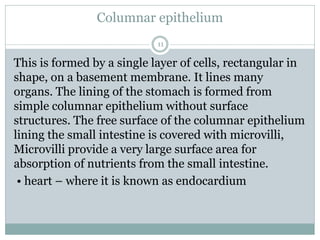
Columnar epithelium
11
This isformed by a single layer of cells, rectangular in
shape, on a basement membrane. It lines many
organs. The lining of the stomach is formed from
simple columnar epithelium without surface
structures. The free surface of the columnar epithelium
lining the small intestine is covered with microvilli,
Microvilli provide a very large surface area for
absorption of nutrients from the small intestine.
• heart – where it is known as endocardium
12.
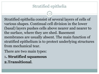
Stratified epithelia
12
Stratified epitheliaconsist of several layers of cells of
various shapes. Continual cell division in the lower
(basal) layers pushes cells above nearer and nearer to
the surface, where they are shed. Basement
membranes are usually absent. The main function of
stratified epithelium is to protect underlying structures
from mechanical tear.
There are two main types:
1. Stratified squamous
2.Transitional.
13.
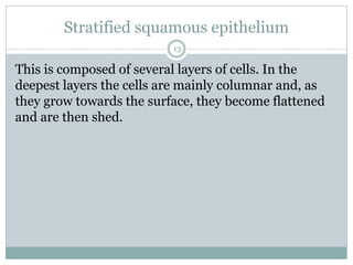
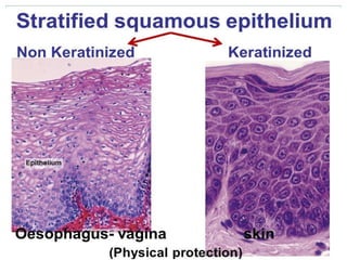
Stratified squamous epithelium
13
Thisis composed of several layers of cells. In the
deepest layers the cells are mainly columnar and, as
they grow towards the surface, they become flattened
and are then shed.
14.
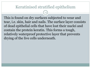
Keratinised stratified epithelium
14
Thisis found on dry surfaces subjected to wear and
tear, i.e. skin, hair and nails. The surface layer consists
of dead epithelial cells that have lost their nuclei and
contain the protein keratin. This forms a tough,
relatively waterproof protective layer that prevents
drying of the live cells underneath.
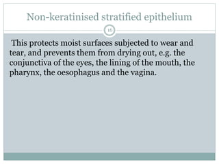
Non-keratinised stratified epithelium
16
Thisprotects moist surfaces subjected to wear and
tear, and prevents them from drying out, e.g. the
conjunctiva of the eyes, the lining of the mouth, the
pharynx, the oesophagus and the vagina.
17.
Transitional epithelium
17
This iscomposed of several layers of pear-shaped cells.
It lines several parts of the urinary tract including the
bladder and allows for stretching as the bladder fills.
18.
Connective tissue
18
Connective tissueis the most abundant tissue in the
body. The connective tissue cells are more widely
separated from each other than in epithelial tissues,
and intercellular substance (matrix) is present in
considerably larger amounts. Most types of connective
tissue have a good blood supply. Major functions of
connective tissue are:
• binding and structural support
• protection
• transport
• insulation.
19.
Cells in connectivetissue
19
The different types of cell involved include:
1.Fibroblasts
2.fat cells
3.Macrophages
4.leukocytes
5.mast cells
20.
Fibroblasts
20
Fibroblasts are largecells with irregular processes
They manufacture collagen and elastic fibres and a
matrix of extracellular material. Fibroblasts are
particularly active in tissue repair (wound healing)
where they may bind together the cut surfaces of
wounds.
21.
Fat cells
21
Also knownas adipocytes, these cells occur singly or in
groups in many types of connective tissue and are
especially abundant in adipose tissue They vary in size
and shape according to the amount of fat they contain.
22.
Macrophages
22
These are largeirregular-shaped cells with granules in
the cytoplasm. Some are fixed, i.e. attached to
connective tissue fibres, and others are motile.
They are an important part of the body’s defence
mechanisms because they are actively phagocytic,
engulfing and digesting cell debris, bacteria and other
foreign bodies.
23.
Leukocytes
23
White blood cellsare normally found in small numbers
in healthy connective tissue but neutrophils migrate in
significant numbers during infection when they play
an important part in tissue defence.
24.
Mast cells
24
These aresimilar to basophil leukocyte. They are
found in loose connective tissue, under the fibrous
capsule of some organs, e.g. liver and spleen, and in
considerable numbers round blood vessels. Their
cytoplasm is packed with granules containing
heparin, histamine and other substances, which are
released when the cells are damaged by disease or
injury.
25.
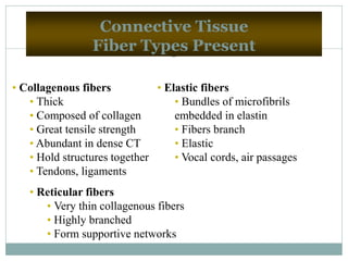
25
Connective Tissue
Fiber TypesPresent
• Collagenous fibers
• Thick
• Composed of collagen
• Great tensile strength
• Abundant in dense CT
• Hold structures together
• Tendons, ligaments
• Elastic fibers
• Bundles of microfibrils
embedded in elastin
• Fibers branch
• Elastic
• Vocal cords, air passages
• Reticular fibers
• Very thin collagenous fibers
• Highly branched
• Form supportive networks
26.
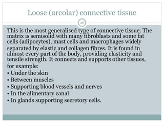
Loose (areolar) connectivetissue
26
This is the most generalised type of connective tissue. The
matrix is semisolid with many fibroblasts and some fat
cells (adipocytes), mast cells and macrophages widely
separated by elastic and collagen fibres. It is found in
almost every part of the body, providing elasticity and
tensile strength. It connects and supports other tissues,
for example:
• Under the skin
• Between muscles
• Supporting blood vessels and nerves
• In the alimentary canal
• In glands supporting secretory cells.
27.
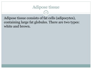
Adipose tissue
27
Adipose tissueconsists of fat cells (adipocytes),
containing large fat globules. There are two types:
white and brown.
28.
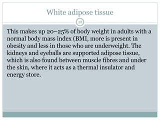
White adipose tissue
28
Thismakes up 20–25% of body weight in adults with a
normal body mass index (BMI, more is present in
obesity and less in those who are underweight. The
kidneys and eyeballs are supported adipose tissue,
which is also found between muscle fibres and under
the skin, where it acts as a thermal insulator and
energy store.
29.
Brown adipose tissue
29
Thisis present in the newborn. It has a more extensive
capillary network than white adipose tissue. When
brown tissue is metabolised, it produces less energy
and considerably more heat than other fat,
contributing to the maintenance of body temperature.
Sometimes small amounts are present in adults.
30.
Reticular tissue
30
Reticular tissuehas a semisolid matrix. It contains
reticular cells and white blood cells (monocytes and
lymphocytes). Reticular tissue is found in lymph nodes
and all organs of the lymphatic system.
31.
Dense connective tissue
31
Thiscontains more fibres and fewer cells than loose
connective tissue.
Fibrous tissue
This tissue is made up mainly of closely packed
bundles of collagen fibres with very little matrix.
Fibrocytes are few in number and lie in rows between
the bundles of fibres.
32.
32
Fibrous tissue formingligaments, which bind bones
together as an outer protective covering for bone,
called periosteum
• As an outer protective covering of some organs,
e.g. the kidneys, lymph nodes and the brain
• Forming muscle sheaths, called muscle fascia.
Fibrous tissue
33.
Elastic tissue
33
Elastic tissueis capable of considerable extension and
recoil. There are few cells and the matrix consists mainly
of masses of elastic fibres secreted by fibroblasts. It is
found in organs where stretching or alteration of shape is
required, e.g. in large blood vessel walls, the trachea and
bronchi, and the lungs.
Blood
This is a fluid connective tissue.
34.
Cartilage
34
Cartilage is firmerthan other connective tissues. The
cell (chondrocytes) are sparse and lie embedded in
matrix reinforced by collagen and elastic fibres. There
are three types:
➢ Hyaline cartilage
➢ Fibrocartilage
➢ Elastic cartilage.
35.
Hyaline cartilage
35
Hyaline cartilageis a smooth bluish-white tissue. The
chondrocytes are arranged in small groups within cell
nests and the matrix is solid and smooth. Hyaline
cartilage provides flexibility, support and smooth
surfaces for movement at joints. It is found:
✓ On the ends of long bones that form joints
✓ Forming the costal cartilages, which attach the ribs
to the sternum
✓ Forming part of the larynx, trachea and bronchi.
36.
Fibrocartilage
36
This consists ofdense masses of white collagen fibres in a
matrix similar to that of hyaline cartilage with the cells
widely dispersed. It is a tough, slightly flexible, supporting
tissue found:
✓ As pads between the bodies of the vertebrae, the
intervertebral discs
✓ Between the articulating surfaces of the bones of the
knee joint, called semilunar cartilages
✓ On the rim of the bony sockets of the hip and shoulder
joints, deepening the cavities without restricting
movement.
37.
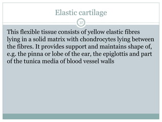
Elastic cartilage
37
This flexibletissue consists of yellow elastic fibres
lying in a solid matrix with chondrocytes lying between
the fibres. It provides support and maintains shape of,
e.g. the pinna or lobe of the ear, the epiglottis and part
of the tunica media of blood vessel walls
38.
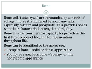
Bone
38
Bone cells (osteocytes)are surrounded by a matrix of
collagen fibres strengthened by inorganic salts,
especially calcium and phosphate. This provides bones
with their characteristic strength and rigidity.
Bone also has considerable capacity for growth in the
first two decades of life, and for regeneration
throughout life.
Bone can be identified by the naked eye:
✓ Compact bone – solid or dense appearance
✓ Spongy or cancellous bone –’spongy’ or fine
honeycomb appearance.
39.
Function:
Bone supports andprotects (by enclosing); provides levers
for the muscles to act on; stores calcium and other minerals
and fat; marrow inside bones is the site for blood cell
formation (hematopoiesis).
39
40.
Muscle tissue
40
This tissueis able to contract and relax, providing
movement within the body and of the body itself.
Muscle contraction requires a blood supply that will
provide sufficient oxygen, calcium and nutrients and
remove waste products. There are three types of
specialised contractile cells, also known as fibres:
➢Skeletal muscle
➢Smooth muscle and
➢Cardiac muscle
41.
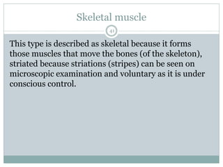
Skeletal muscle
41
This typeis described as skeletal because it forms
those muscles that move the bones (of the skeleton),
striated because striations (stripes) can be seen on
microscopic examination and voluntary as it is under
conscious control.
42.
Smooth muscle
42
Smooth muscleis also described as non-striated, visceral
or involuntary. It does not have striations and is not under
conscious control. Some smooth muscle has the intrinsic
ability to initiate its own contractions (automaticity), e.g.
Peristalsis. It is innervated by the autonomic nervous
system. It is found in the walls of hollow organs:
➢ Regulating the diameter of blood vessels and parts of
the respiratory tract
➢ Propelling contents along, e.g. the ureters, ducts of
glands and the alimentary tract.
43.
Cardiac muscle
43
This isonly found only in the heart wall. It is not under
conscious control but, when viewed under a
microscope, cross-stripes (striations) characteristic of
skeletal muscle can be seen.
44.
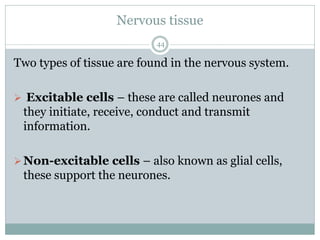
Nervous tissue
44
Two typesof tissue are found in the nervous system.
➢ Excitable cells – these are called neurones and
they initiate, receive, conduct and transmit
information.
➢Non-excitable cells – also known as glial cells,
these support the neurones.
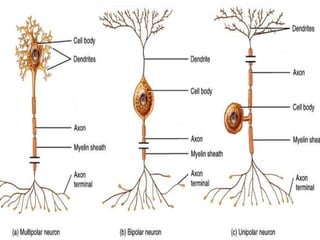
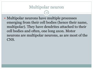
Multipolar neuron
46
Multipolarneurons have multiple processes
emerging from their cell bodies (hence their name,
multipolar). They have dendrites attached to their
cell bodies and often, one long axon. Motor
neurons are multipolar neurons, as are most of the
CNS.
47.
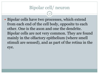
Bipolar cell/ neuron
47
Bipolar cells have two processes, which extend
from each end of the cell body, opposite to each
other. One is the axon and one the dendrite.
Bipolar cells are not very common. They are found
mainly in the olfactory epithelium (where smell
stimuli are sensed), and as part of the retina in the
eye.
48.
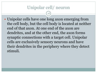
Unipolar cell/ neuron
48
Unipolar cells have one long axon emerging from
the cell body, but the cell body is located at neither
end of that axon. At one end of the axon are
dendrites, and at the other end, the axon forms
synaptic connections with a target cell. Unipolar
cells are exclusively sensory neurons and have
their dendrites in the periphery where they detect
stimuli.
49.
Neuroglial cells
49
Neuroglialcells usually referred to simply as glial
cells or glia are quite different from nerve cells.
The major distinction is that glia do not participate
directly in synaptic interactions and electrical
signalling, although their supportive functions help
define synaptic contacts and maintain the
signalling abilities of neurons.
Astrocytes
51
One cellproviding support to neurons of the CNS is
the astrocyte, so named because it appears to be star-
shaped under the microscope (astro- = “star”).
Astrocytes have many processes extending from their
main cell body (not axons or dendrites like neurons,
just cell extensions). Those processes extend to
interact with neurons, blood vessels, or the
connective tissue covering the CNS. Generally, they
are supporting cells for the neurons in the central
nervous system.
52.
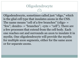
Oligodendrocyte
52
Oligodendrocyte, sometimes calledjust “oligo,” which
is the glial cell type that insulates axons in the CNS.
The name means “cell of a few branches” (oligo- =
“few”; dendro- = “branches”; -cyte = “cell”). There are
a few processes that extend from the cell body. Each
one reaches out and surrounds an axon to insulate it in
myelin. One oligodendrocyte will provide the myelin
for multiple axon segments, either for the same axon
or for separate axons.
53.
Microglia
53
Microglia are,as the name implies, smaller than
most of the other glial cells. Microglia are the cells
in the CNS that can do this in normal, healthy
tissue, and they are therefore also referred to as
CNS-resident macrophages.
54.
Ependymal cells
54
Ependymalcells filter blood to make cerebrospinal
fluid (CSF), the fluid that circulates through the
CNS. In each of the brain cavities (ventricles),
ependymal cells come in contact with blood vessels
to filter and absorb specific components of the blood.
These choroid plexuses produce enough
cerebrospinal fluid everyday to fill a pint glass!
55.
Satellite cell.
55
Oneof the two types of glial cells found in the PNS is
the satellite cell. Satellite cells surround the cell
bodies of neurons in the PNS. They provide support,
performing similar functions in the periphery as
astrocytes do in the CNS—except, of course, for
establishing the BBB.
56.
Schwann cell
56
Thesecond type of glial cell is the Schwann cell,
which insulate axons with myelin in the periphery.
Schwann cells are different than oligodendrocytes
in that a Schwann cell wraps around a portion of
only one axon segment and no others.
Oligodendrocytes have processes that reach out to
multiple axon segments, whereas the entire
Schwann cell surrounds just one axon segment.


![PERI-PROSTHETIC FRACTURE NAIL-PLATE CONSTRUCT [NPC].pptx](https://cdn.slidesharecdn.com/ss_thumbnails/drarunkumardrmohamedashrafperiprostheticfrasturenail-plateconstructnpc-260209164459-7e9d15a1-thumbnail.jpg?width=640&height=640&fit=bounds)


![ONFH[AVN HIP] -TRIPLE REGIME -A NOVAL SURGICAL CONCEPT .pptx](https://cdn.slidesharecdn.com/ss_thumbnails/onfhavnhip2026koaconcalicutdrgokuldevdrmashraf-260210064517-213ec005-thumbnail.jpg?width=640&height=640&fit=bounds)


