Ms. Ankita A. More
Paleobotany
Calamites
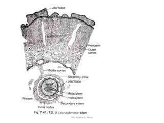
Lepidodendron
Lyginopteris
Pentoxylon
Contribution of Birbal Sahni
-Ms. Ankita Avinash More
GATE, SET
Ms. Ankita A. More
Ms. Ankita A. More
Calamites
Ms. Ankita A. More
Paleobotany is the scientific study of ancient plants, using plant fossils found in
sedimentary rocks.
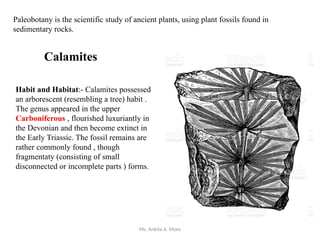
Calamites
Habit and Habitat:- Calamites possessed
an arborescent (resembling a tree) habit .
The genus appeared in the upper
Carboniferous , flourished luxuriantly in
the Devonian and then become extinct in
the Early Triassic. The fossil remains are
rather commonly found , though
fragmentaty (consisting of small
disconnected or incomplete parts ) forms.
Ms. Ankita A. More
External Features of Calamites:
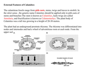
The calamitean fossils range from pith casts, stems, twigs and leaves to strobili. In
the strict sense , the generic name Calamites should be applied only to pith casts of
stems and branches The stem is known as Calamities, leafy twigs are called
Annularia, and fructification is known as Calamostachys. The plant body of
Calamites was a tall tree growing to a height of 20-30 metres.
The plant had an underground prostrate rhizome. The rhizome was differentiated into
nodes and internodes and had a whorl of adventitious roots at each node. From the
upper surface of the rhizome arose a number of aerial shoots.
Ms. Ankita A. More
Some of these aerial shoots produced roots at some of the lower nodes, indicating that
rhizome grew at some distance below the surface of the soil. Erect shoots were
constricted at the point of their junction to the rhizome. The branching was
conspicuous in the aerial shoots which had whorls of branches at the nodal region.
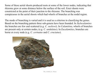
The mode of branching is varied and it is used as a criterion in classifying the genus.
Based on the branching pattern three sub-genera have been founded. In Stylocalamites
the branches are few and scattered (e.g. C. suckowi). In Calamitina, whorls of branches
are present only at certain nodes, (e.g. C. undulatus). In Eucalamities, branches are
borne at every node (e.g. C. cerinatus and C. cruciatus).
Ms. Ankita A. More
The lateral branches of the aerial shoots in some cases persisted for a long time and were
as thick as the main axis giving the familiar appearance of the present day Equisetum, but
much larger in size. The surface of the stem had longitudinal ribs alternating with the
grooves. But in some instances the ribs of the successive internodes were directly above
instead of alternating with one another.
The external surface of the stem of Calamites exhibited ribs and furrows , as are noted in
Equisetum.
Ms. Ankita A. More
Internal Structure of Calamites:
Anatomically the stem of Calamites showed an epidermis, cortex and stele. In the young
stems the cortex had an outer sclerotic zone and an inner thin welled parenchymatous
zone. There were no Vallecular canals as in Equisetum. The stele was siphonostelic. In the
central region there was a parenchymatous pith at the nodes, but represented by a
central cavity at the internodes.
The vascular bundles had collaterally arranged xylem and phloem. The xylem was
endarch. The tracheids had scalariform thickenings in metaxylcm but annular and spiral
thickenings in the protoxylem. In each vascular bundle there was a carinal canal formed
by the dissolution of the protoxylem elements
Ms. Ankita A. More
Secondary growth in the stem took place by the activity of a cambium. The cylinder
attained a thickness of 6 cm. or more due to the secondary growth. The secondary wood
did not show the differentiation of annual rings.
annual rings of tree
Ms. Ankita A. More
These point out that the plant had an evergreen foliage and there was probably no seasonal
variation in a year which is responsible for the annual ring formation. Secondary xylem
had scalariform and pitted tracheids. Secondary growth also took place in the cortex
producing periderm of several centimeters in thickness.
Ms. Ankita A. More
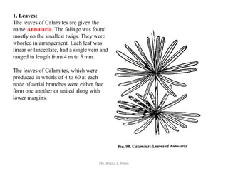
1. Leaves:
The leaves of Calamites are given the
name Annularia. The foliage was found
mostly on the smallest twigs. They were
whorled in arrangement. Each leaf was
linear or lanceolate, had a single vein and
ranged in length from 4 m to 5 mm.
The leaves of Calamites, which were
produced in whorls of 4 to 60 at each
node of aerial branches were either free
form one another or united along with
lower margins.
Ms. Ankita A. More
2. Roots:
Roots of Calamites are called Astromyelon. Internally they had a direct triarch xylem
with centripetal protoxylem. A cambium was present in the root, but produced only a
limited quantity of secondary wood.
Ms. Ankita A. More
3. Strobili:
The strobili associated with the Calamitean stem are given the name Calamostachys. In
this, the strobilus had a central axis bearing whorls of sporangiophores. Alternating with
the whorls of sporangiophores were whorls of sterile appendages called bracts. The
sporangiophores had cruciately (cross shaped) branched apices.
The tip of each branch recurved towards the strobilar axis and had a sporangium at the tip.
The genus Calamostachys had both homosporous and heterosporous species.
The strobili or cones of Calamites showed wide arrangements , the majority of them ,
however, belonging to either of the two genera, Calamostachys and Palaeostachya. The
sporangiophores in the former genus stood out at right angles to the axis of the strobilus ,
while in the later case , they stood out at an angle of near about 45º. The sporangia may be
either homosporous or heterosporous even in the different species of the same genus.
Further in Calamostachys , the leaf bracts and sporangiophores appear in alternate whorl,
while in Palaeostachya , the sporangiophores appear in the axils of the bracts.
Ms. Ankita A. More
Lepidodendron
Ms. Ankita A. More
Habit and Habitat:-
The genus Lepidodendron with its 100 species (approximately ) appeared in the Late
Devonian , flourished in the Carboniferous and then underwent degeneration most
probably during the Late Permian . The plants were mostly tall trees.
The plant body (Sporophyte):-
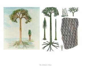
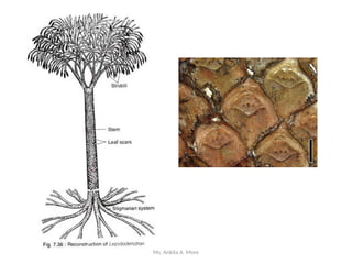
The plant was large tree with a typical Stigmarian root system , a main trunk , which
remained straight and unbranched for a considerable height and then showed a regular
dichotomy in its branches. The stellar organization of the stem and branches of
Lepidodendron was usually of a protostelic nature. The leaves named as the form genus
Lepidophyllum, were circular to linear in shape and simple and they were borne spirally
on the branches as well as in alternating whorls. The persistent leaf bases were pyramidal
in outline and formed peculiar leaf cushions.
Ms. Ankita A. More
On either side of the solitary median vascular strand of the leaf base there was a strand
of parenchyma cells giving rise to the parichnos , each of which ultimately lost its
identity in the mesophyll tissue.
Lepidostrobus:- The name of the form genus of the cone or strobilus of
Lepidodendron is Lepidostrobus which is an elliptical or cylindrical body which
contained both microsporophyll, each bearing one microsporangium with numerous
microspores and megasporophylls bear one mega sporangium containing lesser number
of megaspores. The former lie towards the apex and the later towards the strobilus.
Ms. Ankita A. More
Lepidodendron, extinct genus of tree-sized lycopsid plants lived during the Carboniferous
Period (about 359 million to 299 million years ago).
Lepidodendron and its relatives Lepidophloios, Bothrodendron, and Paralycopodites—
were related to modern club mosses. They grew up to 40 metres (130 feet) in height and 2
metres (about 7 feet) in diameter.
During their juvenile stages, these plants grew as unbranched trunks with a stock of long,
thin leaves that sprouted near the growing tip. They branched at later stages, either in even
dichotomies at the growing tip or in lateral branches that were later shed. After branching,
the leaves became shorter and awl-shaped. As the plant grew, it shed leaves from older
parts of the stem that left diamond-shaped leaf bases.
Ms. Ankita A. More
The Lepidodendrales were the most elaborate and diversified of all the lycopsids, and
dominated the Carboniferous, but with the drying of the climate during the latest
Carboniferous and early Permian they went into a steep decline. By the middle
Permian, they were all gone. Inefficient movement of water and nutrients through the
stems of these tall plants, resulting from a lack of secondary xylem is cited as one of
the reasons for their extinction.
Ms. Ankita A. More
Stems were characterized by a slender central strand of wood and a thick bark. Since
Stigmaria—the underground parts of the plant—resembled stems, they are not
considered true roots. The shape of leaf bases and the arrangement of their vascular
strands distinguish the different genera within the group of arborescent lycopsids.
Ms. Ankita A. More
The Stigmaria had spirally arranged roots coming from them. Attached to the Stigmaria
of Lepidodendron was a long pole-like trunk which had no branches for most of its
length. T
he trunk terminated in a crown of simple branches which were covered with spirally
arranged grass-like leaves, called Lepidophyllum.
At the end of the branches were reproductive cones, called Lepidostrobus, which
contained spores. These different names (called "form genera") have been applied to
different parts of Lepidodendron because they were originally discovered and
scientifically described as separate parts. It was only later when more complete
specimens were found that it was realized that the separately described parts in fact
belonged to the same plant. In some cases the form genus turns out to belong to different
families when the complete plant is considered. For example Stigmaria may belong to
genera assigned to Lepidodendraceae, Sigillariaceae, or Lepidocarpaceae
Ms. Ankita A. More
Roots
Lepidodendron and similar great trees grew in the hot humid swampland of the
Carboniferous period. It possessed branching rooting organs, called Stigmaria, by
which it was anchored in shallow soil.
In all the members of Lepidodendrales, the root-bearing underground axes are called
rhizomorph and the detached rhizomorph and their roots are called Sigmaria which are
mostly found as siliceous casts or molds.
Stigmaria ficoides, the commonest species of Stigmaria, was a large trunk base that
divided dichotomously into four large massive descending axes
Ms. Ankita A. More
Stem
The stem form-genus is called Lepidodendron which has been reported mostly as casts
or compressions. In most species, the trunks attained a height up to 98-115 ft (30-35 m),
because the first branching at a distal end appeared up to 30-35 m in height. At the base,
the trunks are known to be 3.3 ft (1 m) in diameter. Numerous leaf cushions arrange
spirally on the stem surface.
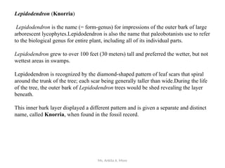
The leaf cushions-are rhomboidal in shape and broader in their vertical dimension than
their transverse length. A leaf scar is situated just above the middle line of the cushion.
The leaf scar comprised of a vascular bundle scar at the centre.
Ms. Ankita A. More
Ms. Ankita A. More
Ms. Ankita A. More
Ms. Ankita A. More
Many of the larger lepidodendrid trees had monosporangiate cones, with highly
specialized megasporangia that mimic the seed habit. A good example is Lepidocarpon.
In Lepidocarpon cones, there is a single megaspore present that remains within the
megasporangium, and the megasporangium remains tucked inside a leafy sporophyll
that encloses it.
The entire cone breaks up and these units thus serve as propagules. Because plants
bearing this type of cone were living in a wet coal swamp, some paleobotanists have
suggested that this dispersal unit acted as a small boat capable of aquatic dispersal.
Ms. Ankita A. More
Lepidodendron (Knorria)
Lepidodendron is the name (= form-genus) for impressions of the outer bark of large
arborescent lycophytes.Lepidodendron is also the name that paleobotanists use to refer
to the biological genus for entire plant, including all of its individual parts.
Lepidodendron grew to over 100 feet (30 meters) tall and preferred the wetter, but not
wettest areas in swamps.
Lepidodendron is recognized by the diamond-shaped pattern of leaf scars that spiral
around the trunk of the tree; each scar being generally taller than wide.During the life
of the tree, the outer bark of Lepidodendron trees would be shed revealing the layer
beneath.
This inner bark layer displayed a different pattern and is given a separate and distinct
name, called Knorria, when found in the fossil record.
Ms. Ankita A. More
Reproduction
Lepidodendron and its relatives reproduced by spores, with megaspores giving rise to the
female (egg-producing) gametophyte and microspores giving rise to the male (sperm-
producing) gametophyte.
Lepidophloios wrapped its megasporangium in a layer of tissue much like that of the
seed plants. This feature, however, was independently derived in the lycopsid lineage. In
some genera, spore-bearing cones were produced at the tips of branches, suggesting that
the plants could reproduce only once in their lifetime.
Lepidodendron and its relatives lived in the extensive peat forming swamps and became
extinct when these swamps disappeared
Ms. Ankita A. More
Reproductive Structure
Lepidodendron formed bisporangiate cones called Flemingites that were borne
terminally. The sporophylls were helically attached to the central cone axis. The
microsporophylls bearing microsporangia were usually borne in the apical portion,
while megasporophylls bearing megasporangia occupied the basal portion of the cones.
Morphologically, both the sporophylls were identical, except for their spore content.
The microspores were small, about 25 mm in diameter, with smooth or granular exine.
The cones containing only microspores are assigned to the form genus Lepidostrobus,
possibly a monsporangiate cone of Lepidodendron
Ms. Ankita A. More
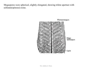
Megaspores were spherical, slightly elongated, showing trilete aperture with
echinate(spinous) exine.
Ms. Ankita A. More
Lepidocarpon: a False Seed
The female gametophyte of Lepidodendron is called Lepidocarpon. Like a
spermatophyte (especially gymnosperm), the megagametophyte (Lepidocarpon) is
retained within the megasporangiun and the sides of the pedicel were extended to
lorm lateral laminae, called integuments —which completely enveloped the
sporangium.
Seed characteristics of Lepidocarpon (similarity with an ovule of gymnosperm):
1. Formation of a single functional megaspore in the megasporangium.
2. Retention of functional megaspore in the megasporangium.
3. Formation of endosporic megagametophyte within megasporangium.
4. Formation of an integument that delimited a microphyle.
All of the above characteristics show that Lepidocarpon apparently possessed the
attributes of a seed habit. However, it did not reach the level of a seed (ovule),
because the basic characteristics of a seed (ovule) are totally absent in Lepidocarpon.
Ms. Ankita A. More
Ms. Ankita A. More
Contribution of birbal
sahni, birbal sahni
institute of paleobotany
lucknow
Ms. Ankita A. More
A very distinguished scientist who laid the foundation for the study of Botany in
India is Prof. Birbal Sahni who had special interest in Paleobotany.
He was born in the year 1891 and his hometown was Bhera in the state of Punjab.
His father was Prof. Ruchi Ram Sahni who specialized in Chemistry.
After graduating from Lahore in 1911, he continued his studies at the University of
Cambridge.
Ms. Ankita A. More
Early Life and career
After his studies at Cambridge, in 1914 he started his research in Botany under Prof.
A. C. Steward.
He was given the responsibility of revising the “Textbook of Botany” by Lawson
which showed his profound knowledge of Indian plants.
In 1919 he was awarded the Doctor of Science by the University of London for his
studies on Paleobotany.
On returning to India in the same year he was in-charge of Botany Departments at the
Banaras and Punjab Univerisites.
In 1921, he was given charge of the Department of Botany at Lucknow University as
its first professor and later became Dean of sciences in 1933.
Ms. Ankita A. More
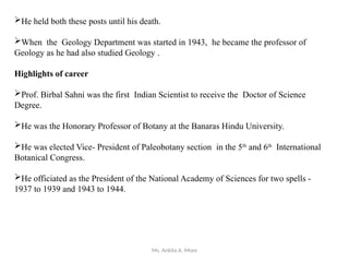
He held both these posts until his death.
When the Geology Department was started in 1943, he became the professor of
Geology as he had also studied Geology .
Highlights of career
Prof. Birbal Sahni was the first Indian Scientist to receive the Doctor of Science
Degree.
He was the Honorary Professor of Botany at the Banaras Hindu University.
He was elected Vice- President of Paleobotany section in the 5th
and 6th
International
Botanical Congress.
He officiated as the President of the National Academy of Sciences for two spells -
1937 to 1939 and 1943 to 1944.
Ms. Ankita A. More
He was President of Botany Section at the Indian science Congress in 1921 and
1938 and held the position of General President at the Science Congress held in 1940.
He was also elected the President of the International Botanical Congress to be held
at Stockholm in 1950 but he suddenly passed away in 1949 before the event could
take place/
He was designated by the Government of India to visit Research Laboratories in
Europe, The United Kingdom and The United States of America to observe and set up
research laboratories in India after Independence in 1947.
He was awarded Fellowship of most of the learned Societies in India.
Ms. Ankita A. More
Awards and Honours
He received the Barclay Medal by the Royal Asiatic Society of Bengal for
outstanding researches in Biological sciences.
He also received the Sir C. R. Reddy National prize for Natural Sciences in 1947.
He was awarded the Honorary Doctorates by the Universities of Patna and
Allahabad.
He received the Nelson Wright Medal of the Numismatic Society of India in 1945
for his article on the art of casting coins in Ancient India.
Ms. Ankita A. More
Important Contributions of Prof. Birbal Sahni
Though a Paleobotanist, most of his work was concerned with living plants
Between 1915 and 1936 he did a lot of work on morphology and Phylogeny of
Pteridophytes and Gymnosperms like Nephrolepis, Tmesipteris, Taxus,
Cephalotaxus and Ginkgo.
He contributed important landmark articles like “Observations on the branching in
Filicales” (1917) and “The Ontogeny of Vascular Plants and Theory of Recapitulation”
(1925)/
He studied the age of the Saline series of Punjab Salt Range and Deccan traps -his
conclusion that they are of tertiary age is still accepted.
He published his researches on the Zygopteridaceae in 1918.
He collaborated with Prof. Seward and revised the Indian Gondwana plants and gave
important data on Indian fossil conifers.
Ms. Ankita A. More
Between 1918 and 1949 he published articles on every aspect of Paleobotany
He described many fossil materials from India.
He started and edited the Research Bulletin called “Paleobotany in India”.
He contributed his observations that solved the problems related to Paleographics
and Geology of Permo-Carboniferous life provinces.
He contributed his ideas to explain Wegner’s theory of Continental drift and
Himalayan Uplift.
The fossiliferous areas that interested Prof. Sahni were the RajMahal Hills of Bihar,
The deccan series and the Salt range of Punjab.
Ms. Ankita A. More
He described a number of fossil forms including Homoxylon rajmahalense,
Rajmahalia paradoxa and Williamsonia sewardiana.
His notable contribution was the institution of a new group of extinct plants –the
Pentoxyleae.
He was instrumental in setting up the Institute of Paleobotany at Lucknow which is
called the Birbal Sahni Institute of Paleobotany.
Ms. Ankita A. More
Lyginopteris
Ms. Ankita A. More
Morphological Features
The stem Lyginopteris was slender and covered with large scaly leaves. Near the base of
the plant adventitious roots developed. The plant seems to have been a climber.
Lyginopteris oldhamia also known as Calymatotheca hoeninghausi was described in detail
by Williamson, Scott, Brongniart, Binney, Potonie, and Oliver and Scott. It was found
abundantly in the horizon of Lancashire and Yorkshire [England].
Ms. Ankita A. More
Anatomical Features
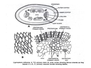
The Primary structure was an ectophloic siphonostele
with large pith round a number of primary mesarch
bundles. Older plants showed normal secondary
growth. In some specimens, however, the xylem
portion of primary vascular bundles was in a
continuous ring. In some there was an abnormal type
of secondary growth.
This abnormality was of two forms, either there was
an inner ring of secondary phloem developed or it was
that the cambium appeared in strips found separately
in vascular bundles giving rise to a polystelic
appearance.
Ms. Ankita A. More
Ms. Ankita A. More
Ms. Ankita A. More
Ms. Ankita A. More
Reproductive Structure of Lyginopteris:
Some of these Palaeozoic leaves bore microsporangia on them. The fertile pinnules were
more or less peltate in form and on their underside they bore usually six sporangia. These
sporangia are usually bilocular. Such a type has been described as Crossotheca type.
The microspores seem to have formed a male prothallus. The male spores seem to have
been of like those of present-day cycads.
Ms. Ankita A. More
Seed of Lyginopteris
The best known seed has been described under the name Lagenostoma. These seeds were
small in size, only about 1/4 but they were highly organized. It was barrel shaped and whole
seed enclosed in cupule. This cupule opened out when seed was mature.
Each seed was borne at the tip of stalk. The cupule rised from the base of the seed but not
fused with it. The cupule was in three main lobes. These lobes were divided in the upper
parts of the seeds. The seed or ovule was orthrotropus and of cycadian type. It was radially
symmetrical.
The cupule was separated from the seed along its entire length. The seed itself has an
integument which surrounded the nucellus. The integument and nucellus were fused except
at the top. The integument formed nine projections. In each of these projections there was a
vascular bundle present.
Ms. Ankita A. More
In some the megaspore membrane is very well seen. In the centre of the seed there was a
tissue but so far neither any archegonium nor any embryo has been found in these
Palaeozoic seeds.
The seeds were borne at the tips of the stalks. They were not organized to form cover. In
addition to this Lagenostoma there are many other Palaeozoic seeds which have been
described some simpler other more complex than Lagenostoma.
A peculiar feature of all Palaeozoic seeds which so for has not been explained is the
absence of any embryo in them. Pollen grains have been found in pollen chambers. Some
of them had even showed their germ tube, but so far no seed is discovered in which
embryo was developed. It is possible that all the seeds described might have not preserved
or they preserved before embryo formation.
Ms. Ankita A. More
Pentoxylon
Ms. Ankita A. More
Pentoxylon
The pentoxylales are a group of seed plants that lived during middle to late
Mesozoic period. Stems, leaves and male and female reproductive organs of this
group have been described under following names:
Stems: Pentoxylon and Nipanioxylon
Leaves: Nipaniophyllum
Pollen-bearing organs: Sahnia
Seed-bearing organs: Carnoconites.
They had long narrow leaves and wood in a characteristic five wedge pattern (penta
– xylon) around the primary xylem..
Ms. Ankita A. More
Habit
The plant was probably a shrub or very small trees.
The stem was dimorphic.
Long and short shoots were covered with spirally arranged scale, foliage bases and
terminally located reproductive organs.
Leaves were thick, simple, lanceolate, had diploxylic leaf trace. The anatomy of stem
revealed five to six steles which were closely aggregated.
Ms. Ankita A. More
The stems of Pentoxylon sahnii attained a diameter from 3mm to 2 cm.
Each stele had its own cambium.
The cambium was uniformly active in the young stems, but at maturity more secondary
tissue developed towards the centre, and thus the secondary wood appeared eccentric.
Primary phloem and primary xylem were present towards outer and inner sides of the
cambium, respectively.
Ms. Ankita A. More
The wood was pycnoxylic (It is compact and strong wood. It has a large number of
xylem tracheids, a small amount of pith, and cortex, and very little parenchyma)
made of compact tracheids.
Ovules were sessile.
Female reproductive organs were like stalked mulberry, consisting of about 20 sessile
seeds attached to central receptacle and surrounded by stony layer and then fleshy
outer layer of integument uniting them.
Male reproductive organs or microsporophyll’s form whorl of branched micro-
sporangiophores. The micro-sporangiophores were fused basally into a disc-like
structure.