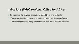
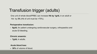
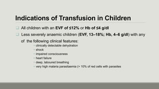
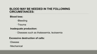
ANEMIA & BLOOD TRANSFUSION
BY KPEHE JIG MAIMIE (INTERN)
Outline
Anemia
1. Definition
2. WHO Threshold of Anemia
3. Symptoms & Physical Findings
4. Severe Anemia
5. Etiology
6. Classification
Blood Transfusion
1. Definition
2. History
3. Indications
4. Types of Donors
5. Criteria for Donation
6. Blood Collection & Screening
7. Physician Responsibility
8. Transfusion Checklist
9. Labs
10. Transfusion Protocol
11. Transfusion Reaction
Anemia
Definition
is ↓ Hgb below the reference level for
a) Age
b) Sex and
c) Pregnancy state
WHO Threshold of Anemia
CHILDREN (6 months to under 5 years): - 11 gm/dl
CHILDREN (5 years to under 12 years): - 11.5 gm/dl
CHILDREN ( 12 years to under 15 years): - 12 gm/dl
NON-PREGNANT WOMEN (15 years and over): - 12 gm/dl
PREGNANT WOMEN: - 11 gm/dl
MEN (15 years and over): - 13 gm/dl
Symptoms
 Depends on
a) Acuteness
b) Severity of anemia.
 Most patients are asymptomatic
Physical Findings
GENERAL
 Pallor of mucus membranes
 Signs of hyperdynamic circulation (tachycardia, bounding pulse)
SPECIFIC
 Koilonychias (ridging and spoon shape nails in iron deficiency anemia)
 Jaundice (hemolytic anemia)
 Bone deformities (thalassemia)
 Leg ulcers (sickle cell disease)
 Splenomegaly, petechaie/purpura (bleeding disorder)
 Glossitis (iron, folate, vitamin B12 deficiencies)
 Neurologic abnormalities (vitamin B12 deficiency)
SEVERE ANEMIA
 Adult is defined by a hemoglobin of < 7 g/Dl
 All children with an EVF of ≤12% or Hb of ≤4 g/dl
Or
 Less severely anaemic children (EVF, 13–18%; Hb, 4–6 g/dl) with any of the
following clinical features:
clinically detectable dehydration
shock
impaired consciousness
heart failure
deep, laboured breathing
very high malaria parasitaemia (> 10% of red cells with parasites
*Severely anemic Patients requires Blood Transfusion
Etiology
Decreased production Increased destruction or loss
Nutritional deficiencies
 iron deficiency*
 vitamin B12 deficiency
 folate deficiency*
Bone marrow suppression
Infections: HIV*, tuberculosis*, malaria,
Schistosomiasis*, hookworm*, hepatitis
Drugs: Isoniazid, chloramphenicol, alcohol,
zidovudine, 5-FU, hydroxyurea
Chronic disease: Renal and liver disease,
Rheumatologic diseases, hypothyroidism
Hemoglobinopathies
Thalassemias
Blood loss (Hemorrhage)
 trauma
 acute or chronic GI bleeding*
 menstrual bleeding*
 Hemolysis
 Malaria
 G6PD deficiency*
 DIC, eclampsia, HELLP
 Hereditary spherocytosis
 Sickle cell disease*
 Paroxysmal nocturnal hemoglobinuria
 Hypersplenism
Classification of Anemia by Size
o Microcytic anemia
o Normocytic Anemia
o Macrocytic Anemia
Microcytic Anemia
Iron deficiency anemia: most common worldwide.
Clinically: brittle nails, atrophy of papillae of tongue, brittle hair
Etiology: Poor diet
Chronic blood loss
(schistosomiasis, worms, GI loss from esophageal varices, peptic ulcer)
Diagnosis: MCV < 80, Blood smear shows hypochromic red cells, pencil shaped
(poikilocytosis), Target cells
Treatment: Iron replacement ferrous sulfate 200 mg tds
Normocytic Anemia
Anemia of chronic disease: Assoc. w/ chronic inflammatory or
Malignant dz.
ETIOLOGIES:
Infectious (TB, lung abscess, pneumonia, endocaritis) or
Non-infectious (rheumatoid arthritis, lupus), malignancy.
Clinical features: Reduced serum iron and TIBC, normal or raised ferritin.
Treatment is treat underlying cause
Macrocytic Anemia
Vitamin B12 deficiency:
ETIOLOGIES
a) Malnutrition (alcoholics, vegetarians),
b) Pernicious anemia decreased absorption (celiac sprue, Crohn’s disease),
c) Increased competition (fish tapeworm, intestinal bacterial overgrowth)
Clinical features neurologic changes
- (numbness, paresthesia's, decreased vibratory and positional sense, ataxia).
- Smear shows hypersegmented neutrophils.
Treatment: Replacement of B12
Macrocytic Anemia
Folate deficiency:
ETIOLOGIES
o Malnutrition (alcoholics, elderly)
o Decreased absorption (sprue)
o Impaired metabolism (methotrexate, trimethoprim, antimalarials)
Treatment: Folate repletion
Hemolytic Anemia:
o Due to red cell destruction and increased red cell turnover
o Bone marrow is able to compensate 5 times the normal rate.
Clinical features: Jaundice, Hepatosplenomegaly, Dark urine.
Labs: ↑ Reticulocyte count, Indirect hyperbilirubinemia, shistocytes on
blood smear
Treatment: Treatment underlying cause of hemolysis
Blood Transfusion
Definition
 Involves the transfer of blood or blood components from the donor
to the recipients. (WHO regional Office for Africa)
Blood Components
LIBERIA: Whole Blood Only
OTHER COUNTRIES:
Packed Cells
Fresh Frozen Plasma
Platelets
Cryoprecipitate
History of Blood Transfusion
1492 - Pope Innocent VIII suffers a stroke and is made to drink blood from three 10-year-old
boys (paid a ducat each). All three boys died, as did the pope later that year
1665 Richard Lower in Oxford conducts the first successful canine transfusions
1667 Jean-Baptiste Denis reports successful sheep–humant ransfusions
1670 Animal–human transfusions are banned in France because of the poor results
1829 James Blundell performs the first successful documented human transfusion in a
woman suffering post-partum haemorrhage.
1900 Karl Landsteiner discovers the ABO system
1914 The Belgian physician Albert Hustin performs the first non-direct transfusion, using
sodium citrate as an anti-coagulant
1926 The British Red Cross institutes the first blood transfusion service in the world
1939 The Rhesus system is identified and recognised as the major cause of transfusion
reactions
Indications (WHO regional Office for Africa)
 To increase the oxygen capacity of blood by giving red cells.
 To restore the blood volume to maintain effective tissue perfusion.
 To replace platelets, coagulation factors and other plasma proteins
Transfusion trigger (adults)
One unit of whole blood/PRBC can increase Hb by 1g/dL in an adult or
Hct by 3% (Hb of unit must be >75%).
Perioperative transfusion:
– 8g/dL for patient undergoing cardiovascular surgery, orthopaedics and
acute GI bleeding.
Chronic anaemia:
– 7g/dL in adults.
Acute blood loss:
– 30% of volume of blood.
Indications of Transfusion in Children
 All children with an EVF of ≤12% or Hb of ≤4 g/dl
 Less severely anaemic children (EVF, 13–18%; Hb, 4–6 g/dl) with any
of the following clinical features:
 clinically detectable dehydration
 shock
 impaired consciousness
 heart failure
 deep, laboured breathing
 very high malaria parasitaemia (> 10% of red cells with parasites
BLOOD MAY BE NEEDED IN THE FOLLOWING
CIRCUMSTANCES:
Blood loss:
Bleeding
Trauma
Inadequate production:
 Diseases such as thalassemia, leukaemia
Excessive destruction of cells:
Disease
Mechanical
Types of Donors
 Altruistic Voluntary, Unpaid Donors “Safest Donors”
 Relatives of Patients (Liberia)
 Preoperative autologous Blood Deposit for Elective Surgery
Avoid High Risk Donors
 Commercial Sex Workers,
 IV Drug Abusers
Criteria for Blood Donation Eligibility
General appearance: Good physical and mental health.
Age: b/w 18 and 60 years of age.
Haemoglobin: > 12.5 g/dL for males and 11.5 g/dL for
females.
Weight: minimum 45 kg.
Blood pressure: (systolic: 100‐140 mm Hg and
(diastolic: 60‐90 mm Hg is recommended)
Temperature: Oral temp. not > 37.5C/99.5 F.
Pulse: b/w 60 and 100 b/m per minute and regular.
Donation interval: b/w 3 to 4 months
Blood Collection
 The donor must not fast before donation.
 If the last meal was > than 4hrs, the donor must be made eat
something
 Blood flowing into the bag is mixed with anticoagulant in a ratio of 1:7
(anticoagulant : blood).
 Total collection volume is from 405‐495 mL
A volume of 450 mL blood is donated, this being approximately 12% of total
blood volume or 10.5 mL/kg body weight.
Blood Screening
Donors blood must screen before Collection
WHO recommends the following:
 HIV
 HEPATITIS B, HEPATITIS C
 SYPHILIS
 MALARIA
 CHAGAS DISEASE (WHERE APPROPRIATE)
 HTLV1 AND HTLV2
 WISE TO SCREEN DONORS BEFORE COLLECTION
 NAT TESTING IS COST EFFECTIVE
Responsibility of the Physician
Assess patient’s clinical need for blood, and when required.
 Inform patient and/or relatives
 Record indications for transfusion in patient’s notes.
 Select blood product and quantity required (i.e. whole
blood/PRBC/FFP/PC) and complete request form accurately and
legibly.
Enter the reason for transfusion on the form,
Obtain and correctly label a blood sample for compatibility testing.
Send the blood request form and blood sample to the blood bank.
Responsibility of the Physician Cont’d
 Don’t Store blood, transfuse it as soon as it arrives
 If blood product is not used immediately, store it under
the correct storage conditions.
 Cross check the identity of the patient and the blood product:
Patient and documentation.
Blood / blood products.
Transfusion Checklist
1. What improvement in the patients condition do I want to achieve?
2. Can blood loss be minimized to reduce the need for transfusion?
3. Are there any other treatments I should give before making the decision to transfuse, such as
intravenous replacement fluids and oxygen?
4. What are the specific clinical or laboratory indications for transfusion in this patient?
5. What are the risk for transmitting HIV, hepatitis, syphilis or other infectious agents through the
blood products that are available for this patient?
6. Do the benefit of transfusion outweigh the risks for this patient?
7. What other options are there if no blood is available on time?
8. Will a trained personel monitor this patient and respond immediately if transfusion reaction
occur?
9. If this blood was for me or my child, will I accept the transfusion under these circumstances?
Blood Sample
 A 5 mL blood sample collected into a dry test tube & Correctly labelled with
detailed patient’s information
Patient’s full name, age and sex.
Registration number.
Ward/bed number.
Date and time specimen taken.
Phlebotomist’s signature/initials
Use positive patient identification to identify the patient.
NEVER pre‐label the sample tube before phlebotomy.
The signature of the individual who took the sample must appear on the specimen label.
Laboratory Performed
 ABO and RhD grouping on patient and donors.
 Antibody screening on patient.
 Cross matching between serum of patient and red cells of donor.
Purpose
To select blood components that will not harm the recipient
Compatibility tests will confirm ABO compatibility between component & recipient
Will detect the most clinically significant unexpected antibodies.
Compatibility (cross match) must be performed before Blood Transfusion
 Incompatible Cross match = Reaction blw patient’s serum and donor’s red cells.
Performing Transfusion
Adults Rate
 Whole blood 150‐200 mL/hour
 PRBC 100‐150 mL/hour
 Platelets / plasma 150‐300 mL/hour
Paediatric patients Rate
 Whole blood / PRBC 2‐5 mL/kg/hour
 Platelets / plasma 1‐2 mL/minute
Discard unit if this period is exceeded
BLOOD PRODUCTS START TRANSFUSION COMPLETE TRANSFUSION
Whole blood / PRBC Within 30 minutes of
removing from
refrigerator
≤4 hours
Platelet concentrate Immediately Within 30 minutes
FFP As soon as possible Within 30 minutes
Cryoprecipitate As soon as possible Within 30 minutes
Transfusion Duration
Guidelines for recognition and management
of acute transfusion reactions
Mild reactions
Signs Symptoms Possible cause
Localized cutaneous:
Urticaria
Rash
Pruritus Hypersensitivity
(mild)
Immediate management of Category 1: Mild reactions
 Slow the transfusion.
 Administer antihistamine IM.
 If no clinical improvement within 30 minutes or if signs and symptoms worsen,
treat as Moderate Reactions .
 If improved, restart transfusion slowly.
Moderately Severe reactions
Signs Symptoms Possible cause
Flushing
Urticaria
Rigors
Fever
Restlessness
Tachycardia
Anxiety
Pruritus
Palpitations
Mild dyspnoea
Headache
Hypersensitivity
Immediate management: Moderate reactions
 Stop the transfusion and keep IV line open with normal saline in another site.
 Return the blood unit with transfusion administration set, freshly collected
urine and new blood samples (1 clotted and 1 anticoagulated), drawn from a vein
opposite to the transfusion site, to the blood transfusion centre for laboratory
investigations.
 Administer antihistamine IM and oral or rectal antipyretic.
 Avoid aspirin in thrombocytopenic patients
Immediate management: Moderate reactions
 Give IV corticosteroids and bronchodilators if there are anaphylactoid features
(e.g. broncho‐spasm, stridor).
 If clinical improvement occurs, restart transfusion slowly with new blood unit
and observe carefully.
 If no clinical improvement within 15 minutes or if signs and symptoms worsen,
treat as Category 3.
 Collect urine for next 24 hours for evidence of haemolysis and send for laboratory
investigations
 If available, a leucocyte reduction filter (WBC filter) may be used in repeated
transfusion.
Severe Reactions
Signs Symptoms Possible cause
Rigor
Fever
Restlessness
Hypotension (fall of 20% in
systolic BP)
Tachycardia (rise of 20% in
heart rate)
Haemoglobinuria (Hb in urine)
Unexplained bleeding (DIC)
Anxiety
Chest pain
Pain along the transfusion line
Respiratory distress/shortness
of breath
Loin/back pain
Headache
Dyspnoea
Acute intravascular haemolysis
(mismatched blood transfusion)
Bacterial contamination and
septic shock
Fluid overload
Anaphylaxis
Transfusion related acute lung
injury (TRALI)
Immediate management: Severe Reactions
 Stop the transfusion .
 Infuse normal saline to maintain systolic BP.
 Maintain airway and give high flow oxygen by mask.
 Give adrenaline (as 1:1000 solution) 0.01 mg/kg body weight by slow
intramuscular injection.
 Give IV corticosteroids and bronchodilators if there are anaphylactoid features.
 Give diuretic: e.g. frusemide 1 mg/kg IV or equivalent.
 Check a fresh urine specimen visually for signs of haemoglobinuria.
 Notify the superior or senior doctor attending the patient, and the blood centre
immediately.
 Send blood unit with transfusion set, fresh urine sample and new blood with the
appropriate request form to the blood transfusion centre for investigation.
References
1. Norman S. Williams et al, Bailey & Love Short Practice of Surgery, 25th Edition
2. Clinical Transfusion Practice, Guideline for Interns (WHO)
3. F. Charles Brunicardi et al, Schwartz Principles of Surgery, 2014 Edition
4. Pocket Book of Hospital Care for Children (WHO), 2013 Edition
Thank you!

Anemia &amp; transfusion

  • 1.
    ANEMIA & BLOODTRANSFUSION BY KPEHE JIG MAIMIE (INTERN)
  • 2.
    Outline Anemia 1. Definition 2. WHOThreshold of Anemia 3. Symptoms & Physical Findings 4. Severe Anemia 5. Etiology 6. Classification Blood Transfusion 1. Definition 2. History 3. Indications 4. Types of Donors 5. Criteria for Donation 6. Blood Collection & Screening 7. Physician Responsibility 8. Transfusion Checklist 9. Labs 10. Transfusion Protocol 11. Transfusion Reaction
  • 3.
  • 4.
    Definition is ↓ Hgbbelow the reference level for a) Age b) Sex and c) Pregnancy state
  • 5.
    WHO Threshold ofAnemia CHILDREN (6 months to under 5 years): - 11 gm/dl CHILDREN (5 years to under 12 years): - 11.5 gm/dl CHILDREN ( 12 years to under 15 years): - 12 gm/dl NON-PREGNANT WOMEN (15 years and over): - 12 gm/dl PREGNANT WOMEN: - 11 gm/dl MEN (15 years and over): - 13 gm/dl
  • 6.
    Symptoms  Depends on a)Acuteness b) Severity of anemia.  Most patients are asymptomatic
  • 7.
    Physical Findings GENERAL  Pallorof mucus membranes  Signs of hyperdynamic circulation (tachycardia, bounding pulse) SPECIFIC  Koilonychias (ridging and spoon shape nails in iron deficiency anemia)  Jaundice (hemolytic anemia)  Bone deformities (thalassemia)  Leg ulcers (sickle cell disease)  Splenomegaly, petechaie/purpura (bleeding disorder)  Glossitis (iron, folate, vitamin B12 deficiencies)  Neurologic abnormalities (vitamin B12 deficiency)
  • 8.
    SEVERE ANEMIA  Adultis defined by a hemoglobin of < 7 g/Dl  All children with an EVF of ≤12% or Hb of ≤4 g/dl Or  Less severely anaemic children (EVF, 13–18%; Hb, 4–6 g/dl) with any of the following clinical features: clinically detectable dehydration shock impaired consciousness heart failure deep, laboured breathing very high malaria parasitaemia (> 10% of red cells with parasites *Severely anemic Patients requires Blood Transfusion
  • 9.
    Etiology Decreased production Increaseddestruction or loss Nutritional deficiencies  iron deficiency*  vitamin B12 deficiency  folate deficiency* Bone marrow suppression Infections: HIV*, tuberculosis*, malaria, Schistosomiasis*, hookworm*, hepatitis Drugs: Isoniazid, chloramphenicol, alcohol, zidovudine, 5-FU, hydroxyurea Chronic disease: Renal and liver disease, Rheumatologic diseases, hypothyroidism Hemoglobinopathies Thalassemias Blood loss (Hemorrhage)  trauma  acute or chronic GI bleeding*  menstrual bleeding*  Hemolysis  Malaria  G6PD deficiency*  DIC, eclampsia, HELLP  Hereditary spherocytosis  Sickle cell disease*  Paroxysmal nocturnal hemoglobinuria  Hypersplenism
  • 10.
    Classification of Anemiaby Size o Microcytic anemia o Normocytic Anemia o Macrocytic Anemia
  • 11.
    Microcytic Anemia Iron deficiencyanemia: most common worldwide. Clinically: brittle nails, atrophy of papillae of tongue, brittle hair Etiology: Poor diet Chronic blood loss (schistosomiasis, worms, GI loss from esophageal varices, peptic ulcer) Diagnosis: MCV < 80, Blood smear shows hypochromic red cells, pencil shaped (poikilocytosis), Target cells Treatment: Iron replacement ferrous sulfate 200 mg tds
  • 12.
    Normocytic Anemia Anemia ofchronic disease: Assoc. w/ chronic inflammatory or Malignant dz. ETIOLOGIES: Infectious (TB, lung abscess, pneumonia, endocaritis) or Non-infectious (rheumatoid arthritis, lupus), malignancy. Clinical features: Reduced serum iron and TIBC, normal or raised ferritin. Treatment is treat underlying cause
  • 13.
    Macrocytic Anemia Vitamin B12deficiency: ETIOLOGIES a) Malnutrition (alcoholics, vegetarians), b) Pernicious anemia decreased absorption (celiac sprue, Crohn’s disease), c) Increased competition (fish tapeworm, intestinal bacterial overgrowth) Clinical features neurologic changes - (numbness, paresthesia's, decreased vibratory and positional sense, ataxia). - Smear shows hypersegmented neutrophils. Treatment: Replacement of B12
  • 14.
    Macrocytic Anemia Folate deficiency: ETIOLOGIES oMalnutrition (alcoholics, elderly) o Decreased absorption (sprue) o Impaired metabolism (methotrexate, trimethoprim, antimalarials) Treatment: Folate repletion
  • 15.
    Hemolytic Anemia: o Dueto red cell destruction and increased red cell turnover o Bone marrow is able to compensate 5 times the normal rate. Clinical features: Jaundice, Hepatosplenomegaly, Dark urine. Labs: ↑ Reticulocyte count, Indirect hyperbilirubinemia, shistocytes on blood smear Treatment: Treatment underlying cause of hemolysis
  • 16.
  • 17.
    Definition  Involves thetransfer of blood or blood components from the donor to the recipients. (WHO regional Office for Africa) Blood Components LIBERIA: Whole Blood Only OTHER COUNTRIES: Packed Cells Fresh Frozen Plasma Platelets Cryoprecipitate
  • 18.
    History of BloodTransfusion 1492 - Pope Innocent VIII suffers a stroke and is made to drink blood from three 10-year-old boys (paid a ducat each). All three boys died, as did the pope later that year 1665 Richard Lower in Oxford conducts the first successful canine transfusions 1667 Jean-Baptiste Denis reports successful sheep–humant ransfusions 1670 Animal–human transfusions are banned in France because of the poor results 1829 James Blundell performs the first successful documented human transfusion in a woman suffering post-partum haemorrhage. 1900 Karl Landsteiner discovers the ABO system 1914 The Belgian physician Albert Hustin performs the first non-direct transfusion, using sodium citrate as an anti-coagulant 1926 The British Red Cross institutes the first blood transfusion service in the world 1939 The Rhesus system is identified and recognised as the major cause of transfusion reactions
  • 19.
    Indications (WHO regionalOffice for Africa)  To increase the oxygen capacity of blood by giving red cells.  To restore the blood volume to maintain effective tissue perfusion.  To replace platelets, coagulation factors and other plasma proteins
  • 20.
    Transfusion trigger (adults) Oneunit of whole blood/PRBC can increase Hb by 1g/dL in an adult or Hct by 3% (Hb of unit must be >75%). Perioperative transfusion: – 8g/dL for patient undergoing cardiovascular surgery, orthopaedics and acute GI bleeding. Chronic anaemia: – 7g/dL in adults. Acute blood loss: – 30% of volume of blood.
  • 21.
    Indications of Transfusionin Children  All children with an EVF of ≤12% or Hb of ≤4 g/dl  Less severely anaemic children (EVF, 13–18%; Hb, 4–6 g/dl) with any of the following clinical features:  clinically detectable dehydration  shock  impaired consciousness  heart failure  deep, laboured breathing  very high malaria parasitaemia (> 10% of red cells with parasites
  • 22.
    BLOOD MAY BENEEDED IN THE FOLLOWING CIRCUMSTANCES: Blood loss: Bleeding Trauma Inadequate production:  Diseases such as thalassemia, leukaemia Excessive destruction of cells: Disease Mechanical
  • 23.
    Types of Donors Altruistic Voluntary, Unpaid Donors “Safest Donors”  Relatives of Patients (Liberia)  Preoperative autologous Blood Deposit for Elective Surgery Avoid High Risk Donors  Commercial Sex Workers,  IV Drug Abusers
  • 24.
    Criteria for BloodDonation Eligibility General appearance: Good physical and mental health. Age: b/w 18 and 60 years of age. Haemoglobin: > 12.5 g/dL for males and 11.5 g/dL for females. Weight: minimum 45 kg. Blood pressure: (systolic: 100‐140 mm Hg and (diastolic: 60‐90 mm Hg is recommended) Temperature: Oral temp. not > 37.5C/99.5 F. Pulse: b/w 60 and 100 b/m per minute and regular. Donation interval: b/w 3 to 4 months
  • 25.
    Blood Collection  Thedonor must not fast before donation.  If the last meal was > than 4hrs, the donor must be made eat something  Blood flowing into the bag is mixed with anticoagulant in a ratio of 1:7 (anticoagulant : blood).  Total collection volume is from 405‐495 mL A volume of 450 mL blood is donated, this being approximately 12% of total blood volume or 10.5 mL/kg body weight.
  • 26.
    Blood Screening Donors bloodmust screen before Collection WHO recommends the following:  HIV  HEPATITIS B, HEPATITIS C  SYPHILIS  MALARIA  CHAGAS DISEASE (WHERE APPROPRIATE)  HTLV1 AND HTLV2  WISE TO SCREEN DONORS BEFORE COLLECTION  NAT TESTING IS COST EFFECTIVE
  • 27.
    Responsibility of thePhysician Assess patient’s clinical need for blood, and when required.  Inform patient and/or relatives  Record indications for transfusion in patient’s notes.  Select blood product and quantity required (i.e. whole blood/PRBC/FFP/PC) and complete request form accurately and legibly. Enter the reason for transfusion on the form, Obtain and correctly label a blood sample for compatibility testing. Send the blood request form and blood sample to the blood bank.
  • 28.
    Responsibility of thePhysician Cont’d  Don’t Store blood, transfuse it as soon as it arrives  If blood product is not used immediately, store it under the correct storage conditions.  Cross check the identity of the patient and the blood product: Patient and documentation. Blood / blood products.
  • 29.
    Transfusion Checklist 1. Whatimprovement in the patients condition do I want to achieve? 2. Can blood loss be minimized to reduce the need for transfusion? 3. Are there any other treatments I should give before making the decision to transfuse, such as intravenous replacement fluids and oxygen? 4. What are the specific clinical or laboratory indications for transfusion in this patient? 5. What are the risk for transmitting HIV, hepatitis, syphilis or other infectious agents through the blood products that are available for this patient? 6. Do the benefit of transfusion outweigh the risks for this patient? 7. What other options are there if no blood is available on time? 8. Will a trained personel monitor this patient and respond immediately if transfusion reaction occur? 9. If this blood was for me or my child, will I accept the transfusion under these circumstances?
  • 30.
    Blood Sample  A5 mL blood sample collected into a dry test tube & Correctly labelled with detailed patient’s information Patient’s full name, age and sex. Registration number. Ward/bed number. Date and time specimen taken. Phlebotomist’s signature/initials Use positive patient identification to identify the patient. NEVER pre‐label the sample tube before phlebotomy. The signature of the individual who took the sample must appear on the specimen label.
  • 31.
    Laboratory Performed  ABOand RhD grouping on patient and donors.  Antibody screening on patient.  Cross matching between serum of patient and red cells of donor. Purpose To select blood components that will not harm the recipient Compatibility tests will confirm ABO compatibility between component & recipient Will detect the most clinically significant unexpected antibodies. Compatibility (cross match) must be performed before Blood Transfusion  Incompatible Cross match = Reaction blw patient’s serum and donor’s red cells.
  • 33.
    Performing Transfusion Adults Rate Whole blood 150‐200 mL/hour  PRBC 100‐150 mL/hour  Platelets / plasma 150‐300 mL/hour Paediatric patients Rate  Whole blood / PRBC 2‐5 mL/kg/hour  Platelets / plasma 1‐2 mL/minute
  • 34.
    Discard unit ifthis period is exceeded BLOOD PRODUCTS START TRANSFUSION COMPLETE TRANSFUSION Whole blood / PRBC Within 30 minutes of removing from refrigerator ≤4 hours Platelet concentrate Immediately Within 30 minutes FFP As soon as possible Within 30 minutes Cryoprecipitate As soon as possible Within 30 minutes Transfusion Duration
  • 35.
    Guidelines for recognitionand management of acute transfusion reactions Mild reactions Signs Symptoms Possible cause Localized cutaneous: Urticaria Rash Pruritus Hypersensitivity (mild) Immediate management of Category 1: Mild reactions  Slow the transfusion.  Administer antihistamine IM.  If no clinical improvement within 30 minutes or if signs and symptoms worsen, treat as Moderate Reactions .  If improved, restart transfusion slowly.
  • 36.
    Moderately Severe reactions SignsSymptoms Possible cause Flushing Urticaria Rigors Fever Restlessness Tachycardia Anxiety Pruritus Palpitations Mild dyspnoea Headache Hypersensitivity Immediate management: Moderate reactions  Stop the transfusion and keep IV line open with normal saline in another site.  Return the blood unit with transfusion administration set, freshly collected urine and new blood samples (1 clotted and 1 anticoagulated), drawn from a vein opposite to the transfusion site, to the blood transfusion centre for laboratory investigations.  Administer antihistamine IM and oral or rectal antipyretic.  Avoid aspirin in thrombocytopenic patients
  • 37.
    Immediate management: Moderatereactions  Give IV corticosteroids and bronchodilators if there are anaphylactoid features (e.g. broncho‐spasm, stridor).  If clinical improvement occurs, restart transfusion slowly with new blood unit and observe carefully.  If no clinical improvement within 15 minutes or if signs and symptoms worsen, treat as Category 3.  Collect urine for next 24 hours for evidence of haemolysis and send for laboratory investigations  If available, a leucocyte reduction filter (WBC filter) may be used in repeated transfusion.
  • 38.
    Severe Reactions Signs SymptomsPossible cause Rigor Fever Restlessness Hypotension (fall of 20% in systolic BP) Tachycardia (rise of 20% in heart rate) Haemoglobinuria (Hb in urine) Unexplained bleeding (DIC) Anxiety Chest pain Pain along the transfusion line Respiratory distress/shortness of breath Loin/back pain Headache Dyspnoea Acute intravascular haemolysis (mismatched blood transfusion) Bacterial contamination and septic shock Fluid overload Anaphylaxis Transfusion related acute lung injury (TRALI)
  • 39.
    Immediate management: SevereReactions  Stop the transfusion .  Infuse normal saline to maintain systolic BP.  Maintain airway and give high flow oxygen by mask.  Give adrenaline (as 1:1000 solution) 0.01 mg/kg body weight by slow intramuscular injection.  Give IV corticosteroids and bronchodilators if there are anaphylactoid features.  Give diuretic: e.g. frusemide 1 mg/kg IV or equivalent.  Check a fresh urine specimen visually for signs of haemoglobinuria.  Notify the superior or senior doctor attending the patient, and the blood centre immediately.  Send blood unit with transfusion set, fresh urine sample and new blood with the appropriate request form to the blood transfusion centre for investigation.
  • 40.
    References 1. Norman S.Williams et al, Bailey & Love Short Practice of Surgery, 25th Edition 2. Clinical Transfusion Practice, Guideline for Interns (WHO) 3. F. Charles Brunicardi et al, Schwartz Principles of Surgery, 2014 Edition 4. Pocket Book of Hospital Care for Children (WHO), 2013 Edition
  • 41.