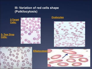
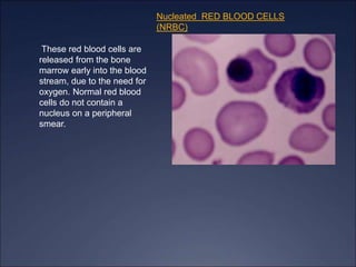
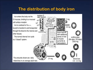
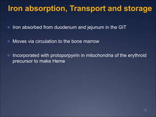
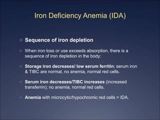
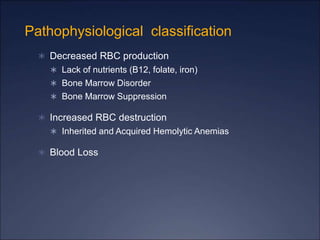
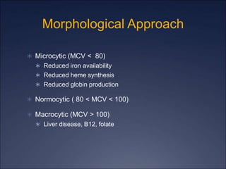
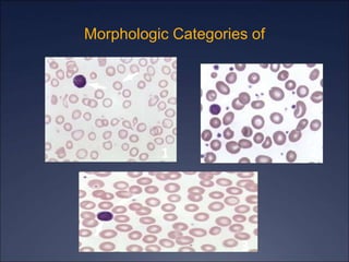
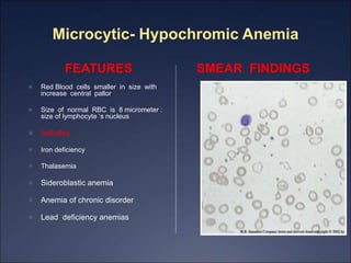
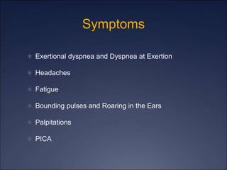
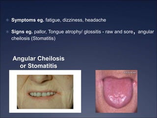
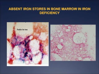
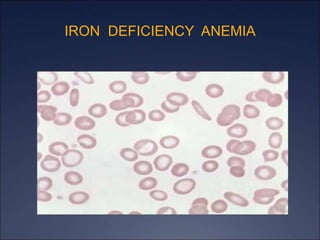
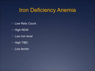
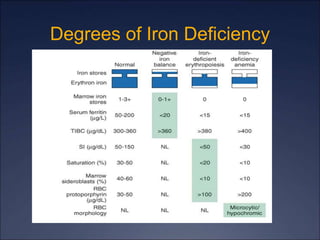
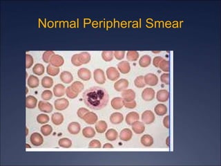
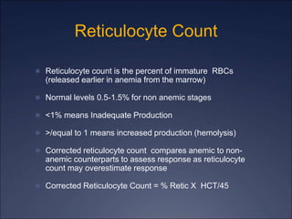
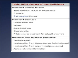
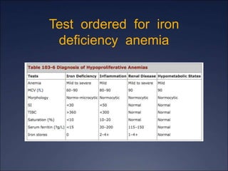
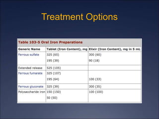
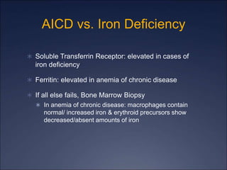
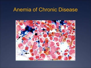
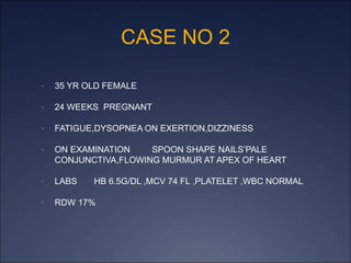
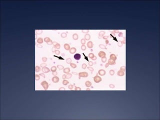
2. Objectives
Learn about iron deficiency anemia(IDA)
Learn about Erythropoiesis
Learn about Iron Cycle
Learn about lab diagnosis of IDA
3. What is Anemia?
Anemia is defined by reduction in Hg Concentration,
Hematocrit (Hct) Concentration or RBC count
Or defined as 2 standard deviations below the mean
WHO criteria is Hg < 13 in men and Hg < 12 in
women
Revised WHO criteria for patient’s with malignancy Hg
< 14 in men and Hg < 12 in women
Pregnant females lower limits were 10.5 g/dl
4.
5.
6.
7. RED BLOOD CELL MORPHOLOGY
DEFINITIONS PERIPHERAL SMEAR
Abnormal erythrocyte
morphology is found in
pathological states that may
be :
- abnormalities in size
(anisocytosis).
- In shape (poikilocytosis).
-In hemoglobin content or the
presence of inclusion bodies
in erythrocyte.
l
8. RED BLOOD CELL MORPHOLOGY
Hypochromic: A descriptive term applied to a red
blood cell with a decreased concentration of
hemoglobin.
Normochromic:
A descriptive term applied to a red blood cell with a
normal concentration of hemoglobin.
Normocytic
A descriptive term applied to normal size of RBC
MacrocyticA descriptive term applied to a larger than
normal red blood cell.
9. 1-Variation in erythrocyte size
(anisocytosis)
Most erythrocytes
presented in the picture
are microcytes
(compare with the small
lymphocyte). The
degree of
hemoglobinization is
sufficient. Normal
platelets and single
ovalocytes are present.
10. 2-Macrocytosis
Morphology
Increase in the size of a red cell.
Red cells are larger than 9µm in
diameter. May be round or oval
in shape, the diagnostic
significance being different.
Found in:
- Folate and B12 deficiencies
(oval)
- Ethanol (round)
- Liver disease (round)
- Reticulocytosis (round)
11. III- Variation of red cells shape
(Poikilocytosis)
2-Target
Cells:
Ovalocytes
Elliptocytosis
5- Tear Drop
Cells:
12. Nucleated RED BLOOD CELLS
(NRBC)
These red blood cells are
released from the bone
marrow early into the blood
stream, due to the need for
oxygen. Normal red blood
cells do not contain a
nucleus on a peripheral
smear.
15. 15
Iron absorption, Transport and storage
Iron absorbed from duodenum and jejunum in the GIT
Moves via circulation to the bone marrow
Incorporated with protoporpyirin in mitochondria of the erythroid
precursor to make Heme
16. IRON TRANSPORT
There are three proteins important for
transporting
1.Transferrin
2.Transferrin receptors
3.Ferritin
Transport
Transferrin: transports iron from the plasma to the
erythroblasts in the marrow for erythropoiesis
The transferrin will bind to transferrin receptor on the
erythrocyte membrane
17. Storage
Hgb contains about two third of the body iron
At the end of their life, RBCs are broken down in the
macrophage of reticuloendothelial system and then
iron is released from Hgb enters plasma and
provided to transferrin.
Some stored in reticuloendothelial cell as ferritin
soluble protein – iron complex) and hemosiderin
(37%) (degraded form of ferritin insoluble)
iron is also found in muscles as myoglobin and in
other cells as iron containing enzymes
19. Iron Deficiency Anemia (IDA)
Sequence of iron depletion
When iron loss or use exceeds absorption, there is a
sequence of iron depletion in the body:
Storage iron decreases/ low serum ferritin; serum iron
& TIBC are normal, no anemia, normal red cells.
Serum iron decreases/TIBC increases (increased
transferrin); no anemia, normal red cells.
Anemia with microcytic/hypochromic red cells = IDA.
20. Pathophysiological classification
Decreased RBC production
Lack of nutrients (B12, folate, iron)
Bone Marrow Disorder
Bone Marrow Suppression
Increased RBC destruction
Inherited and Acquired Hemolytic Anemias
Blood Loss
23. Microcytic- Hypochromic Anemia
FEATURES
Red Blood cells smaller in size with
increase central pallor
Size of normal RBC is 8 micrometer :
size of lymphocyte ‘s nucleus
Includes
Iron deficiency
Thalasemia
Sideroblastic anemia
Anemia of chronic disorder
Lead deficiency anemias
SMEAR FINDINGS
24. Symptoms
Exertional dyspnea and Dyspnea at Exertion
Headaches
Fatigue
Bounding pulses and Roaring in the Ears
Palpitations
PICA
26. Symptoms eg. fatigue, dizziness, headache
Signs eg. pallor, Tongue atrophy/ glossitis - raw and sore, angular
cheilosis (Stomatitis)
Angular Cheilosis
or Stomatitis
27. Diagnosis of anemia
Lab investigations
A complete blood count, CBC
RBC count
Hematocrit (Hct) or packed cell volume
Hemoglobin determination
RBC indices calculation
Reticulocyte count
Blood smear examination to evaluate:
PLeukocytes or Platelets abnormalities
poikilocytosis
28. Lab investigation
A bone marrow smear and biopsy to observe:
Maturation of RBC and WBC series
Ratio of myeloid to erythroid series
Abundance of iron stores (ringed sideroblasts)
Presence or absence of granulomas or tumor cells
Red to yellow ratio
Presence of megakaryocytes
32. Iron Deficiency Anemia
RBC morphology
Hypochromia
Anisocytosis : Variation in size of RBC
Microcytosis : Decrease in size of RBC
Poikilocytosis : Variation in shapes of RBC
Pencil cells (cigar cells)
Target cells
no RBC inclusions
Iron parameters
Low serum iron,
Low serum Ferritin
High TIBC,
33. Lab Investigation cont’d
Serum iron level
measures the amount of iron bound to transferrin
Does not include the free form of iron
Total Iron Binding Capacity (TIBC)
Is an indirect measure of the amount of transferrin protein in the serum
Inversely proportional to the serum iron level
If serum iron is decreased, total iron binding capacity
of transferrin increased (transferrin has more empty
space to carry iron)
34. Lab Investigation cont’d
Serum ferritin
indirectly reflects storage iron in tissues
found in trace amount in plasma
It is in equilibrium with the body stores
Variation in the quantity of iron in the storing compartment is reflected
by plasma ferritin concentration
e.g. Plasma ferritin is decreases in IDA
Plasma ferritin increases in ACD
Limitation: During infection or inflammation Serum Ferritin
increases like other acute phase proteins, and then it is not an
accurate indicator in such situations.
35. Bone marrow iron (Tissue iron)
Tissue biopsy of bone marrow
Prussian blue stain
Type of iron is hemosiderin
37. Labs
Information can be gleaned from good history taking and a
physical exam (pallor, jaundice, etc)
CBC With Diff
Leukopenia with anemia may suggest aplastic anemia
Increased Neutrophils may suggest infection
Increased Monocytes may suggest Myelodysplasia
Thrombocytopenia may suggest hypersplenism, marrow
involvement with malignancy, autoimmune destruction,
folate deficiency
Reticulocyte Count
Peripheral Smear
42. Reticulocyte Count
Reticulocyte count is the percent of immature RBCs
(released earlier in anemia from the marrow)
Normal levels 0.5-1.5% for non anemic stages
<1% means Inadequate Production
>/equal to 1 means increased production (hemolysis)
Corrected reticulocyte count compares anemic to non-
anemic counterparts to assess response as reticulocyte
count may overestimate response
Corrected Reticulocyte Count = % Retic X HCT/45
45. So now that it’s iron deficiency….
What Causes Iron Deficiency?
Blood Loss (occult or overt): PUD, Diverticulosis, Colon
Cancer
Decreased Iron Absorption: achlorhydria, atrophic
gastritis, celiac disease
Foods and Medications: phytate, calcium, soy protein,
polyphenols decrease iron absorption
Uncommon causes: intravascular hemolysis, pulmonary
hemosiderosis, EPO, gastric bypass
Decreased Intake (rare)
46.
47. Who needs a GI work-up?
All men, all women without menorrhagia, women
greater than 50 with menorrhagia
If UGI symptoms, EGD
If asymptomatic, colonoscopy
Women less than 50 plus menorrhagia: consider GI
workup based upon symptoms
50. Gold Standard for Diagnosis
Bone Marrow Biopsy
Prussian Blue staining shows lack of iron in erythroid
precursors and macrophages
However, it is invasive and costly
52. AICD vs. Iron Deficiency
Soluble Transferrin Receptor: elevated in cases of
iron deficiency
Ferritin: elevated in anemia of chronic disease
If all else fails, Bone Marrow Biopsy
In anemia of chronic disease: macrophages contain
normal/ increased iron & erythroid precursors show
decreased/absent amounts of iron
54. Treatment
Treat the underlying cause
And Treat the Underlying Cause!
Consider co-existent iron deficiency as well
If underlying disease state requires it, consider EPO
injection
56. CASE NO 2
• 35 YR OLD FEMALE
• 24 WEEKS PREGNANT
• FATIGUE,DYSOPNEA ON EXERTION,DIZZINESS
• ON EXAMINATION SPOON SHAPE NAILS’PALE
CONJUNCTIVA,FLOWING MURMUR AT APEX OF HEART
• LABS HB 6.5G/DL ,MCV 74 FL ,PLATELET ,WBC NORMAL
• RDW 17%
57. CASE NO 1
• 18 YEAR OLD FEMALE
• FATIGUE, FAILS TO CONCENTRATE,URGE TO EAT ICE
• HISTORY OF HEAVY MENSTRUAKL BLEEDING EVERY MONTH
• PHYSICAL EXAMINATION SHOWS PALE CONJUNCTIVA,SMOOTH TONGUE ,BRITTLE NAILS
• LABS SHOWS CBC :Hg 7g/dl ,MCV 70FL ,MCHC 28 G/DL ,MCH 20G/DL,hct 19.8fl
• Retic count 1.5% N 0-5-2 %
• RDW 18 % N 16%
• TIBC 600 N 250-400 MG/DI
• Serum Ferritin 23 ng/ml N 32-100
• Peripheral smear show microcytic hypochromasia,pencil cells,target cells
58.
59. QUERRY
What could be the most
likely diagnosis?
What is differential
diagnosis ?
Iron deficiency anemia
Aplastic anemia
Megaloblastic anemia
Thalessemia
Anemia of chronic disorder
60. References
Harrison’s Principles of Internal Medicine
Adamson JW. Chapter 103. Iron Deficiency and Other Hypoproliferative
Anemias. In: Longo DL, Fauci AS, Kasper DL, Hauser SL, Jameson JL,
Loscalzo J, eds. Harrison's Principles of Internal Medicine. 18th ed. New York:
McGraw-Hill; 2012.
http://www.accessmedicine.com/content.aspx?aID=9117223. Accessed
December 7, 2011
Wians, F.H. and Urban JE. “Discriminating between Anemia of Chronic
disease Using Traditional Indices of Iron Status v. Transferring Receptor
Concentration”. 2001. American Journal of Clinical Pathology. Volume 115.
UptoDate
Schrier, SL. Approach to the adult patient with anemia. In: UpToDate, Landaw,
SA(ED). UptoDate, Waltham, MA. 2012.
Schrier, SL. Causes and diagnosis of anemia due to iron deficiency. In:
UpToDate. Landaw, SA.(ED). Uptodate, Waltham, MA. 2012.
Editor's Notes
The arrows show that the RBCs are smaller and paler
When give 200-300 mg of elemental iron, only 50-60 mg/day is absorbed
Iron Tolerance Test: two iron tablets given on an empty stomach and the serum iron level measured over the next two hours. Normal absorption will result in increase of serum iron level by 100 ug/dL
Pts may not take due to side effects (GI Side Effects)
Parenteral Formulations
When patient cannot tolerate PO due to side effects
When patient needs iron on an ongoing basis
Side Effect: 0.7% of iron dextran causes anaphylaxis so usually a small tester dose is given to monitor response