This document lists important anatomical landmarks and structures associated with each vertebral level from cervical (C3-C7) to sacral (S2-S3). Some key points include:
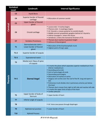
- At C6, the larynx ends and the trachea begins, the pharynx ends and the esophagus begins. The inferior laryngeal nerve enters the larynx.
- At T4-5, the plane that separates the superior and inferior mediastinum. The arch of the aorta begins and ends. The descending aorta begins.
- At L1, landmarks include the pylorus of the stomach, beginning of the duodenum, and course of splenic vessels along the pancreas
![M.A.A.Al-Maqrashi
T9-L3 Costal Margin ▪ Costal slips of diaphragm
T10
▪ Esophagus passes through diaphragm behind 7th
-8th
costal
cartilage just to the left [1-2cm] of median plane and Terminates
at the cardiac orifice of the stomach
T12
▪ Aorta, azygos v. & thoracic duct pass through diaphragm
▪ Upper border of the kidneys
Lumbar
L1
Trans-Pyloric plane
[found at midpoint b/w
jugular notch & pubic
symphysis]
[tip of 9th costal cartilage]
▪ Pyloris of stomach immediately above and to the right of the
midline.
▪ Beginning of duodenum
▪ Duodenojejunal flexure to the left of midline and immediately
below it
▪ Course of splenic vessels along Pancreas
▪ Root of transverse mesocolon
▪ Position of renal pelvis
▪ Hilum of kidneys: left is above, and right is below.
▪ R. renal a. originate just below this line & L. renal a. is just above
▪ Celiac a. originates just above
▪ Origin of Superior Mesenteric artery
▪ Termination of spinal cord
L2
▪ Thoracic duct begins
▪ Azygos & hemiazygos v. begin
L3
Sub-Costal Plane
[Through 10th costal
cartilage]
▪ Lowest point of costal margin
▪ Lower border of the kidneys
L4
Supra-Cristal Plane
[Through the highest point
of Iliac Crest]
▪ Bifurcation of aorta to R & L common iliac a.
▪ Inf. Vena cava begins.
▪ Important to locate 4th
, 3rd
or 2nd
IV space for lumbar puncture
▪ Spinous process of L3 lies just above → identifying vertebral
levels
L5
Trans-Tubercular Plane
[passes through the iliac
tubercles, small elevations
found in the iliac crest of the
iliac bone.]
▪ Continuation of cecum as ascending colon
Sacral
S2
Posterior Superior iliac spine
[dimple]
▪ End of sural sac
▪ Middle of sacroiliac joint
S3 Posterior inferior iliac spine
▪ End of sigmoid colon → rectum starts
[imp. For recto-sigmoid carcinoma surgery]](https://image.slidesharecdn.com/importantvertebrallevel-180226234837/85/Anatomy-Important-vertebral-level-2-320.jpg)


![Vascular anatomy of head, [autosaved]](https://cdn.slidesharecdn.com/ss_thumbnails/vascularanatomyofheadautosaved-181218123734-thumbnail.jpg?width=640&height=640&fit=bounds)


