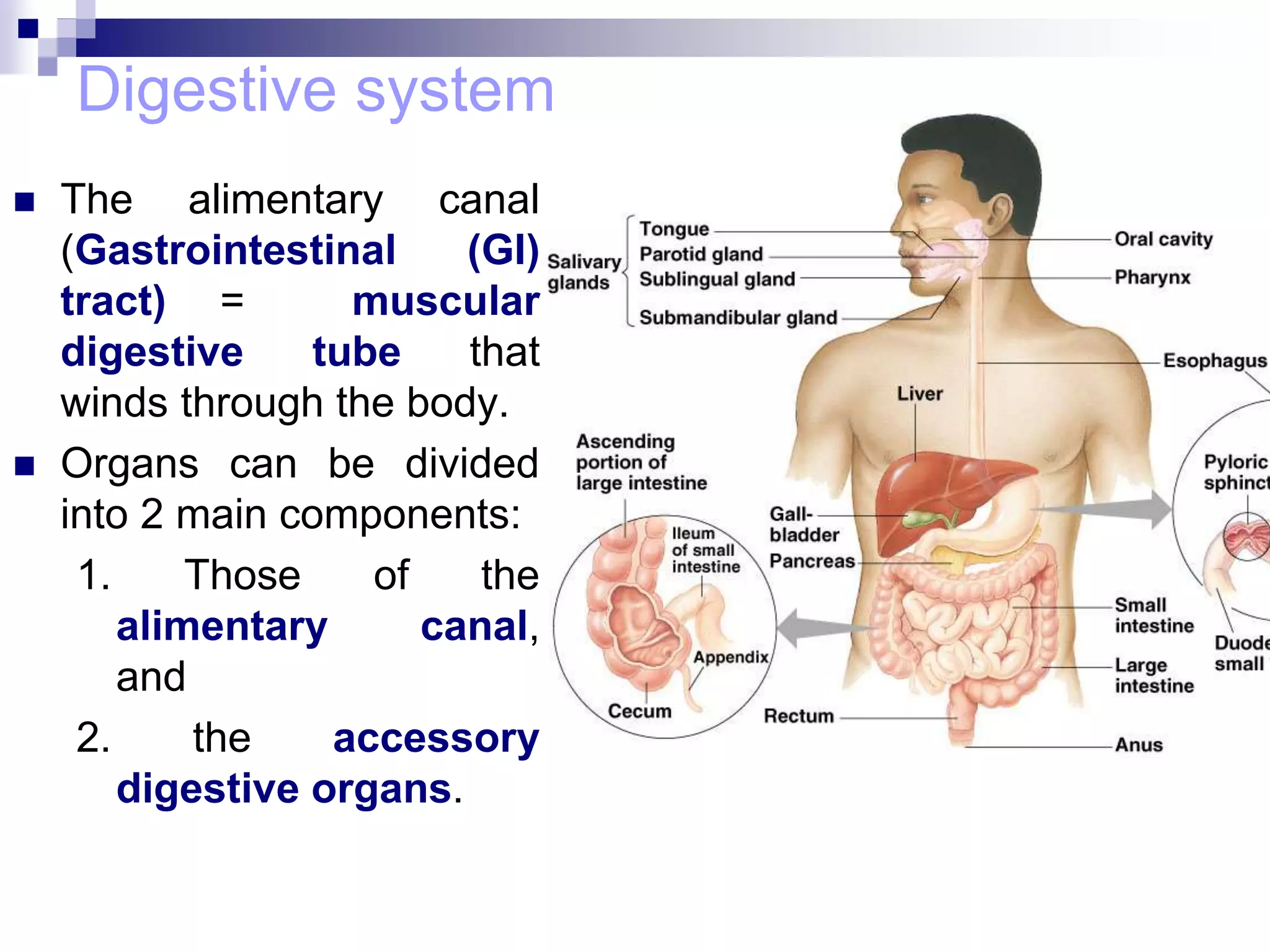
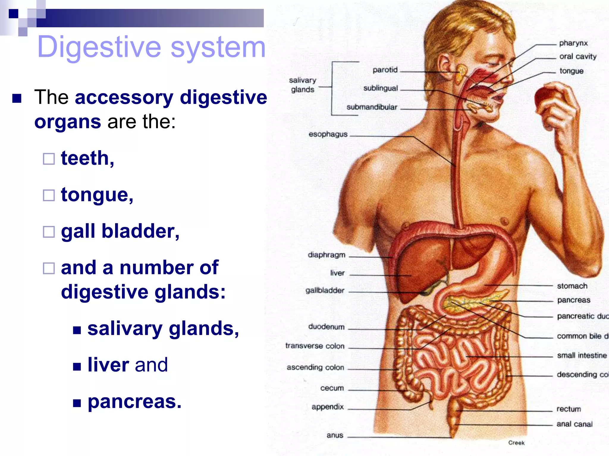
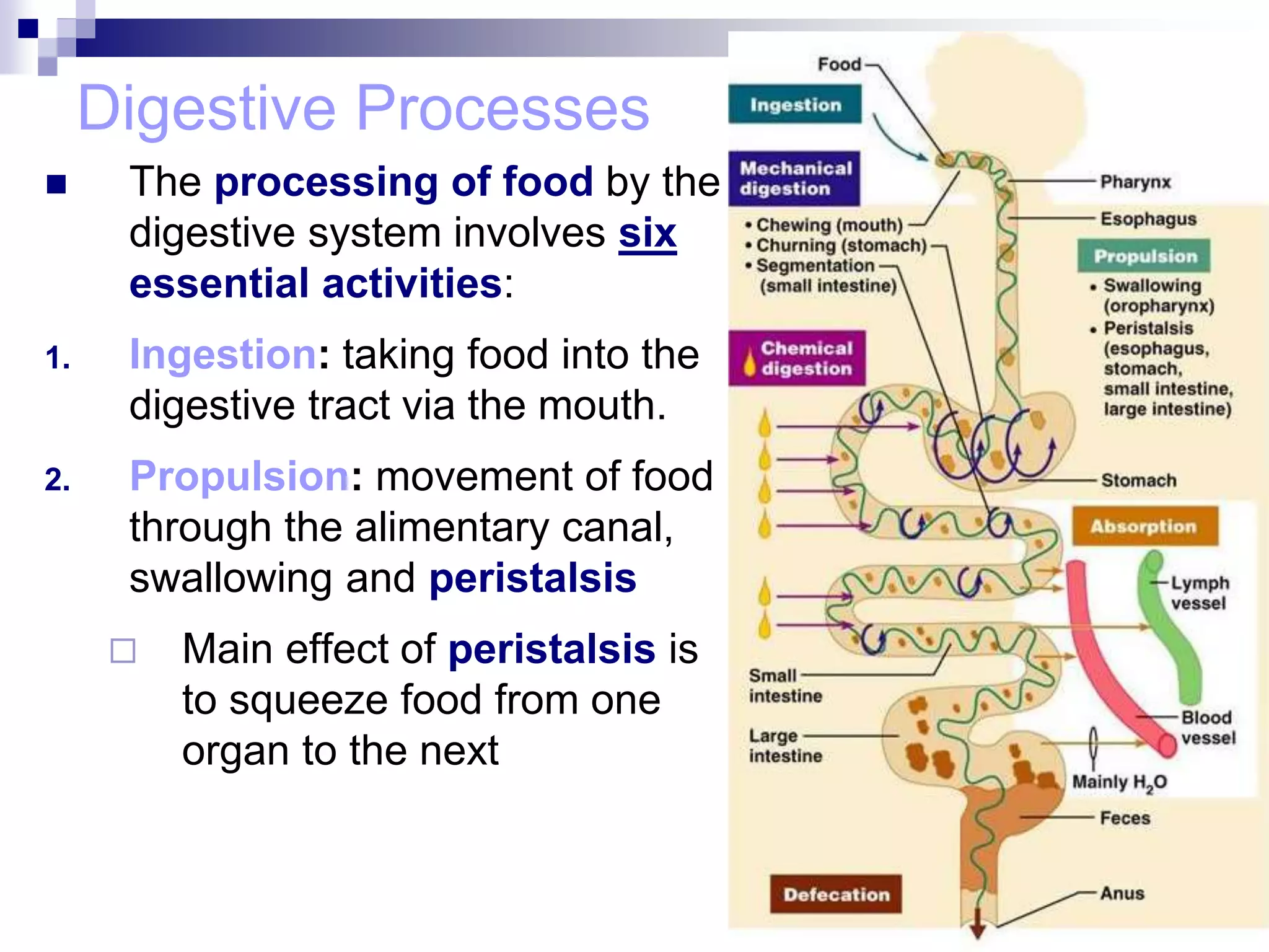
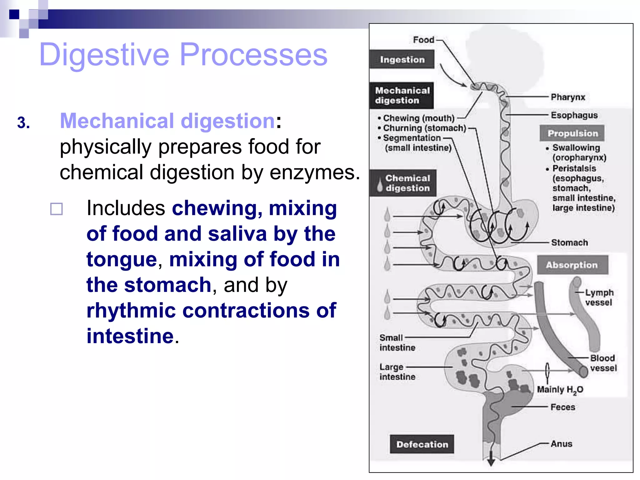
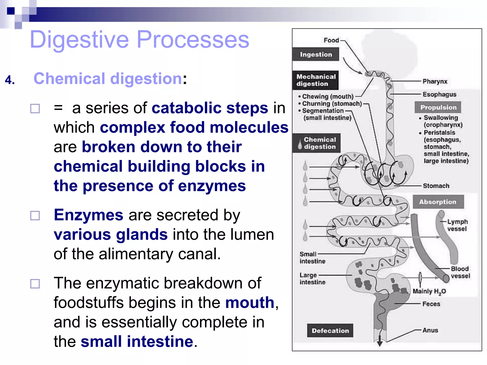
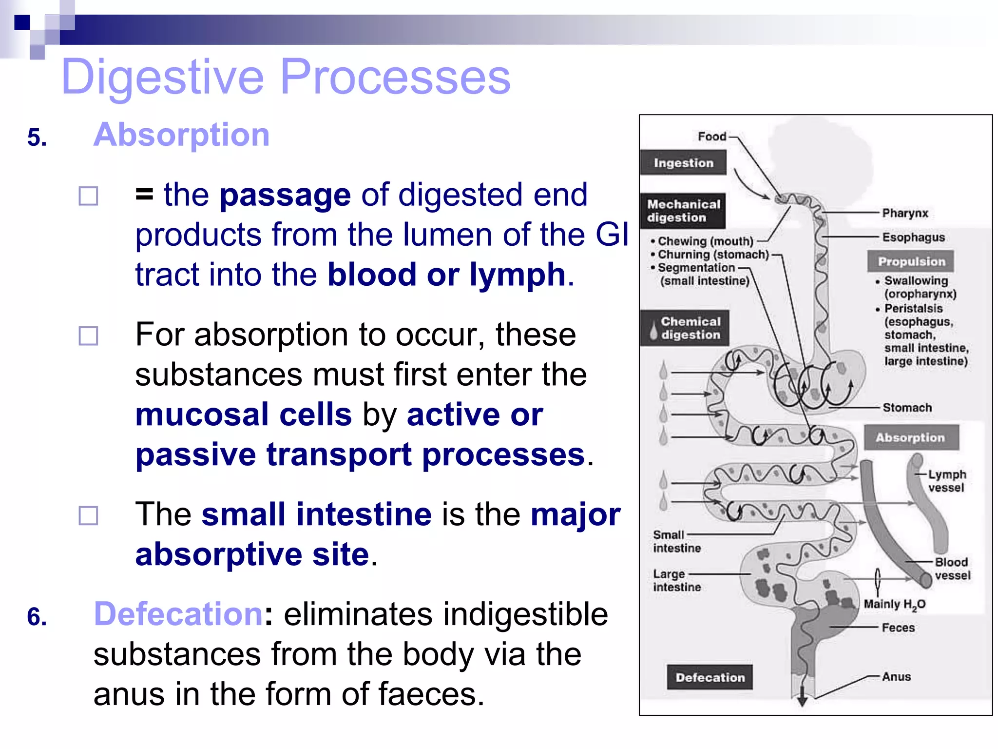
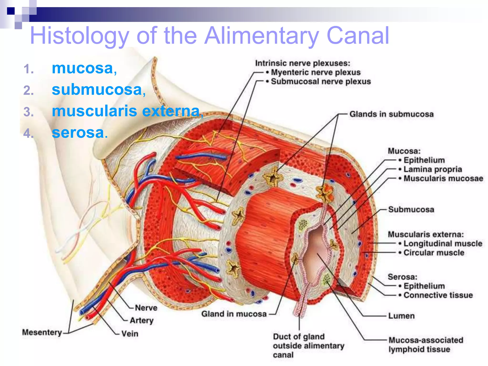
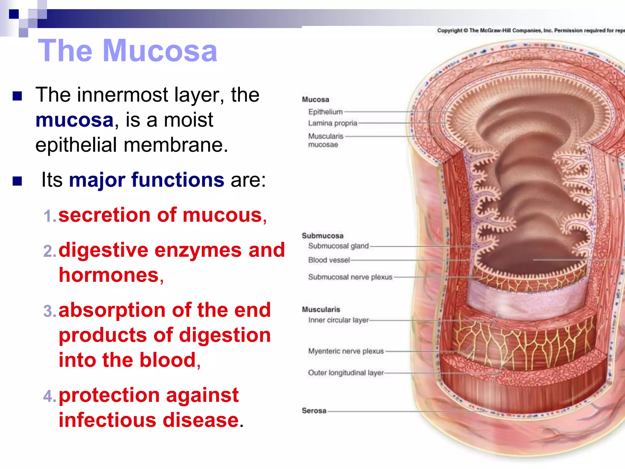
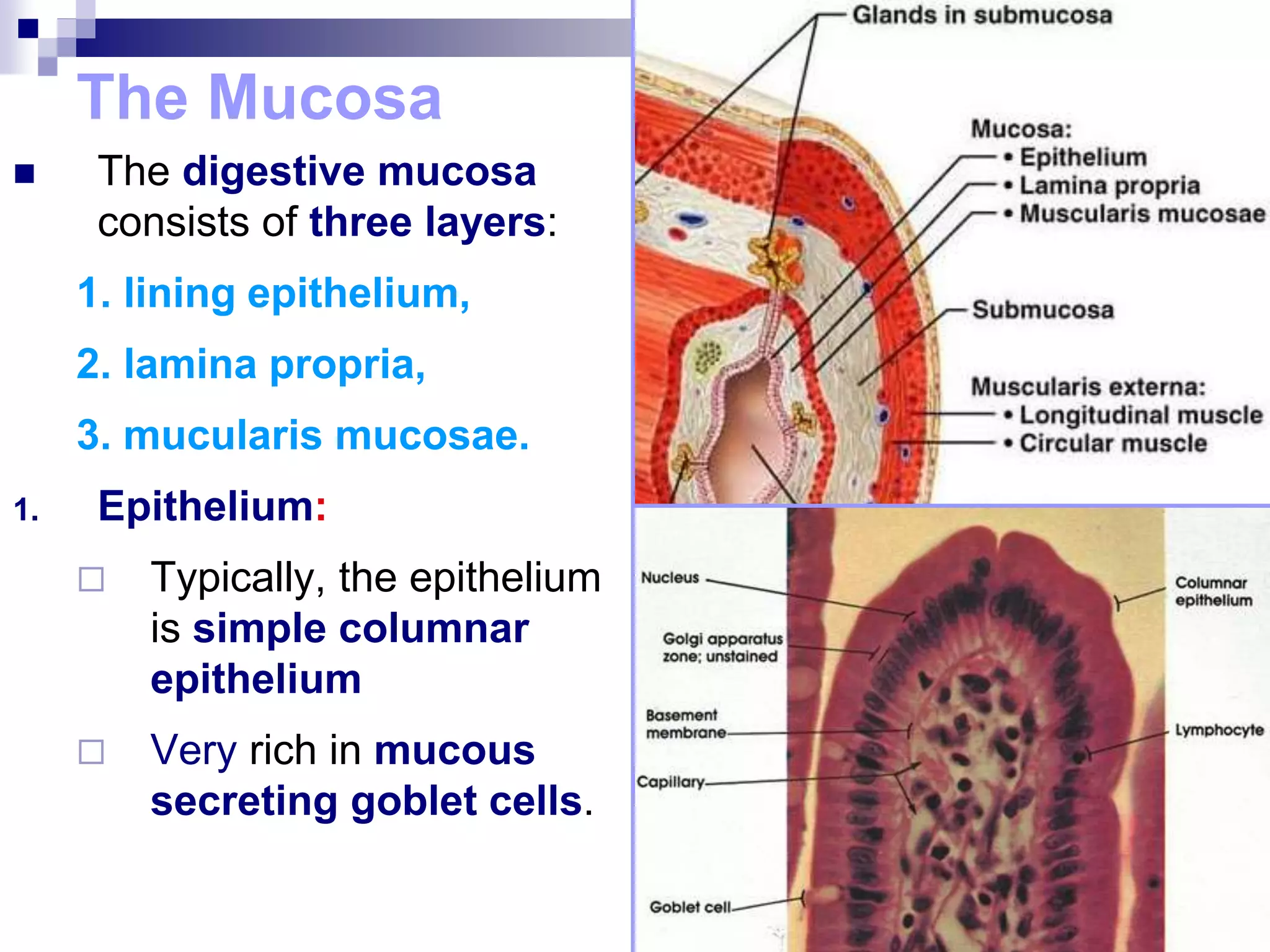
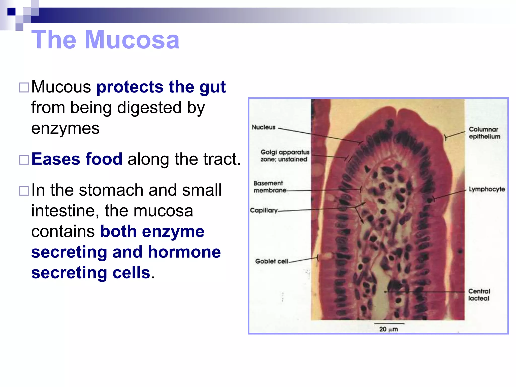
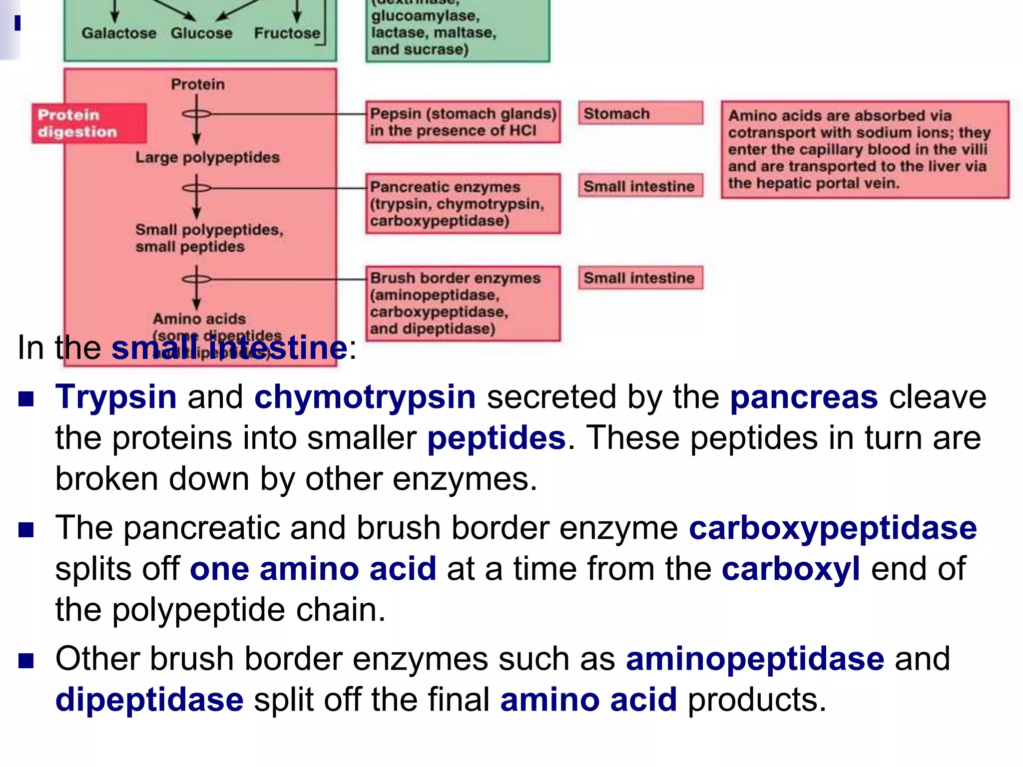
The digestive system consists of the alimentary canal and accessory organs. The alimentary canal includes the mouth, pharynx, esophagus, stomach, small intestine, and large intestine. Accessory organs include the teeth, tongue, liver, gallbladder, and pancreas. Food is ingested and moves through the digestive tract, where it undergoes mechanical and chemical digestion to break nutrients into smaller molecules that can be absorbed and used by the body. Enzymes produced in glands aid in digestion at different points along the tract.
This PowerPoint presentation details out the anatomy of the human digestive system. Their are general terminologies that involves the topic but over-all this work focuses on how digestion takes place in the human body. The details coming from this presentation are combined from four different and liable sources/references including Biology (Thomson Asian Edition). I can say that this presentation is brief and well-organized so I hope this could help you in your class or seminars. Thanks.
In Class 11 Biology, excretory products and their elimination are important topics typically covered under the unit "Excretory System."
For more information, visit- www.vavaclasses.com
Digestive system Mouth Buccal cavity Tongue Teeth Salivary glands Pharynx Oesophagus Stomach Small intestine Large intestine Rectum Anus Liver Gall bladder Pancreas Absorption Digestion
The Impact of Artificial Intelligence on Modern Society.pdfssuser3e63fc
Just a game Assignment 3
1. What has made Louis Vuitton's business model successful in the Japanese luxury market?
2. What are the opportunities and challenges for Louis Vuitton in Japan?
3. What are the specifics of the Japanese fashion luxury market?
4. How did Louis Vuitton enter into the Japanese market originally? What were the other entry strategies it adopted later to strengthen its presence?
5. Will Louis Vuitton have any new challenges arise due to the global financial crisis? How does it overcome the new challenges?Assignment 3
1. What has made Louis Vuitton's business model successful in the Japanese luxury market?
2. What are the opportunities and challenges for Louis Vuitton in Japan?
3. What are the specifics of the Japanese fashion luxury market?
4. How did Louis Vuitton enter into the Japanese market originally? What were the other entry strategies it adopted later to strengthen its presence?
5. Will Louis Vuitton have any new challenges arise due to the global financial crisis? How does it overcome the new challenges?
This comprehensive program covers essential aspects of performance marketing, growth strategies, and tactics, such as search engine optimization (SEO), pay-per-click (PPC) advertising, content marketing, social media marketing, and more
2. Digestive system
The alimentary canal
(Gastrointestinal (GI)
tract) = muscular
digestive tube that
winds through the body.
Organs can be divided
into 2 main components:
1. Those of the
alimentary canal,
and
2. the accessory
digestive organs.
3. Digestive system
The organs of the
alimentary canal
are the:
mouth,
pharynx,
oesophagus,
stomach,
small intestine
and large
intestine.
4. Digestive system
The accessory digestive
organs are the:
teeth,
tongue,
gall bladder,
and a number of
digestive glands:
salivary glands,
liver and
pancreas.
5. Digestive Processes
The processing of food by the
digestive system involves six
essential activities:
1. Ingestion: taking food into the
digestive tract via the mouth.
2. Propulsion: movement of food
through the alimentary canal,
swallowing and peristalsis
Main effect of peristalsis is
to squeeze food from one
organ to the next
6. Digestive Processes
3. Mechanical digestion:
physically prepares food for
chemical digestion by enzymes.
Includes chewing, mixing
of food and saliva by the
tongue, mixing of food in
the stomach, and by
rhythmic contractions of
intestine.
7. Digestive Processes
4. Chemical digestion:
= a series of catabolic steps in
which complex food molecules
are broken down to their
chemical building blocks in
the presence of enzymes
Enzymes are secreted by
various glands into the lumen
of the alimentary canal.
The enzymatic breakdown of
foodstuffs begins in the mouth,
and is essentially complete in
the small intestine.
8. Digestive Processes
5. Absorption
= the passage of digested end
products from the lumen of the GI
tract into the blood or lymph.
For absorption to occur, these
substances must first enter the
mucosal cells by active or
passive transport processes.
The small intestine is the major
absorptive site.
6. Defecation: eliminates indigestible
substances from the body via the
anus in the form of faeces.
9. Histology of the Alimentary Canal
From the oesophagus down, the alimentary canal wall has
the same four basic layers, or tunics
From the lumen outward these layers are the:
1. mucosa,
2. submucosa,
3. muscularis externa,
4. serosa.
10. Histology of the Alimentary Canal
1. mucosa,
2. submucosa,
3. muscularis externa,
4. serosa.
11. The Mucosa
The innermost layer, the
mucosa, is a moist
epithelial membrane.
Its major functions are:
1.secretion of mucous,
2.digestive enzymes and
hormones,
3.absorption of the end
products of digestion
into the blood,
4.protection against
infectious disease.
12. The Mucosa
The digestive mucosa
consists of three layers:
1. lining epithelium,
2. lamina propria,
3. mucularis mucosae.
1. Epithelium:
Typically, the epithelium
is simple columnar
epithelium
Very rich in mucous
secreting goblet cells.
13. The Mucosa
Mucous protects the gut
from being digested by
enzymes
Eases food along the tract.
In the stomach and small
intestine, the mucosa
contains both enzyme
secreting and hormone
secreting cells.
14. The Mucosa
2. Lamina propria: consists of loose areolar connective tissue.
Contains capillaries + central lacteal + lymphoid tissues
and variety of glands
Capillaries absorb digested nutrients + nourish epithelium.
15. The Mucosa
3. Muscularis mucosae:
Is a thin layer of
smooth muscle cells
that produces local
movements of the
mucosa.
16. The Submucosa
Located just outside the
mucosa, consists of dense
connective tissue
Contains blood and
lymphatic vessels, lymph
nodules, and nerve fibres.
Rich supply of elastic fibres:
enables stomach to regain
its normal shape after
temporarily storing a large
meal.
17. Muscularis Externa
Double layer of smooth
muscle cells:
inner circular layer and
outer longitudinal layer
In some places the circular
layer thickens to form
sphincters that act as valves
to prevent backflow and
control food passage from
part to the next
Responsible for peristalsis, i.e
it mixes and pushes
foodstuff along the digestive
tract.
18. Serosa
A protective outermost layer
Consists of areolar
connective tissue covered
with mesothelium, a single
layer of squamous epithelial
cells .
19. Functional Anatomy of the Digestive
System
We will now consider the special structural and
functional capabilities of each organ in the digestive
system.
20. 1. Mouth and Associated Organs
The mouth, also called the oral cavity or buccal cavity, is
the only part of the GI involved with ingestion: the taking in
of food into the body.
Most digestive functions associated with the mouth reflect
the activity of related accessory organs, such as the
teeth, salivary glands and tongue.
Its boundaries are:
the lips in front,
the cheeks on the sides,
the palate on top,
the tongue on the bottom.
21. 1. Mouth and Associated Organs
At the back the mouth connects to the oropharynx.
The walls of the mouth have to withstand considerable
friction, thus are lined with abrasion resistant stratified
squamous epithelium.
22. Mouth and Associated Organs
The Lips and Cheeks
The lips (labia) and the cheeks have a core of skeletal
muscle covered externally by skin.
They help to keep food in the mouth and between the teeth
during chewing.
23. Mouth and Associated Organs
The Palate
Forms the roof of the mouth,
2 distinct parts:
1.hard palate in front
2.soft palate at the back.
Hard palate forms a rigid hard surface against which the
tongue forces food during chewing.
Mucosa on either side of the raphe, a midline ridge, is
corrugated, which helps to create friction.
Soft palate rises reflexively to close off the nasopharynx
when we swallow.
24.
25. The Tongue
The tongue is comprised mainly of
muscle: 8 individual muscles
During chewing it grips the food and
constantly repositions it between the
teeth.
Tongue movements also mix the food
with saliva and form it into a compact
mass called a bolus, and
initiates swallowing by forcing the bolus
to the back of the mouth.
26. The Tongue
Dorsal side of the tongue bears three types of papillae:
1. fungiform,
2. filiform and
3. circumvallate
27. The Tongue
Filiform papillae: conical and are the smallest and most
abundant type, and aligned in parallel rows
Give the tongue surface a roughness that aids in licking
semi-solid foods e.g. cat tongue
Also provide friction for manipulating food in the mouth.
28. The Tongue
Mushroom shaped fungiform papillae are scattered
widely over the surface of the tongue.
10-12 large circumvallate papillae are located in a V-
shaped row at the back of the tongue.
Fungiform and circumvallate papillae house the taste
buds, which are crucial during the tasting process.
29. The Salivary Glands
Salivary glands inside and outside the oral cavity produce
and secrete saliva.
The functions of saliva are:
1. To cleanse the mouth,
2. dissolve food chemicals so that they can be tasted,
3. moisten food and aid in compacting it into a bolus,
and
4. begin the chemical breakdown of starchy foods by
releasing enzymes.
30. The Salivary Glands
2 types of salivary glands:
1. Three pairs of extrinsic
salivary glands that lie
outside of the oral cavity,
but empty their secretions
into it:
1.parotid,
2.submandibular
3.sublingual glands.
2. Intrinsic salivary glands,
also called buccal glands,
scattered throughout the
oral mucosa.
31. The Salivary Glands
Parotid gland: Large gland just in front of the ear, its duct
opening near the back upper molars.
Submandibular gland: lies beneath the tongue and opens
at the base of the tongue.
Sublingual gland: small gland that lies in front of the
submandibular gland under the tongue.
32. The Salivary Glands
Salivary glands composed of two types of secretory cells:
mucous and serous.
Serous cells produce a watery secretion containing
enzymes and a small amount of mucin,
Mucous cells produce mucous, a viscous solution.
3 glands differ in cell complement and thus secretion:
Parotid glands contain only serous cells
Submandibular (and buccal glands) contain equal
numbers of serous and mucous cells,
Sublingual glands contain mostly mucous cells.
33. The Teeth
Teeth lie in sockets in the mandible and maxilla bones
The role of teeth is to chew, or masticate, the food.
34. Pharynx
From the mouth food passes into
oropharynx and then to the
laryngopharynx.
Both are common
passageways for food and air.
Constrictor muscles help to
push food toward oesophagus.
35. The Oesophagus
Muscular tube about 25 cm long
Collapsed when not involved in
movement of food.
passes through diaphragm at
oesophageal hiatus.
Joins stomach at cardiac orifice.
Cardiac orifice is surrounded by cardiac
or gastro-oesophageal sphincter,
This acts as a valve to seal stomach,
preventing flow of digestive juices up the
oesophagus.
36. Oesophagus
Unlike mouth and pharynx, oesophagus wall has all four
basic alimentary canal layers.
Mucosa contains non-keratinised stratified squamous
epithelium.
At oesophagus-stomach junction this changes abruptly
to the simple columnar epithelium of the stomach,
which is specialised for secretion
37. The Stomach
Eosophagus expands to form the
stomach, a temporary 'storage
tank'
Food is converted to a creamy
paste called chyme.
15-25 cm long, but diameter and
volume depend on how much
food it contains
When empty, its mucosa and
submucosa show large,
longitudinal folds called rugae.
38. Anatomy of stomach
The major regions of the
stomach are:
The small cardiac region
surrounds the cardiac orifice
through which food enters
from the oesophagus.
fundus is the dome shaped
part.
The body is the mid-portion
of the stomach, and this
leads into the pyloric
region.
The wider and more superior
part of the pyloric region, the
pyloric antrum, narrows to
form the pyloric canal,
which ends in the pylorus.
39. Anatomy of stomach
Pylorus is continuous with
duodenum through pyloric
sphincter, which controls
emptying of the stomach (pylorus
means gatekeeper).
Convex surface - greater
curvature
Concave surface - lesser
curvature.
Extending from these curvatures
are two mesenteries called
omenta that help to attach the
stomach to other digestive
organs and the body wall.
40. Microscopic Anatomy: Stomach
4 layers, but muscularis and
mucosa are modified.
Mucosa is a simple columnar
epithelium composed entirely
of goblet cells, which produce a
protective coat of alkaline
mucous.
Muscularis externa has
additional inner oblique
smooth muscle layer
Allows the stomach not only to
move food along the tract, but
also to churn and mix the food,
breaking it into smaller pieces.
41. Microscopic Anatomy
Wall is dotted with deep
gastric pits which lead into
gastric glands that
collectively produce the
stomach secretion called
gastric juice.
Cells forming the walls of
the gastric pits are
primarily goblet cells, but
those composing the
gastric glands vary in
different proportions in
the stomach
42. Microscopic Anatomy
Cardiac and pylorus: primarily mucous
secreting,
Pyloric antrum: mostly produce
stimulatory hormone called gastrin.
Fundus and body, where most
chemical digestion occurs, are larger
and produce the majority of the
stomach secretions.
The glands in these regions contain a
variety of cells:
43. Microscopic Anatomy
1. Mucous neck cells, found
in the upper or 'neck'
regions of the glands
produce a acidic mucous
44. Microscopic Anatomy
2. Parietal (oxyntic) cells, middle region
of the glands, secrete HCl and intrinsic
factor.
HCl makes the stomach contents
extremely acidic (pH 1.5-3.5),
Low pH condition needed for
activation and optimal activity of
pepsin (a protein digesting enzyme),
and
harsh enough to kill many of the
bacteria ingested with foods.
Intrinsic factor is a glycoprotein
required for absorption of vitamin B12
in the small intestine
45. Microscopic Anatomy
3. Chief, or zymogenic, cells produce
pepsinogen, the inactive form of the
protein digesting enzyme pepsin.
Occur mainly in the basal regions of
the gastric glands.
When chief cells are stimulated, the
first pepsinogen molecules they
release are activated by HCl acid
encountered in the apical region of
the gland
Once pepsin is present, it also
catalyses the conversion of
pepsinogen to pepsin.
46. Microscopic Anatomy
4. Enteroendocrine cells release a
variety of hormones or hormone-
like products.
These products, including gastrin,
histamine, endorphins, serotonin,
cholecystokinin and somatostatin
diffuse into the blood capillaries
Function is to influence several
digestive system organs
47. Hormone Site of Production Stimulus for production Target organ Activity
Gastrin Stomach mucosa Food (particularly partially digested
proteins) in stomach (chemical
stimulation); acetylcholine released
by nerve fibres
Stomach
Small intestine
ileocaecal valve
Large intestine
Causes gastric glands to
increase secretory
activity; most pronounced
effect is on HCl secretion
Stimulates gastric
emptying
Stimulates contraction of
intestinal wall
Relaxes ileocaecal valve
Stimulates mass
movements
Serotonin Stomach mucosa Food in stomach Stomach Causes contraction of
stomach muscle
Histamine Stomach mucosa Food in stomach Stomach Activates parietal cells to
release HCl
Somatostatin Stomach mucosa and
duodenal mucosa
Food in stomach; stimuulation of
sympathetic nerve fibres
Stomach
Pancreas
Small intestine
Gallbladder
Inhibits gastric secretion
of all products; inhibits
gastric motility and
emptying
Inhibits secretion
Inhibits GI blood flow;
thus inhibits intestinal
absorption
Inhibits contraction and
bile release
Intestinal gastrin Duodenal mucosa Acidic and partially digested foods
in duodenum
Stomach Stimulates gastric glands
and motility
Secretin Duodenal mucosa Acidic chyme (also partially
digested proteins and fats)
Stomach
Pancreas
Liver
Inhibits gastric gland
secretion and gastric
motility during gastric
phase of secretion
Increases output of
pancreatic juice rich in
bicarbonate ions
Increases bile output
48. Microscopic Anatomy
How does the stomach protect itself from the harsh
environment in the stomach? By creating a mucosal
barrier in 4 ways:
1. A thick coating of bicarbonate-rich mucous is built
up on the stomach wall.
2. Mucosa epithelial cells are joined together by tight
junctions to prevent gastric juice from leaking into
the underlying tissues.
3. Deep in the gastric glands, where the protective
alkaline mucous is absent, the external faces of the
glandular cells are impermeable to HCl.
4. Damaged epithelial mucosal cells are shed and
quickly replaced every 3-6 days.
49. Digestive Processes in the Stomach
Stomach has 3 primary functions:
1. Serves as a storage area for ingested food.
2. Mechanical digestion process started in the mouth is
continued
3. Chemical digestion:
Protein digestion is initiated via the release of the
enzyme pepsin
This is the only type of enzymatic digestion that
occurs in the stomach
Intrinsic factor, which is required for intestinal
absorption of vitamin B12
50. Regulation of Gastric Secretion
Gastric glands secretes 3 l of gastric juice per day
Secretion controlled by both neural and hormonal
mechanisms.
Neural control: When nerves actively stimulate the
stomach, secretion of almost all glands increases.
Hormonal control: controlled by gastrin, which stimulates
secretion of both enzymes and HCl,
51. Regulation of Gastric Secretion
Secretion of Gastric juice controlled by stimuli received
from 3 sites - the head, stomach and small intestine
These all stimulate or inhibit activity of gastric glands
Thus, there are 3 phases of gastric secretion:
1. Cephalic Phase:
occurs before food enters the stomach.
triggered by aroma, sight, taste and thought of food.
CNS prepares stomach for digestive duties
52. Regulation of Gastric Secretion
2. Gastric phase:
Initiated by local neural and hormonal mechanisms as
soon as food reaches the stomach
provides about 66% of the gastric juice that is released.
Most important stimuli for initiating this phase
are stomach stretching,
entrance of peptides into the stomach, and
low acidity conditions in the stomach.
53. Regulation of Gastric Secretion
3. Intestinal Phase:
Has 2 components:
1. Excitatory component initiated as partially digested
food begins to fill initial part of duodenum.
This stimulates intestinal mucosal cells to release a
hormone called intestinal gastrin to encourage the
gastric glands to continue their secretory activity
54. Regulation of Gastric Secretion
2. Inhibitory component:
Intestinal mechanism stimulate
intestinal gastrin secretion only
briefly.
As the intestine begins to fill
with chyme, inhibitory
component is triggered in the
form of the enterogastric
reflex.
This causes pyloric sphincter
to tighten and thus prevent
the further entry of food into
the duodenum.
As a result gastric secretory
activity declines.
56. Small Intestine
Body's major digestive organ:
digestion is completed
virtually all absorption occurs
here.
twisted tube extending from pyloric
sphincter to ileocaecal valve
longest part of alimentary system:
2-4 m long, with a diameter of about
2.5 cm
Three subdivisions: duodenum,
jejunum and ileum
57. 2 ducts join duodenum:
1.Bile duct from liver,
2.Pancreatic duct
from pancreas,
These unite in a bulb-
like point called
hepatopancreatic
ampulla
entry is controlled by a
muscular valve called
the hepatopancreatic
sphincter
Small Intestine
58. Small Intestine: Microscopic Anatomy
Modifications for Absorption:
Modified to create
enormous surface area
for absorption - total
surface area is 200 m2
Its length alone provides a
huge surface area
Surface area further
increased by 3 structural
modifications –
1. Plicae circularis,
2. Villi
3. Microvilli
59. Microscopic Anatomy
Villi:
finger-like projections of the mucosa,
Only about 1 mm high,
epithelial cells mainly absorptive
columnar cells called enterocytes.
in the centre of each villus is a dense
capillary bed
Also contains a wide lymph capillary
called lacteal.
Are largest in the duodenum, and
gradually become smaller and smaller
along the length of the small intestine.
60. Microscopic Anatomy
Microvilli:
tiny projections of plasma
membrane of mucosal cells
Increases the absorptive surface
area,
Also, plasma membranes of
microvilli contain enzymes, known
as brush border enzymes.
These enzymes complete final
stages of digestion of
carbohydrates and proteins in the
small intestine.
62. Histology of Intestine wall
The four layers of GI tract are present,
but the mucosa and submucosa are
modified.
Mucosa modifications:
Epithelium is simple columnar
absorptive cells covered with large
numbers of microvilli.
Other cells making up mucosa include:
Many mucous secreting goblet
cells,
interspersed T cells called intra-
epithelial lymphocytes (important
for immunology), and
scattered enteroendocrine cells.
63. Histology of Intestinal wall
Mucosa studded with pits that lead to
intestinal glands called crypts of
Lieberkühn.
The cells of these glands secrete intestinal
juice, a watery mixture containing
mucous that serves as a carrier fluid for
absorption of nutrients from chyme.
Located deep in crypts are specialised
secretory cells called Paneth cells
These cells release lysozyme, an
antibacterial enzyme to increase the small
intestines defences against bacteria
64. Histology of Intestinal wall
Submucosa:
Contains lymphoid
follicles called Peyers
patches which has an
immune function
Also contains mucous
secreting glands that
secrete an alkaline
mucous that helps to
neutralise acidic chyme
from stomach.
65. The Large Intestine
The large intestine
extends from the
ileocaecal valve to the
anus.
Major function:
to absorb water from
indigestible food
residues
eliminate indigestible
food from the body as
semisolid faeces.
66. Gross Anatomy
Exhibits 3 features not seen
elsewhere in the intestine:
1. The longitudinal muscle
layer of the muscularis is
only 3 bands of smooth
muscle called teniae coli.
2. They pull the wall of the
large intestine together into
pocket-like sacks called
haustra.
3. Epiploic appendages are
small, fat filled pouches of
visceral peritoneum that
hang from its surface
67. Subdivisions
The large intestine has 5
subdivisions:
1. The sack-like cecum is the
first part of the large
intestine
2. Attached to it is the
vermiform appendix,
which contains lymphoid
tissue, and plays an
important role in
immunity.
69. Subdivisions
5. Rectum
6. Anal canal is last
segment
Anal canal has two
sphincters:
involuntary internal
anal sphincter
composed of smooth
muscle
voluntary external
sphincter composed
of skeletal muscle.
70. Microscopic Anatomy
The wall of the large intestine differs in several ways from
that of the small intestine.
Mucosa is simple columnar epithelium, except in the
anal canal.
71. Microscopic Anatomy
Because no food is absorbed in the large intestine, there are
no circular folds, no villi, and virtually no cells that
secrete digestive enzymes.
However, its mucosa is thicker, its crypts are deeper,
Crypts contain mainly goblet cells
73. Bacterial Flora
Large intestine also contains millions
of bacteria called the bacterial flora.
Bacteria make up to 60% of the dry
mass of feces.
Functions:
1. Fermenting indigestible
carbohydrate (e.g. cellulose),
releasing acids and gases.
2. Synthesises B complex vitamins
and most of the vitamin K
3. Immune function by protecting the
body against certain kinds of
infection.
74. Test
Thursday 16 Oct
Time: 9h30
Venue: Chapel
Scope: all work up to end of today’s
lecture
75. The Liver and Gallbladder
The liver and gallbladder are accessory organs.
Liver:
largest gland in the body - about 1.4kg
has many metabolic and regulatory roles.
Its main digestive function is to produce bile.
Gallbladder is chiefly an organ for the storage of bile
produced by the liver.
76.
77. Liver is composed of structural and functional units called
liver lobules.
Each lobule is 6-sided, consisting of plates of
hepatocytes, organised like bricks in a wall.
The Liver: Microscopic structure
78. Hepatocyte plates radiate out
from a central vein in the
centre of the lobule.
At each of the six corners is a
group of 3 tubes, the portal
triad:
1. hepatic artery - supplying
oxygen rich arterial blood to
the liver,
2. hepatic portal vein - carrying
venous blood laden with
nutrients from the digestive
tract,
3. bile duct.
The Liver: Microscopic structure
79. Between the hepatocyte plates
are large, very leaky capillaries,
the sinusoids.
Blood flows over plates towards
central vein.
Hepatocytes remove
metabolites and nutrients
from the blood, which are then
processed and stored
Hepatocytes produce bile,
which flows though tiny canals
called cannaliculi toward the
bile duct branches in the portal
triads.
The Liver:
80. The Liver and Gallbladder
Bile is a yellow-green alkaline solution containing bile
salts and bile pigments.
Bile is a fat emulsifier - that is, it breaks up fats into tiny
particles so that they are more accessible to digestive
enzymes.
Large fat globules entering small intestine are broken into
millions of small fat droplets that provide a large surface
area for the fat digesting enzymes to work on.
81. Regulation of Bile Release
Although the liver makes bile continuously, bile does not
enter the small intestine until the gallbladder contracts.
When digestion is not occurring, the hepatopancreatic
sphincter is tightly closed.
82. Regulation of Bile Release
Bile release and gall bladder contraction is controlled via
cholecystokinin, an intestinal hormone
Cholecystokinin is released when acidic, fatty chyme
enters the duodenum.
Cholecystokinin has two other important effects:
1. It stimulates secretion of pancreatic juice, and
2. It relaxes the hepatopancreatic sphincter so that bile
and pancreatic juice can enter the duodenum.
83. Regulation of Bile Release
Bile release and gall
bladder contraction
is controlled via
cholecystokinin, an
intestinal hormone
84. The Pancreas
Pancreas is a gland that is critical to digestion
Located within the folds of the small intestine
Produces a collection of enzymes called pancreatic juice
containing digestive enzymes that breaks down all
categories of nutrients.
85. The Pancreas
The pancreas is actually 2
glands in 1:
1.Exocrine function: clusters
of secretory cells, called
acini, responsible for
secreting the pancreatic
juice.
2.Endocrine function:
Contains Islets of
Langerhans that release
insulin and glucosin
(carbohydrate metabolism)
86. Composition of Pancreatic Juice
Pancreatic juice consists mainly of water but also contains
a variety of digestive enzymes including trypsinogen,
chymotrypsinogen, carboxypeptidase, pancreatic
lipase, and amylase
Pancreatic proteases are produced and released in
inactive forms, which are then activated in the
duodenum where they do their work
This prevents the pancreas from self-digesting.
Has a high pH which neutralises the acid chyme entering
the small intestine and provides the optimal environment
for the activity of intestinal and pancreatic enzymes.
87. Activation of pancreatic proteases in the small intestine. Pancreatic proteases are
secreted in an inactive form and are activated in the duodenum. Enterokinase, a
membrane bound (brush border) intestinal enzyme, activates trypsinogen to the active
trypsin form. Trypsin, itself a proteolytic enzyme, then activates procarboxypeptidase and
chymotrypsinogen
88. Chemical Digestion and Absorption
Chemical digestion of food begins almost immediately
after food enters the mouth, but most of it occurs in the
stomach and small intestine.
Chemical digestion is a catabolic process in which large
food molecules are broken down to their monomers or
building blocks
e.g. proteins amino acids
Chemical digestion is accomplished by enzymes released
by both gut and accessory glands.
89. Enzymatic breakdown is generally
called hydrolysis as it involves the
addition of a water molecule to
each molecular bond to be broken
(lysed).
91. CHEMICAL DIGESTION OF SPECIFIC
FOODS: Carbohydrates
Monomers of carbohydrates are monosaccharides (simple
sugars), which are absorbed immediately.
3 are common in our diet:
glucose, fructose and galactose.
Chemical digestion of starch begins in the mouth, where
salivary amylase splits starch into oligosaccharides,
smaller fragments of two to eight linked monosaccharides
Lets look at the further details of carbohydrate digestion:
92. Carbohydrates
Carbohydrates not broken down by salivary amylase are
acted on by pancreatic amylase in the small intestine.
This converts starch to oligosaccharides first, then
disaccharides and then to monosaccharides.
93. Carbohydrates
Brush border enzymes are important in this process:
dextrinase and glucoamylase: act on oligosaccharides
composed of more than three simple sugars,
disaccharide enzymes maltase, sucrase and lactase:
which hydrolyse maltose, sucrose and lactose into their
monosaccharides.
94. Proteins
Proteins are digested to their monomers: amino acids
Protein digestion only begins in the stomach as pepsinogen
secreted by the chief cells is quickly activated to pepsin.
Pepsin functions best in the pH range 1.5-2.5.
proteins are broken into polypeptides and then into a small
number of free amino acids.
95. In the small intestine:
Trypsin and chymotrypsin secreted by the pancreas cleave
the proteins into smaller peptides. These peptides in turn are
broken down by other enzymes.
The pancreatic and brush border enzyme carboxypeptidase
splits off one amino acid at a time from the carboxyl end of
the polypeptide chain.
Other brush border enzymes such as aminopeptidase and
dipeptidase split off the final amino acid products.
96. Lipids
The small intestine is the only site for
lipid digestion because the pancreas is
the only site where fat digesting enzymes,
the lipases, are produced.
Most abundant fats in diets, i.e.
triglycerides and triacylglycerols are
insoluble in water, thus fats need special
‘pre-treatment’ with bile salts to be
digested.
In water (stomach), triglycerides aggregate
to form large fat globules that are very
difficult for lipase enzymes to digest.
But, as soon as fats enter the duodenum,
they are coated with bile salts.
97. Lipids
This breaks down the large fatty globule into millions of
small fatty droplets.
This does not break chemical bonds, just reduces the
attraction between fat molecules and thus increases the
surface area exposed to enzymes,
The pancreatic lipases break down lipids into fatty acids
and monoglycerides.
98. Absorption
Proteins and carbohydrates are
absorbed through mucosa of the
villi in small intestine by active
transport processes.
They then enter the capillaries in
the villi and are transported via the
hepatic portal vein to the liver.
Lipids are the exception, which are
absorbed passively by diffusion.
Inside the cell they form a
chylomicron and then enter the
lacteal in the villus and are then
carried to the blood via
lymphatic fluid.
99. Absorption
Because epithelial cells are
joined at their apical surfaces by
tight junctions, substances
cannot move between the cells.
Consequently, materials must
pass through the epithelial
cells and into the interstitial fluid
if they are to enter the blood
capillaries = transepithelial
transport.
101. Test 2 results
SZOL122 TEST 2
0
2
4
6
8
10
12
14
16
18
20
0-5
5-10
10-15
15-20
20-25
25-30
30-35
35-40
40-45
45-50
50-55
55-60
60-65
65-70
70-75
75-80
80-85
85-90
90-95
95-100
Mark
Frequency
0%
10%
20%
30%
40%
50%
60%
70%
80%
90%
100%
Average 41%
Pass rate: only 30%
102. Metabolism
Metabolic processes are either anabolic
(synthesise) or catabolic (degrade).
Anabolism includes reactions in which larger
molecules or structures are built from smaller
ones.
An example is the bonding of amino acids to
make proteins and of proteins and lipids to form
cells membranes.
Catabolism involves processes that break down
complex substances to simpler ones.
Hydrolysis of foods in the digestive system is
catabolic.
103. Metabolism
So too is the group of reactions called cellular
respiration
During this process food fuels are broken down
within cells and some of the energy released is
captured to form ATP, the cells energy currency.
ATP then serves as the energy that links energy
releasing catabolic reactions to cellular work.
104. Metabolism
Reactions driven by ATP are coupled reactions.
ATP is never hydrolysed directly.
Instead enzymes shift its high-energy phosphate
group to other molecules, which are then said to
be phosphorylated (Figure 66).
Phosphorylation energises the molecule to
change in a way that increases its activity,
produces motion, or does work.
For example, many regulatory enzymes that
catalyse key steps in metabolic pathways are
activated by phosphorylation.
105. three major stages are involved:
Stage 1 is digestion, which occurs in the gastrointestinal
tract The absorbed nutrients are then transported by the
blood to the body tissue cells.
In stage 2, which occurs in the cytoplasm of the body cells,
the newly delivered nutrients are
1. Built into cellular molecules (lipids, proteins and glycogen) by
anabolic pathways or
2. Broken down by catabolic pathways to pyruvic acid and acetyl
coenzyme A.
Stage 3, which is almost entirely catabolic, occurs in the
mitochondria of cells, requires oxygen, and completes the
breakdown of newly digested or stored foodstuffs, producing
carbon dioxide and water and harvesting large amounts of
energy.
106. Metabolism
The primary function of cellular respiration,
consisting of glycolysis of stage 2 and all events
of stage three, is to generate ATP, which traps
some of the chemical energy of the original food
molecules in its high energy bonds.
Thus, food fuels, such as glycogen and fats,
store energy in the body, and these stores are
later used to produce ATP for cellular use.
107. CHEMICAL DIGESTION OF SPECIFIC
FOODS: Carbohydrates
The monomers of carbohydrates are monosaccharides
(simple sugars), which are absorbed immediately.
Only three of these are common in our diet:
glucose, fructose and galactose.
Lets look at the details of carbohydrates digestion:
Chemical digestion of starch begins in the mouth, where
salivary amylase splits starch into oligosaccharides,
smaller fragments of two to eight linked
monosaccharides.
Starchy foods and other digestible carbohydrates that
escape being broken down by salivary amylase are acted
on by pancreatic amylase in the small intestine.
108. Carbohydrates
This converts starch to oligosaccharides first, and then
to monosaccharides.
Brush border enzymes are important in this process,
the most important of these being dextrinase and
glucoamylase, that act on oligosaccharides composed
of more than three simple sugars,
Also important are the disaccharide enzymes maltase,
sucrase and lactase, which hydrolyse maltose,
sucrose and lactose into their monosaccharides.
109. Proteins
Pepsin is inactivated by the high pH in the small
intestine, so its activity is restricted to the
stomach only.
Protein fragments entering the small intestine
from the stomach are digested by a number of
proteolytic enzymes.
Trypsin and chymotrypsin secreted by the
pancreas cleave the proteins into smaller
peptides.
These peptides in turn are broken down by
other enzymes.
110. Proteins
Carboxypeptidase then splits off one amino
acid at a time from the end of the polypeptide
chain that bears the carboxyl group.
Aminopeptidase digests a protein, one amino
acid at a time, by working from the amine end.
Dipeptidase then helps to produce the final
amino acid products.