SlideShare a Scribd company logo
The Digestive System
By:
Neha Rani
Assistant Professor
Shantha College of Physiotherapy
Digestive system is important to our body. It is a group of organs working together to convert food into
energy and basic nutrients to feed the entire body.
Food passes through a long tube inside the body known as the alimentary canal or GI tract. The
alimentary canal is made up of the oral cavity, pharynx, oesophagus, stomach, small intestine , and large
intestine.
Function:
• Ingestion: it means taking food into the mouth to the alimentary canal.
• Secretion: it means secret hormone for digestion of ingested food.
• Mixing and movement: it means mixing of ingested food and prepare bolus.
• Digestion: is the break down of large insoluble food molecules into small water- soluble food
molecules so that they can be absorbed into the watery blood plasma. the following secretion helps in
digestion – saliva from the salivary gland , gastric juice from the stomach , intestine juice from the
small intestine, pancreatic juice from the pancreas and bile from the liver.
• Absorption: it is the process in which, absorption of digested food substance take place in the walls of
alimentary canal for metabolism and to get energy; from there they are carried to the liver for metabolism.
• Excretion: food substance that cannot be digested and absorbed are excreted by the bowel as faecal matter.
Classification of food:
Food play important role in life. It gives energy after metabolic process, food rich in protein, vitamin, fat,
carbohydrate. It provides nutrient that the body uses for growth and health.
Carbohydrate:
yet they are essential macronutrient , since they fuel the brain and muscles. They also contain the fiber the gut
need to properly function. Simple carbohydrate contain one or two sugars, while complex carbohydrate are
made of three or more linked sugars.
Protein:
Protein give strucutre to all cells. They also help repair tissue and fight infection. when consumption exceeds
the body’s needs, protein can serve an energy source delivering 4gm per calorie.
Fats:
Like carbohydrates and protein, fats supply energy to fuel the process that keep your body alive. They
generally fall in one of four categories, based on their chemical structure: monosaturated,
polyunsaturated, saturated and trans fats.
Vitamins:
Vitamins are complex organic substances that team up with protein called enzymes to help chemical
reaction takes place in the body. They vary in their specific roles and are either water soluble, such as
vitamin C and B complex vitamins or fat soluble such as A,D, E and k.
Minerals:
Mineral give structure to our bones, teeth and nails .
The Gastro- intestinal tract:
Consist of a long tube through which food passes. It starts at mouth and end at anus. The tract is 8 –
10 meter in length.
• Mouth
• Pharynx
• Oesophagus
• Stomach
• Small Intestine
• LARGE Intestine
• Rectum
• Anus
Accessory organ include teeth and tongue, salivary gland, liver , gallbladder and panaceas. These
glands are situated outside the tract.
The GI Tract is composed of four layers. Each layer has different tissue and functions.
From inside out they :
• Mucosa
• Sub mucosa
• Muscularis
• Serosa
Layer of GI Tract:
The GI tract is composed of four layers. Each layer has different tissue and functions, i.e
• Mucosa
• Sub mucosa
• Serosa
Mucosa:
Is the inner most layer , function is absorption and secretion, on the mucosa layer, small
finger like projection called villi and microvilli help to increase the surface for nutrient
absorption.
Sub mucosa layer:
The sub mucosa is relatively thick, highly vascular and serves the mucosa. The absorbed elements
pass through the mucosa picked up from the blood vessel of the sub mucosa.
Serosa:
The alimentary canal in the abdomen is lined by serous coat i.e. visceral peritoneum.
Peritoneum:
The peritoneum is a continuous transparent membrane which lines the abdominal cavity and covers
the abdominal organs. It act to support the viscera, and provides a pathway for blood vessel and
lymph.
It consist of two layer:
• Parietal peritoneum
• Visceral peritoneum
Parietal peritoneum: lines the internal surface of the abdominal pelvic wall.
Visceral peritoneum: invaginates to cover the majority of the abdominal viscera.
Peritoneal cavity: is the space between the parietal and visceral peritoneum.
Peritoneum is divided into three parts:
• Omentum: greater and lesser
• Mesentery
• Peritoneal ligaments
Omentum:
Is a double layer of peritoneum that extends from the stomach and proximal part of the duodenum to
other abdominal organs.
Mesentry:
A Mesentry is a double layer of visceral peritoneum. It connects an intraperitoneal organ to the
posterior abdominal wall. It provides a pathway for nerves , blood vessel and lymphatics from the
body wall to the viscera.
A peritoneal ligament:
It is a double fold of peritoneum that connects viscera together or connects viscera to the abdominal
wall.
Functions of the peritoneum:
• The abdominal organs are covered partly or completely
• Adipose tissue deposited in omenta and mesentry. They are the fat storage organs.
• The Omentum help to prevent the spread of infection to the rest of the peritoneum
• It has the ability to absorb small amount of fluid.
Parts of the digestive system:
Oral cavity:
The mouth or oral cavity is the first part of the digestive tube. It is divided into mouth proper and a vestibule. The
vestibule is a slit like space between the lips and cheek externally, and the gums and teeth internally. It
communicates through the oral fissure.
Lips:
Lips (labia) are fleshy fold lined externally by skin and internally by mucous membrane. Each lip is composed of
skin , superficial fascia and orbicularis oris muscle, submucosal and mucous membrane.
Cheeks:
Cheeks(buccae) are fleshy flaps, forming a large part of each side of the face. The cheeks are composed of skin,
buccinators muscle, parotid duct , vessels and nerves, mucous glands and mucosa.
Oral cavity proper:
Boundaries:
• Anterolaterally: by teeth, gums and alveolar arches of jaws.
• Roof is formed by hard and soft palate.
• Floor is formed by tongue and sublingual region.
• Posteriorly the oral cavity communicates with the pharynx though the orophyrngeal isthmus which is bounded
by superiorly by soft palate, inferiorly by tongue and on each side by palatoglossal arches.
Gums:
Gums are the soft tissue which cover the alveolar process of the upper and lower jaws and surround the neck of
the teeth. The gums are made up of dense fibrous tissue, covered by stratified squamous epithelium.
Teeth:
Enamel: it is the hardest tissue in the human body covering the crown of the tooth. It is inert, a cellular and
formed by ectoderm. It is supported by the underlying of dentin.
Dentin: it is less calcified, more resilient, vital , hard tissue forming the main bulk of the tooth, it is formed from
and supported by the dental pulp. In crown portion, it is covered by the enamel and in the root portion it is
covered by the cemented , the junction between the enamel and dentin is called dentinoenamel junction.
Cementum: it is less mineralized tissue, covering the radicular portion of the tooth, the junction between enamel
and cementum is known as cervical line and cementoenamel junction.
Pulp: it is the soft, connective tissue in the central part of the tooth enclosed by the dentin ,pulp cavity is known
as pulp chamber and the root portion called pulp canal and root canal.
Function of teeth:
• Helps in mastication
• Helps in articulation and speech
• Give a definite shape and beauty of the face
• May be used for self protection
• Growth and development of jaw are dependent on tooth.
Nerve supply: the teeth of upper jaw supplied by maxillary nerve and lower jaw supplied by mandibular nerve.
Palate:
It forms the arched of roof of the oral cavity and the floor of the nasal cavities. It separate the nasal cavities
and nasophrynx from the oral cavity.
parts: the anterior 2/3rd or bony part called the hard palate and posterior 1/3rd or fibro muscular part known as
soft palate.
Hard palate:
Is formed by palatine process of maxillae and the horizontal process of palatine bones.
Anteriorlly and laterally the hard palate is bounded by alveolar process and gums.
Posterior to the central incisor teeth, there is and incisor canal.
• The hard palate is covered by mucous membrane that is firmly attached to the bone, there are numerous
mucus secreting palatine glands deep to the mucosa.
Soft palate:
• The soft palate is movable, fibro muscular fold, that is attached to the posterior edge of the hard palate.
• The soft palate shows a conical process called uvula (greek word= grapes) which hangs its posterior,
curved free margin.
• Laterally soft palate is continuous with the wall of the pharynx and is joined to the tongue and pharynx by
the palatoglossal and palatopharyngeal arches.
• Nerve supply: palatine nerves ( sensory)
• And motor nerve supplied by some cranial nerve i.e. Accessory nerve, Vagus nerve.
Tongue:
The tongue is highly mobile muscular organ that can vary greatly in shape , it is situated partly in the mouth and
partly in oropharynx.
Parts:
It has a root, tip , a dorsal surface , a ventral surface and two lateral borders.
Muscle of the tongue:
Extrinsic muscle:
• Genioglossus
• Hyoglossus
• Styloglossus
• palatoglossal
Intrinsic muscle:
• Superior longitudinal muscle
• Inferior longitudinal muscle
• Transverse muscle
• Vertical muscle
Nerve supply: hypoglossal nerve and cranial nerve i.e. Vagus nerve.
Function of tongue:
• Help in speech
• Help in chewing and swallowing and mastication
• Secretion of mucin
• Mixes food with saliva
Pharynx:
The pharynx is a wide muscular tube, situated behind the nose, mouth and larynx. It is about 13cm long.its
upper part is wide and lower part is narrowest.
Parts of pharynx:
Nasal part: Nasophrynx
Oral part: Oropharynx
Laryngeal part: Laryngopharynx
Nasopharynx:
• This part is situated behind the nose.
• It resembles the nose structurally and functionally, it is respiratory in function.
• It is lined by ciliated columnar epithelium.
• Anteriorly it communicates with the nasal cavities and posterior through the nasal aperture.
• The lateral wall of the Nasopharynx shows the pharyngeal opening of the auditory tube or Eustachian
tube
Oropharynx:
• It is the middle part of the pharynx situated behind the oral cavity.
• Superiorly it communicates with oral cavity through the orophyrngeal isthmus.
• Inferiorly it opens the laryngopharynx.
• The lateral wall of the oropharynx presents the palatine tonsil, lying between the palatoglossal and
palatopharyngeal arches.
Laryngopharynx:
• It is situated behind the larynx.
• It extends the upper border of epiglottis to the lower border of the cricoid cartilage.
• Lateral wall of laryngopharynx shows a depression called pisiform fossa or sinus on each side of the inlet of
larynx.
Salivary glands:
The secretion of these glands help to keep the mouth moist and provide a protective and lubricant coat of
mucus.
Classification of salivary gland:
Major glands are those glands which discharge their secretions into the oral cavity through a duct.
Major salivary gland are Parotid, Submandibular, Sublingual.
Minor glands are these glands which open directly into oral cavity, minor glands are labial gland
( lips), buccae glands (cheeks), lingual glands( tongue), Palatine glands( palate).
Ducts are: stensen’s duct, Wharton’s duct, ducts of rivinus
Nerve supplied by parasympathetic and sympathetic nerve supplied.
Ducts of major salivary gland:
Parotid gland:
• Parotid glands are paired gland.
• They are the largest among the salivary gland.
• This gland is wedged between mandible and strenoclaidomastoid muscle, it occupies the side of the face
anterior and inferior to the auricle.
Submandibular gland:
• It is a large salivary gland, roughly j shaped, situated along the body of the mandible.
• It opens the floor of the mouth then summit the sublingual papilla at the side of the frenulum of the tongue.
Sublingual gland:
• This is the smallest of the three major salivary glands. It weigh is about 3-4 g , this gland is lined by the
mucosa of the floor of the mouth . Most of them open directly into the floor od the mouth.
Saliva:
Total amount of saliva secreted by 1500ml/day.
Contribution of salivary glands are:
• Parotid glands : 25%
• Sub maxillary gland : 70%
• Sublingual gland: 5%
The chemical composition of 98 percent water and 2 percent other substances such as electrolyte or minerals (
Na , k), mucus ( mucopolysacchrides and glycoprotein) and enzymes. Saliva is a frothy substances that is
produced and secreted from the salivary gland referred as mouth is blood
Composition of saliva:
Saliva contain 99% water and 1 % solids. It include organic and inorganic constituents are:
Organic constituents are:
• The enzyme ptyalin or salivary amylase secreted from the parotid gland.
• Lingual lipase is another enzyme present in the saliva which acts on triglycerides.
• Inorganic constituents are Na+, K+, Ca++, Hco3- and Cl-
FUNCTIONS OF SALIVA:
• Lubrication and speech: saliva easily help in swallowing of food. As it lubricates the food.
• Appreciation of taste: only food dissolves in the saliva can stimulate the taste bud.
Digestive function: saliva contains salivary amylase or ptyalin which mainly acts on cooked starch
and convert into maltose. After chewing the food mixed with ptyalin is swallowed
Regulation of salivary secretion:
There can be spontaneous as well as stimulated secretion of saliva:
Spontaneous secretion of saliva occurs without any stimulus and it is always present. It is in small
quantity (0.5ml/min.).
Stimulated secretion of saliva is controlled by nervous stimulation via secret motors nerves.
Two types of reflex secretion:
• Unconditioned reflex
• Conditioned reflex
Unconditioned reflex: this is present since birth, here salivation occurs when food is placed inside
the mouth the receptors involved in the mechanoreceptors and taste receptors in the oral cavity , the
afferent nerves involved are the V,VII, IX and X cranial nerves constitute the superior and inferior
salivary nuclei. And efferent nerve (VII and XI cranial nerve) from the salivary center stimulate the
salivary gland to secrete saliva.
Conditioned reflex: this reflex is developed by previous experiences of the subject, sight smell or
even thought of familiar food substances can initiate salivary secretion.
Esophagus:
Extent: the esophagus is a narrow part of the alimentary canal extending from the lower end of the
pharynx( at the level of 6th cervical vertebrae) to the cardiac orifice of the stomach.
It length is 25cm and diameter 1.5cm.
Parts: Three parts:
• Cervical: posterior : vertebral column, lateral: carotid sheath, anterior: trachea
• Thoracic: posteriorly: vertebral column, anterior: trachea
• Abdominal: short segment of the esophagus lies in the groove on the posterior surface of the left lobe of
the liver.
It is a tubular strucutre which remains collapsed anteroposteriorly.
Nerve supply: vagus nerve and splanchnic nerve.
Stomach:
Stomach is the most dilated part of the alimentary tract. It extends from the cardiac notch end to pyloric
end, upper end is continuous with esophagus and lower end is continuous with the duodenum.
Position:
Position:
In the supine position, the stomach occupies parts of the epigastric, umbilical and left hypochondric
regions.
Shape:
J shape , upper border is wider than the lower border, it has two ends upper cardiac and lower pyloric, it
has two surfaces anterior and posterior , two curvatures greater curvature on right side and on left side
lesser curvature .
Capacity:
Newborn infant : hold upto 30 ml of milk, adult hold 2 to 3 liters of food.
Parts of stomach:
• Cardiac part
• Fundus
• Body of the stomach
• Pyloric region
• Greater curvature and lesser curvature
Cardiac part: The cardiac part or cardiac lies around the cardiac orifice, which receives the opening of the
abdominal part of the esophagus.
Fundus: The fundus of the stomach is the dilated portion, to the left and superior to the cardiac orifice, this is
the most superior part of the stomach, which is related to the dome of diaphragm.
Body of the stomach: It is the major portion of the stomach , it lies between the fundus and the pyloric antrum.
Pyloric region: The pyloric region has two parts: antrum and canal . Body of the stomach ends in antrum and
the junction between the body and antrum is marked by an angular notch, antrum is continued as the narrow
canal which is called pyloric canal or pyloric end.
Lesser curvature: It gives attachment to a double fold of peritoneum called the lesser Omentum which stretches
towards the liver.
Greater curvature: It gives attachment to the following peritoneal folds from above downwards. Between the
tow layers of lesser Omentum and lesser curvature is the anastomosis between left and right gastric vessels.
Between the two layer of greater Omentum and greater curvature are the right and left gastroepiploic vessels.
Small intestine:
Extent:
• The small intestine extends from the pylorus to the ileocecal junction.
• It is about 6meters long.
• Its strucutre is adapted for digestion and absorption
Parts: It is divided into:
• An upper fixed part called the duodenum, approximately 25cm long.
• The proximal two -fifth of this long convoluted tube is continuous with the duodenum and is known as the
jejunum.
• The distal three= fifth is called ileum.
Functions of small intestine:
• Segmental contraction in the small intestine help in mixing the chime coming from stomach with pancreatic
juice, bile and intestinal juice for proper digestion.
• Activation of trypsinogen occurs by enterokinase secreted by small intestine.
• A large number of hormones are secreted by the APUD cells present in the small intestine . Enterogastrones
secretin and CCK control the secretory activity of pancreas, small intestine and also control gastrointestinal
movements.
• Final products of digestion, vitamins, minerals, water etc. are absorbed in the small intestine
Small intestine juice( Succus ENTERICUS):
It is the characteristics alkaline fluid secreted by the crypts of liberkulin. About 2L of fluid is secreted per day
which is rapidly reabsorbed by the intestinal villi. The digestive enzymes in the Succus entericus are present in
the brush border or microvilli of the epithelial cells.
Enzymes are:
• Disaccharides: they are sucrose, maltase, lactase enzymes , they are act on carbohydrates and split in the
disaccharides into monosacchrides.
• Proteolytic enzymes or peptidase:
• They bring about the final breakdown of polypeptides into amino acids.
• Intestinal lipase: it splits into neural fats into diglycerides, monoglycerides, fatty acids and glycerol.
Duodenum:
The duodenum is the shortest, widest and most fixed part of the small intestine, approx. 10 inches.
Extent:
it extends from the pylorus to the duodenojejunal flexure.
It is closely related to the head of the pancreas in the form of c letter.
Position:
the duodenum is retroperitoneal lies above the level of the umbilicus opposite to the vertebrae L1,L2,L3.
Parts:
• First part (superior)
• Second part (descending)
• Third part ( horizontal )
• Fourth part ( ascending)
Nerve supply: parasympathetic and sympathetic.
Jejunum and ileum:
• The coils of jejunum and ileum are suspended by the mesentry from the posterior abdominal wall so that they
are freely mobile.
• The jejunum begins at the duodenoileum flexure.
• The head and neck of the pancreas lies inferior to the first part. Medially to the second part and superiorly to
the third part.
Large intestine:
Large intestine extends from the ileocecal junction to the anus. It s about 1.5m long.
Parts of large intestine are cecum, ascending colon, transverse colon, descending colon, sigmoid colon,
rectum and anal canal.
Rectum and anal canal are situated in the pelvis, the remaining parts in the abdomen.
Nerve supply: puborectalis part of the levator ani muscle also
Sourround the anal muscle.
Function of the large intestine:
• Secretory function.
• Absorption of water and electrolyte
• Excretion of heavy materials like hg
• Synthesis of vitamin K, vitamin B12 and folic acid by colonic flora
• Storage of feces.
Accessory organs of digestion
Liver:
The liver is the largest gland in the body, it is situated in the upper part of the abdominal cavity
occupying the greater part of the right hypochondric region, part of epigastric region and extending
the left hypochondric region.
Lobes: anatomically, liver has two lobes , large right lobe and small left lobe. They are separated
from each other by the line of attachment of the falciform ligament
Quadrant lobe: it is seen on the inferior surface of the right lobe.
Caudate lobe: it is chiefly situated on the posterior surface of the right lobe.
Function of the liver:
• Production of bile, which helps carry away waste and break down fats in the small intestine during
digestion.
• Production of certains protein for blood plasma.
• Store and release glucose as needed.
• Clearing the blood of drugs and other harmful substances.
Gall bladder:
The gall bladder is a pear shaped sac attached to the posterior surface of the liver by connective tissue. It
has a fundus, or expanded end, a body or main part and a neck which is continuous with the cystic duct.
Function of bile:
• Reservoir for bile
• Concentration of bile by up 10 to 15 fold by absorption of water through the walls of the gall bladder.
• Released of stored bile. When the muscle wall of the gall bladder contract bile passes through the bile
ducts to the abdomen.
Pancreas:
The pancreas is about 6inches long and sits across the back of the abdomen, behind the stomach. The head
of the pancreas is on the right side of the abdomen and it is connected to the duodenum( is the first section
of the SI) thorugh a small called pancreatic duct.
Pancreas is both exocrine and endocrine gland.
It lies transversely across the posterior abdominal all at the level of L1 and L2 Vertebrae.
Nerve supply: parasympathetic ( vagus) and sympathetic( lower thoracic) fibers innervates the pancreas.
Pancreatic juice: it is secretion of exocrine pancreas. Normally about 1500ml of pancreatic juice is
produced everyday.
Organic constituents are:
• Proteases
• Trypsinogen : trypsin secreted as inactive trypsinogen.
• Chymotrypsinogen: it activate Chymotrypsinogen into chymotrypsin.
• Proelastase: it activate Proelastase into elastase.
• Procarboxypeptidase: it convert into carboxypeptidases.
Lipolytic enzyme: are pancreatic lipase and colipase for triglycerides digestion.
Phospholipase into phospholipid digestion. Chloestrol esterhydrase for cholesterol digestion.
digestive system nursing [Autosaved].pptx

More Related Content

Similar to digestive system nursing [Autosaved].pptx

Digestion and absorption
Digestion and absorptionDigestion and absorption
Digestion and absorptionzoosphere
 
Anatomy & Physiology of GIT
Anatomy & Physiology of GITAnatomy & Physiology of GIT
Anatomy & Physiology of GITpharmacampus
 
Digestive system
Digestive systemDigestive system
Digestive systemTRUMAN COLLEGE
 
ANATOMY AND PHYSIOLOGY OF HUMAN DIGESTIVE SYSTEM
ANATOMY AND PHYSIOLOGY OF HUMAN DIGESTIVE SYSTEMANATOMY AND PHYSIOLOGY OF HUMAN DIGESTIVE SYSTEM
ANATOMY AND PHYSIOLOGY OF HUMAN DIGESTIVE SYSTEMSAYANTANDUTTA49
 
Histology of gastrointestinal tract
Histology of gastrointestinal tract Histology of gastrointestinal tract
Histology of gastrointestinal tract 2015101
 
Chapter22notesChapter22notesdsfsdfsdf.ppt
Chapter22notesChapter22notesdsfsdfsdf.pptChapter22notesChapter22notesdsfsdfsdf.ppt
Chapter22notesChapter22notesdsfsdfsdf.pptAbdelrhman Abooda
 
Anatomy of ailmentary canal
Anatomy of ailmentary canalAnatomy of ailmentary canal
Anatomy of ailmentary canalSELINA SRAVANTHI
 
Digestive system lecture slides.pdf
Digestive system lecture slides.pdfDigestive system lecture slides.pdf
Digestive system lecture slides.pdfSiphamandlaMadikizel1
 
13-Digestive_system_new11.pptx
13-Digestive_system_new11.pptx13-Digestive_system_new11.pptx
13-Digestive_system_new11.pptxHarshikaKDG1
 
DIGESTIVE SYSTEM.pdf
DIGESTIVE SYSTEM.pdfDIGESTIVE SYSTEM.pdf
DIGESTIVE SYSTEM.pdfpriyagarcia1
 
Digestive system
Digestive systemDigestive system
Digestive systemPavithra Pavi
 
21 Digestive System #1
21 Digestive System #121 Digestive System #1
21 Digestive System #1guest334add
 
Histology Of The Oral Cavity
Histology Of The Oral CavityHistology Of The Oral Cavity
Histology Of The Oral CavityMBBS IMS MSU
 
Lab 5 Digestive system Tongue, Esophagus & Stomach.pdf
Lab 5 Digestive system Tongue, Esophagus & Stomach.pdfLab 5 Digestive system Tongue, Esophagus & Stomach.pdf
Lab 5 Digestive system Tongue, Esophagus & Stomach.pdfNoor Muhsen Jawad
 
THE DIGESTIVE SYSTEM [Autosaved].pptx
THE DIGESTIVE SYSTEM [Autosaved].pptxTHE DIGESTIVE SYSTEM [Autosaved].pptx
THE DIGESTIVE SYSTEM [Autosaved].pptxKeyaArere
 
Anatomy of the gastrointestinal system by Dawood Alatefi
Anatomy of the gastrointestinal system by Dawood AlatefiAnatomy of the gastrointestinal system by Dawood Alatefi
Anatomy of the gastrointestinal system by Dawood AlatefiDawood Alatefi
 

Similar to digestive system nursing [Autosaved].pptx (20)

Digestion and absorption
Digestion and absorptionDigestion and absorption
Digestion and absorption
 
Anatomy & Physiology of GIT
Anatomy & Physiology of GITAnatomy & Physiology of GIT
Anatomy & Physiology of GIT
 
Digestive system
Digestive systemDigestive system
Digestive system
 
ANATOMY AND PHYSIOLOGY OF HUMAN DIGESTIVE SYSTEM
ANATOMY AND PHYSIOLOGY OF HUMAN DIGESTIVE SYSTEMANATOMY AND PHYSIOLOGY OF HUMAN DIGESTIVE SYSTEM
ANATOMY AND PHYSIOLOGY OF HUMAN DIGESTIVE SYSTEM
 
Histology of gastrointestinal tract
Histology of gastrointestinal tract Histology of gastrointestinal tract
Histology of gastrointestinal tract
 
Chapter22notesChapter22notesdsfsdfsdf.ppt
Chapter22notesChapter22notesdsfsdfsdf.pptChapter22notesChapter22notesdsfsdfsdf.ppt
Chapter22notesChapter22notesdsfsdfsdf.ppt
 
Anatomy of ailmentary canal
Anatomy of ailmentary canalAnatomy of ailmentary canal
Anatomy of ailmentary canal
 
Digestive system lecture slides.pdf
Digestive system lecture slides.pdfDigestive system lecture slides.pdf
Digestive system lecture slides.pdf
 
Oral region
Oral regionOral region
Oral region
 
ppt on Digestive system akki
ppt on Digestive system akkippt on Digestive system akki
ppt on Digestive system akki
 
GItract.pptx
GItract.pptxGItract.pptx
GItract.pptx
 
13-Digestive_system_new11.pptx
13-Digestive_system_new11.pptx13-Digestive_system_new11.pptx
13-Digestive_system_new11.pptx
 
DIGESTIVE SYSTEM.pdf
DIGESTIVE SYSTEM.pdfDIGESTIVE SYSTEM.pdf
DIGESTIVE SYSTEM.pdf
 
Digestive system
Digestive systemDigestive system
Digestive system
 
Unit VII
Unit VIIUnit VII
Unit VII
 
21 Digestive System #1
21 Digestive System #121 Digestive System #1
21 Digestive System #1
 
Histology Of The Oral Cavity
Histology Of The Oral CavityHistology Of The Oral Cavity
Histology Of The Oral Cavity
 
Lab 5 Digestive system Tongue, Esophagus & Stomach.pdf
Lab 5 Digestive system Tongue, Esophagus & Stomach.pdfLab 5 Digestive system Tongue, Esophagus & Stomach.pdf
Lab 5 Digestive system Tongue, Esophagus & Stomach.pdf
 
THE DIGESTIVE SYSTEM [Autosaved].pptx
THE DIGESTIVE SYSTEM [Autosaved].pptxTHE DIGESTIVE SYSTEM [Autosaved].pptx
THE DIGESTIVE SYSTEM [Autosaved].pptx
 
Anatomy of the gastrointestinal system by Dawood Alatefi
Anatomy of the gastrointestinal system by Dawood AlatefiAnatomy of the gastrointestinal system by Dawood Alatefi
Anatomy of the gastrointestinal system by Dawood Alatefi
 

Recently uploaded

💃Joint ❤89011-83002❤ #ℂALL #gIRLS Ludhiana Escorts by ✔️🍑💃Hotel #cALL #gIRLS...
💃Joint ❤89011-83002❤ #ℂALL #gIRLS Ludhiana Escorts  by ✔️🍑💃Hotel #cALL #gIRLS...💃Joint ❤89011-83002❤ #ℂALL #gIRLS Ludhiana Escorts  by ✔️🍑💃Hotel #cALL #gIRLS...
💃Joint ❤89011-83002❤ #ℂALL #gIRLS Ludhiana Escorts by ✔️🍑💃Hotel #cALL #gIRLS...aunty1x1
 
Integrated Mother and Neonate Childwood Illness Health Care
Integrated Mother and Neonate Childwood Illness  Health CareIntegrated Mother and Neonate Childwood Illness  Health Care
Integrated Mother and Neonate Childwood Illness Health CareASKatoch1
 
Enhancing-Patient-Centric-Clinical-Trials.pdf
Enhancing-Patient-Centric-Clinical-Trials.pdfEnhancing-Patient-Centric-Clinical-Trials.pdf
Enhancing-Patient-Centric-Clinical-Trials.pdfgajendrasinh1303
 
ASSISTING WITH THE USE OF BED PAN BY ANUSHRI SRIVASTAVA.pptx
ASSISTING WITH THE USE OF BED PAN BY ANUSHRI SRIVASTAVA.pptxASSISTING WITH THE USE OF BED PAN BY ANUSHRI SRIVASTAVA.pptx
ASSISTING WITH THE USE OF BED PAN BY ANUSHRI SRIVASTAVA.pptxAnushriSrivastav
 
Master the Art of Yoga with Joga Yoga Training
Master the Art of Yoga with Joga Yoga TrainingMaster the Art of Yoga with Joga Yoga Training
Master the Art of Yoga with Joga Yoga TrainingJoga Yoga Training
 
Antibiotic Stewardship by Anushri Srivastava.pptx
Antibiotic Stewardship by Anushri Srivastava.pptxAntibiotic Stewardship by Anushri Srivastava.pptx
Antibiotic Stewardship by Anushri Srivastava.pptxAnushriSrivastav
 
Occupational Therapy Management for Parkinson's Disease - Webinar 2024
Occupational Therapy Management for Parkinson's Disease - Webinar 2024Occupational Therapy Management for Parkinson's Disease - Webinar 2024
Occupational Therapy Management for Parkinson's Disease - Webinar 2024Phinoj K Abraham
 
What can we really do to give meaning and momentum to equality, diversity and...
What can we really do to give meaning and momentum to equality, diversity and...What can we really do to give meaning and momentum to equality, diversity and...
What can we really do to give meaning and momentum to equality, diversity and...Rick Body
 
Sugar Medicine_ Natural Homeopathy Remedies for Blood Sugar Management.pdf
Sugar Medicine_ Natural Homeopathy Remedies for Blood Sugar Management.pdfSugar Medicine_ Natural Homeopathy Remedies for Blood Sugar Management.pdf
Sugar Medicine_ Natural Homeopathy Remedies for Blood Sugar Management.pdfDharma Homoeopathy
 
The Docs PPG - 30.01.2024.pptx..........
The Docs PPG - 30.01.2024.pptx..........The Docs PPG - 30.01.2024.pptx..........
The Docs PPG - 30.01.2024.pptx..........TheDocs
 
Importance of Diet on Dental Health.docx
Importance of Diet on Dental Health.docxImportance of Diet on Dental Health.docx
Importance of Diet on Dental Health.docxSachin Mittal
 
Demystifying-Gene-Editing-The-Promise-and-Peril-of-CRISPR.pdf
Demystifying-Gene-Editing-The-Promise-and-Peril-of-CRISPR.pdfDemystifying-Gene-Editing-The-Promise-and-Peril-of-CRISPR.pdf
Demystifying-Gene-Editing-The-Promise-and-Peril-of-CRISPR.pdfSasikiranMarri
 
Overcome Your Phobias with Hypnotherapy.pptx
Overcome Your Phobias with Hypnotherapy.pptxOvercome Your Phobias with Hypnotherapy.pptx
Overcome Your Phobias with Hypnotherapy.pptxrenewlifehypnosis
 
Call Girls in Jaipur (Rajasthan) call me [🔝89011-83002🔝] Escort In Jaipur ℂal...
Call Girls in Jaipur (Rajasthan) call me [🔝89011-83002🔝] Escort In Jaipur ℂal...Call Girls in Jaipur (Rajasthan) call me [🔝89011-83002🔝] Escort In Jaipur ℂal...
Call Girls in Jaipur (Rajasthan) call me [🔝89011-83002🔝] Escort In Jaipur ℂal...aunty1x1
 
Myopia Management & Control Strategies.pptx
Myopia Management & Control Strategies.pptxMyopia Management & Control Strategies.pptx
Myopia Management & Control Strategies.pptxRitonDeb1
 
Urinary Elimination BY ANUSHRI SRIVASTAVA.pptx
Urinary Elimination BY ANUSHRI SRIVASTAVA.pptxUrinary Elimination BY ANUSHRI SRIVASTAVA.pptx
Urinary Elimination BY ANUSHRI SRIVASTAVA.pptxAnushriSrivastav
 
R3 Stem Cells and Kidney Repair A New Horizon in Nephrology.pptx
R3 Stem Cells and Kidney Repair A New Horizon in Nephrology.pptxR3 Stem Cells and Kidney Repair A New Horizon in Nephrology.pptx
R3 Stem Cells and Kidney Repair A New Horizon in Nephrology.pptxR3 Stem Cell
 
QA Paediatric dentistry department, Hospital Melaka 2020
QA Paediatric dentistry department, Hospital Melaka 2020QA Paediatric dentistry department, Hospital Melaka 2020
QA Paediatric dentistry department, Hospital Melaka 2020Azreen Aj
 
CHAPTER- 1 SEMESTER - V NATIONAL HEALTH PROGRAMME RELATED TO CHILD.pdf
CHAPTER- 1 SEMESTER - V NATIONAL HEALTH PROGRAMME RELATED TO CHILD.pdfCHAPTER- 1 SEMESTER - V NATIONAL HEALTH PROGRAMME RELATED TO CHILD.pdf
CHAPTER- 1 SEMESTER - V NATIONAL HEALTH PROGRAMME RELATED TO CHILD.pdfSachin Sharma
 
Notify ME 89O1183OO2 #cALL# #gIRLS# In Chhattisgarh By Chhattisgarh #ℂall #gI...
Notify ME 89O1183OO2 #cALL# #gIRLS# In Chhattisgarh By Chhattisgarh #ℂall #gI...Notify ME 89O1183OO2 #cALL# #gIRLS# In Chhattisgarh By Chhattisgarh #ℂall #gI...
Notify ME 89O1183OO2 #cALL# #gIRLS# In Chhattisgarh By Chhattisgarh #ℂall #gI...aunty1x1
 

Recently uploaded (20)

💃Joint ❤89011-83002❤ #ℂALL #gIRLS Ludhiana Escorts by ✔️🍑💃Hotel #cALL #gIRLS...
💃Joint ❤89011-83002❤ #ℂALL #gIRLS Ludhiana Escorts  by ✔️🍑💃Hotel #cALL #gIRLS...💃Joint ❤89011-83002❤ #ℂALL #gIRLS Ludhiana Escorts  by ✔️🍑💃Hotel #cALL #gIRLS...
💃Joint ❤89011-83002❤ #ℂALL #gIRLS Ludhiana Escorts by ✔️🍑💃Hotel #cALL #gIRLS...
 
Integrated Mother and Neonate Childwood Illness Health Care
Integrated Mother and Neonate Childwood Illness  Health CareIntegrated Mother and Neonate Childwood Illness  Health Care
Integrated Mother and Neonate Childwood Illness Health Care
 
Enhancing-Patient-Centric-Clinical-Trials.pdf
Enhancing-Patient-Centric-Clinical-Trials.pdfEnhancing-Patient-Centric-Clinical-Trials.pdf
Enhancing-Patient-Centric-Clinical-Trials.pdf
 
ASSISTING WITH THE USE OF BED PAN BY ANUSHRI SRIVASTAVA.pptx
ASSISTING WITH THE USE OF BED PAN BY ANUSHRI SRIVASTAVA.pptxASSISTING WITH THE USE OF BED PAN BY ANUSHRI SRIVASTAVA.pptx
ASSISTING WITH THE USE OF BED PAN BY ANUSHRI SRIVASTAVA.pptx
 
Master the Art of Yoga with Joga Yoga Training
Master the Art of Yoga with Joga Yoga TrainingMaster the Art of Yoga with Joga Yoga Training
Master the Art of Yoga with Joga Yoga Training
 
Antibiotic Stewardship by Anushri Srivastava.pptx
Antibiotic Stewardship by Anushri Srivastava.pptxAntibiotic Stewardship by Anushri Srivastava.pptx
Antibiotic Stewardship by Anushri Srivastava.pptx
 
Occupational Therapy Management for Parkinson's Disease - Webinar 2024
Occupational Therapy Management for Parkinson's Disease - Webinar 2024Occupational Therapy Management for Parkinson's Disease - Webinar 2024
Occupational Therapy Management for Parkinson's Disease - Webinar 2024
 
What can we really do to give meaning and momentum to equality, diversity and...
What can we really do to give meaning and momentum to equality, diversity and...What can we really do to give meaning and momentum to equality, diversity and...
What can we really do to give meaning and momentum to equality, diversity and...
 
Sugar Medicine_ Natural Homeopathy Remedies for Blood Sugar Management.pdf
Sugar Medicine_ Natural Homeopathy Remedies for Blood Sugar Management.pdfSugar Medicine_ Natural Homeopathy Remedies for Blood Sugar Management.pdf
Sugar Medicine_ Natural Homeopathy Remedies for Blood Sugar Management.pdf
 
The Docs PPG - 30.01.2024.pptx..........
The Docs PPG - 30.01.2024.pptx..........The Docs PPG - 30.01.2024.pptx..........
The Docs PPG - 30.01.2024.pptx..........
 
Importance of Diet on Dental Health.docx
Importance of Diet on Dental Health.docxImportance of Diet on Dental Health.docx
Importance of Diet on Dental Health.docx
 
Demystifying-Gene-Editing-The-Promise-and-Peril-of-CRISPR.pdf
Demystifying-Gene-Editing-The-Promise-and-Peril-of-CRISPR.pdfDemystifying-Gene-Editing-The-Promise-and-Peril-of-CRISPR.pdf
Demystifying-Gene-Editing-The-Promise-and-Peril-of-CRISPR.pdf
 
Overcome Your Phobias with Hypnotherapy.pptx
Overcome Your Phobias with Hypnotherapy.pptxOvercome Your Phobias with Hypnotherapy.pptx
Overcome Your Phobias with Hypnotherapy.pptx
 
Call Girls in Jaipur (Rajasthan) call me [🔝89011-83002🔝] Escort In Jaipur ℂal...
Call Girls in Jaipur (Rajasthan) call me [🔝89011-83002🔝] Escort In Jaipur ℂal...Call Girls in Jaipur (Rajasthan) call me [🔝89011-83002🔝] Escort In Jaipur ℂal...
Call Girls in Jaipur (Rajasthan) call me [🔝89011-83002🔝] Escort In Jaipur ℂal...
 
Myopia Management & Control Strategies.pptx
Myopia Management & Control Strategies.pptxMyopia Management & Control Strategies.pptx
Myopia Management & Control Strategies.pptx
 
Urinary Elimination BY ANUSHRI SRIVASTAVA.pptx
Urinary Elimination BY ANUSHRI SRIVASTAVA.pptxUrinary Elimination BY ANUSHRI SRIVASTAVA.pptx
Urinary Elimination BY ANUSHRI SRIVASTAVA.pptx
 
R3 Stem Cells and Kidney Repair A New Horizon in Nephrology.pptx
R3 Stem Cells and Kidney Repair A New Horizon in Nephrology.pptxR3 Stem Cells and Kidney Repair A New Horizon in Nephrology.pptx
R3 Stem Cells and Kidney Repair A New Horizon in Nephrology.pptx
 
QA Paediatric dentistry department, Hospital Melaka 2020
QA Paediatric dentistry department, Hospital Melaka 2020QA Paediatric dentistry department, Hospital Melaka 2020
QA Paediatric dentistry department, Hospital Melaka 2020
 
CHAPTER- 1 SEMESTER - V NATIONAL HEALTH PROGRAMME RELATED TO CHILD.pdf
CHAPTER- 1 SEMESTER - V NATIONAL HEALTH PROGRAMME RELATED TO CHILD.pdfCHAPTER- 1 SEMESTER - V NATIONAL HEALTH PROGRAMME RELATED TO CHILD.pdf
CHAPTER- 1 SEMESTER - V NATIONAL HEALTH PROGRAMME RELATED TO CHILD.pdf
 
Notify ME 89O1183OO2 #cALL# #gIRLS# In Chhattisgarh By Chhattisgarh #ℂall #gI...
Notify ME 89O1183OO2 #cALL# #gIRLS# In Chhattisgarh By Chhattisgarh #ℂall #gI...Notify ME 89O1183OO2 #cALL# #gIRLS# In Chhattisgarh By Chhattisgarh #ℂall #gI...
Notify ME 89O1183OO2 #cALL# #gIRLS# In Chhattisgarh By Chhattisgarh #ℂall #gI...
 

digestive system nursing [Autosaved].pptx

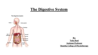
  • 1. The Digestive System By: Neha Rani Assistant Professor Shantha College of Physiotherapy
  • 2. Digestive system is important to our body. It is a group of organs working together to convert food into energy and basic nutrients to feed the entire body. Food passes through a long tube inside the body known as the alimentary canal or GI tract. The alimentary canal is made up of the oral cavity, pharynx, oesophagus, stomach, small intestine , and large intestine. Function: • Ingestion: it means taking food into the mouth to the alimentary canal. • Secretion: it means secret hormone for digestion of ingested food. • Mixing and movement: it means mixing of ingested food and prepare bolus. • Digestion: is the break down of large insoluble food molecules into small water- soluble food molecules so that they can be absorbed into the watery blood plasma. the following secretion helps in digestion – saliva from the salivary gland , gastric juice from the stomach , intestine juice from the small intestine, pancreatic juice from the pancreas and bile from the liver.
  • 3. • Absorption: it is the process in which, absorption of digested food substance take place in the walls of alimentary canal for metabolism and to get energy; from there they are carried to the liver for metabolism. • Excretion: food substance that cannot be digested and absorbed are excreted by the bowel as faecal matter. Classification of food: Food play important role in life. It gives energy after metabolic process, food rich in protein, vitamin, fat, carbohydrate. It provides nutrient that the body uses for growth and health. Carbohydrate: yet they are essential macronutrient , since they fuel the brain and muscles. They also contain the fiber the gut need to properly function. Simple carbohydrate contain one or two sugars, while complex carbohydrate are made of three or more linked sugars. Protein: Protein give strucutre to all cells. They also help repair tissue and fight infection. when consumption exceeds the body’s needs, protein can serve an energy source delivering 4gm per calorie.
  • 4. Fats: Like carbohydrates and protein, fats supply energy to fuel the process that keep your body alive. They generally fall in one of four categories, based on their chemical structure: monosaturated, polyunsaturated, saturated and trans fats. Vitamins: Vitamins are complex organic substances that team up with protein called enzymes to help chemical reaction takes place in the body. They vary in their specific roles and are either water soluble, such as vitamin C and B complex vitamins or fat soluble such as A,D, E and k. Minerals: Mineral give structure to our bones, teeth and nails .
  • 5. The Gastro- intestinal tract: Consist of a long tube through which food passes. It starts at mouth and end at anus. The tract is 8 – 10 meter in length. • Mouth • Pharynx • Oesophagus • Stomach • Small Intestine • LARGE Intestine • Rectum • Anus Accessory organ include teeth and tongue, salivary gland, liver , gallbladder and panaceas. These glands are situated outside the tract.
  • 6. The GI Tract is composed of four layers. Each layer has different tissue and functions. From inside out they : • Mucosa • Sub mucosa • Muscularis • Serosa Layer of GI Tract: The GI tract is composed of four layers. Each layer has different tissue and functions, i.e • Mucosa • Sub mucosa • Serosa Mucosa: Is the inner most layer , function is absorption and secretion, on the mucosa layer, small finger like projection called villi and microvilli help to increase the surface for nutrient absorption.
  • 7. Sub mucosa layer: The sub mucosa is relatively thick, highly vascular and serves the mucosa. The absorbed elements pass through the mucosa picked up from the blood vessel of the sub mucosa. Serosa: The alimentary canal in the abdomen is lined by serous coat i.e. visceral peritoneum.
  • 8. Peritoneum: The peritoneum is a continuous transparent membrane which lines the abdominal cavity and covers the abdominal organs. It act to support the viscera, and provides a pathway for blood vessel and lymph. It consist of two layer: • Parietal peritoneum • Visceral peritoneum Parietal peritoneum: lines the internal surface of the abdominal pelvic wall. Visceral peritoneum: invaginates to cover the majority of the abdominal viscera. Peritoneal cavity: is the space between the parietal and visceral peritoneum. Peritoneum is divided into three parts: • Omentum: greater and lesser • Mesentery • Peritoneal ligaments
  • 9. Omentum: Is a double layer of peritoneum that extends from the stomach and proximal part of the duodenum to other abdominal organs. Mesentry: A Mesentry is a double layer of visceral peritoneum. It connects an intraperitoneal organ to the posterior abdominal wall. It provides a pathway for nerves , blood vessel and lymphatics from the body wall to the viscera. A peritoneal ligament: It is a double fold of peritoneum that connects viscera together or connects viscera to the abdominal wall. Functions of the peritoneum: • The abdominal organs are covered partly or completely • Adipose tissue deposited in omenta and mesentry. They are the fat storage organs. • The Omentum help to prevent the spread of infection to the rest of the peritoneum • It has the ability to absorb small amount of fluid.
  • 10. Parts of the digestive system: Oral cavity: The mouth or oral cavity is the first part of the digestive tube. It is divided into mouth proper and a vestibule. The vestibule is a slit like space between the lips and cheek externally, and the gums and teeth internally. It communicates through the oral fissure. Lips: Lips (labia) are fleshy fold lined externally by skin and internally by mucous membrane. Each lip is composed of skin , superficial fascia and orbicularis oris muscle, submucosal and mucous membrane. Cheeks: Cheeks(buccae) are fleshy flaps, forming a large part of each side of the face. The cheeks are composed of skin, buccinators muscle, parotid duct , vessels and nerves, mucous glands and mucosa. Oral cavity proper: Boundaries: • Anterolaterally: by teeth, gums and alveolar arches of jaws. • Roof is formed by hard and soft palate. • Floor is formed by tongue and sublingual region. • Posteriorly the oral cavity communicates with the pharynx though the orophyrngeal isthmus which is bounded by superiorly by soft palate, inferiorly by tongue and on each side by palatoglossal arches.
  • 11. Gums: Gums are the soft tissue which cover the alveolar process of the upper and lower jaws and surround the neck of the teeth. The gums are made up of dense fibrous tissue, covered by stratified squamous epithelium. Teeth: Enamel: it is the hardest tissue in the human body covering the crown of the tooth. It is inert, a cellular and formed by ectoderm. It is supported by the underlying of dentin. Dentin: it is less calcified, more resilient, vital , hard tissue forming the main bulk of the tooth, it is formed from and supported by the dental pulp. In crown portion, it is covered by the enamel and in the root portion it is covered by the cemented , the junction between the enamel and dentin is called dentinoenamel junction. Cementum: it is less mineralized tissue, covering the radicular portion of the tooth, the junction between enamel and cementum is known as cervical line and cementoenamel junction. Pulp: it is the soft, connective tissue in the central part of the tooth enclosed by the dentin ,pulp cavity is known as pulp chamber and the root portion called pulp canal and root canal.
  • 12.
  • 13. Function of teeth: • Helps in mastication • Helps in articulation and speech • Give a definite shape and beauty of the face • May be used for self protection • Growth and development of jaw are dependent on tooth. Nerve supply: the teeth of upper jaw supplied by maxillary nerve and lower jaw supplied by mandibular nerve. Palate: It forms the arched of roof of the oral cavity and the floor of the nasal cavities. It separate the nasal cavities and nasophrynx from the oral cavity. parts: the anterior 2/3rd or bony part called the hard palate and posterior 1/3rd or fibro muscular part known as soft palate. Hard palate: Is formed by palatine process of maxillae and the horizontal process of palatine bones. Anteriorlly and laterally the hard palate is bounded by alveolar process and gums. Posterior to the central incisor teeth, there is and incisor canal.
  • 14. • The hard palate is covered by mucous membrane that is firmly attached to the bone, there are numerous mucus secreting palatine glands deep to the mucosa. Soft palate: • The soft palate is movable, fibro muscular fold, that is attached to the posterior edge of the hard palate. • The soft palate shows a conical process called uvula (greek word= grapes) which hangs its posterior, curved free margin. • Laterally soft palate is continuous with the wall of the pharynx and is joined to the tongue and pharynx by the palatoglossal and palatopharyngeal arches. • Nerve supply: palatine nerves ( sensory) • And motor nerve supplied by some cranial nerve i.e. Accessory nerve, Vagus nerve.
  • 15. Tongue: The tongue is highly mobile muscular organ that can vary greatly in shape , it is situated partly in the mouth and partly in oropharynx. Parts: It has a root, tip , a dorsal surface , a ventral surface and two lateral borders. Muscle of the tongue: Extrinsic muscle: • Genioglossus • Hyoglossus • Styloglossus • palatoglossal
  • 16. Intrinsic muscle: • Superior longitudinal muscle • Inferior longitudinal muscle • Transverse muscle • Vertical muscle Nerve supply: hypoglossal nerve and cranial nerve i.e. Vagus nerve. Function of tongue: • Help in speech • Help in chewing and swallowing and mastication • Secretion of mucin • Mixes food with saliva
  • 17. Pharynx: The pharynx is a wide muscular tube, situated behind the nose, mouth and larynx. It is about 13cm long.its upper part is wide and lower part is narrowest. Parts of pharynx: Nasal part: Nasophrynx Oral part: Oropharynx Laryngeal part: Laryngopharynx Nasopharynx: • This part is situated behind the nose. • It resembles the nose structurally and functionally, it is respiratory in function. • It is lined by ciliated columnar epithelium. • Anteriorly it communicates with the nasal cavities and posterior through the nasal aperture. • The lateral wall of the Nasopharynx shows the pharyngeal opening of the auditory tube or Eustachian tube
  • 18. Oropharynx: • It is the middle part of the pharynx situated behind the oral cavity. • Superiorly it communicates with oral cavity through the orophyrngeal isthmus. • Inferiorly it opens the laryngopharynx. • The lateral wall of the oropharynx presents the palatine tonsil, lying between the palatoglossal and palatopharyngeal arches. Laryngopharynx: • It is situated behind the larynx. • It extends the upper border of epiglottis to the lower border of the cricoid cartilage. • Lateral wall of laryngopharynx shows a depression called pisiform fossa or sinus on each side of the inlet of larynx. Salivary glands: The secretion of these glands help to keep the mouth moist and provide a protective and lubricant coat of mucus.
  • 19. Classification of salivary gland: Major glands are those glands which discharge their secretions into the oral cavity through a duct. Major salivary gland are Parotid, Submandibular, Sublingual. Minor glands are these glands which open directly into oral cavity, minor glands are labial gland ( lips), buccae glands (cheeks), lingual glands( tongue), Palatine glands( palate). Ducts are: stensen’s duct, Wharton’s duct, ducts of rivinus Nerve supplied by parasympathetic and sympathetic nerve supplied. Ducts of major salivary gland:
  • 20. Parotid gland: • Parotid glands are paired gland. • They are the largest among the salivary gland. • This gland is wedged between mandible and strenoclaidomastoid muscle, it occupies the side of the face anterior and inferior to the auricle. Submandibular gland: • It is a large salivary gland, roughly j shaped, situated along the body of the mandible. • It opens the floor of the mouth then summit the sublingual papilla at the side of the frenulum of the tongue. Sublingual gland: • This is the smallest of the three major salivary glands. It weigh is about 3-4 g , this gland is lined by the mucosa of the floor of the mouth . Most of them open directly into the floor od the mouth.
  • 21. Saliva: Total amount of saliva secreted by 1500ml/day. Contribution of salivary glands are: • Parotid glands : 25% • Sub maxillary gland : 70% • Sublingual gland: 5% The chemical composition of 98 percent water and 2 percent other substances such as electrolyte or minerals ( Na , k), mucus ( mucopolysacchrides and glycoprotein) and enzymes. Saliva is a frothy substances that is produced and secreted from the salivary gland referred as mouth is blood Composition of saliva: Saliva contain 99% water and 1 % solids. It include organic and inorganic constituents are: Organic constituents are: • The enzyme ptyalin or salivary amylase secreted from the parotid gland. • Lingual lipase is another enzyme present in the saliva which acts on triglycerides. • Inorganic constituents are Na+, K+, Ca++, Hco3- and Cl-
  • 22. FUNCTIONS OF SALIVA: • Lubrication and speech: saliva easily help in swallowing of food. As it lubricates the food. • Appreciation of taste: only food dissolves in the saliva can stimulate the taste bud. Digestive function: saliva contains salivary amylase or ptyalin which mainly acts on cooked starch and convert into maltose. After chewing the food mixed with ptyalin is swallowed Regulation of salivary secretion: There can be spontaneous as well as stimulated secretion of saliva: Spontaneous secretion of saliva occurs without any stimulus and it is always present. It is in small quantity (0.5ml/min.). Stimulated secretion of saliva is controlled by nervous stimulation via secret motors nerves.
  • 23. Two types of reflex secretion: • Unconditioned reflex • Conditioned reflex Unconditioned reflex: this is present since birth, here salivation occurs when food is placed inside the mouth the receptors involved in the mechanoreceptors and taste receptors in the oral cavity , the afferent nerves involved are the V,VII, IX and X cranial nerves constitute the superior and inferior salivary nuclei. And efferent nerve (VII and XI cranial nerve) from the salivary center stimulate the salivary gland to secrete saliva. Conditioned reflex: this reflex is developed by previous experiences of the subject, sight smell or even thought of familiar food substances can initiate salivary secretion.
  • 24. Esophagus: Extent: the esophagus is a narrow part of the alimentary canal extending from the lower end of the pharynx( at the level of 6th cervical vertebrae) to the cardiac orifice of the stomach. It length is 25cm and diameter 1.5cm. Parts: Three parts: • Cervical: posterior : vertebral column, lateral: carotid sheath, anterior: trachea • Thoracic: posteriorly: vertebral column, anterior: trachea • Abdominal: short segment of the esophagus lies in the groove on the posterior surface of the left lobe of the liver. It is a tubular strucutre which remains collapsed anteroposteriorly. Nerve supply: vagus nerve and splanchnic nerve. Stomach: Stomach is the most dilated part of the alimentary tract. It extends from the cardiac notch end to pyloric end, upper end is continuous with esophagus and lower end is continuous with the duodenum. Position:
  • 25. Position: In the supine position, the stomach occupies parts of the epigastric, umbilical and left hypochondric regions. Shape: J shape , upper border is wider than the lower border, it has two ends upper cardiac and lower pyloric, it has two surfaces anterior and posterior , two curvatures greater curvature on right side and on left side lesser curvature . Capacity: Newborn infant : hold upto 30 ml of milk, adult hold 2 to 3 liters of food. Parts of stomach: • Cardiac part • Fundus • Body of the stomach • Pyloric region • Greater curvature and lesser curvature
  • 26. Cardiac part: The cardiac part or cardiac lies around the cardiac orifice, which receives the opening of the abdominal part of the esophagus. Fundus: The fundus of the stomach is the dilated portion, to the left and superior to the cardiac orifice, this is the most superior part of the stomach, which is related to the dome of diaphragm. Body of the stomach: It is the major portion of the stomach , it lies between the fundus and the pyloric antrum. Pyloric region: The pyloric region has two parts: antrum and canal . Body of the stomach ends in antrum and the junction between the body and antrum is marked by an angular notch, antrum is continued as the narrow canal which is called pyloric canal or pyloric end. Lesser curvature: It gives attachment to a double fold of peritoneum called the lesser Omentum which stretches towards the liver. Greater curvature: It gives attachment to the following peritoneal folds from above downwards. Between the tow layers of lesser Omentum and lesser curvature is the anastomosis between left and right gastric vessels. Between the two layer of greater Omentum and greater curvature are the right and left gastroepiploic vessels.
  • 27. Small intestine: Extent: • The small intestine extends from the pylorus to the ileocecal junction. • It is about 6meters long. • Its strucutre is adapted for digestion and absorption Parts: It is divided into: • An upper fixed part called the duodenum, approximately 25cm long. • The proximal two -fifth of this long convoluted tube is continuous with the duodenum and is known as the jejunum. • The distal three= fifth is called ileum. Functions of small intestine: • Segmental contraction in the small intestine help in mixing the chime coming from stomach with pancreatic juice, bile and intestinal juice for proper digestion. • Activation of trypsinogen occurs by enterokinase secreted by small intestine. • A large number of hormones are secreted by the APUD cells present in the small intestine . Enterogastrones secretin and CCK control the secretory activity of pancreas, small intestine and also control gastrointestinal movements. • Final products of digestion, vitamins, minerals, water etc. are absorbed in the small intestine
  • 28. Small intestine juice( Succus ENTERICUS): It is the characteristics alkaline fluid secreted by the crypts of liberkulin. About 2L of fluid is secreted per day which is rapidly reabsorbed by the intestinal villi. The digestive enzymes in the Succus entericus are present in the brush border or microvilli of the epithelial cells. Enzymes are: • Disaccharides: they are sucrose, maltase, lactase enzymes , they are act on carbohydrates and split in the disaccharides into monosacchrides. • Proteolytic enzymes or peptidase: • They bring about the final breakdown of polypeptides into amino acids. • Intestinal lipase: it splits into neural fats into diglycerides, monoglycerides, fatty acids and glycerol. Duodenum: The duodenum is the shortest, widest and most fixed part of the small intestine, approx. 10 inches. Extent: it extends from the pylorus to the duodenojejunal flexure. It is closely related to the head of the pancreas in the form of c letter.
  • 29. Position: the duodenum is retroperitoneal lies above the level of the umbilicus opposite to the vertebrae L1,L2,L3. Parts: • First part (superior) • Second part (descending) • Third part ( horizontal ) • Fourth part ( ascending) Nerve supply: parasympathetic and sympathetic. Jejunum and ileum: • The coils of jejunum and ileum are suspended by the mesentry from the posterior abdominal wall so that they are freely mobile. • The jejunum begins at the duodenoileum flexure. • The head and neck of the pancreas lies inferior to the first part. Medially to the second part and superiorly to the third part.
  • 30. Large intestine: Large intestine extends from the ileocecal junction to the anus. It s about 1.5m long. Parts of large intestine are cecum, ascending colon, transverse colon, descending colon, sigmoid colon, rectum and anal canal. Rectum and anal canal are situated in the pelvis, the remaining parts in the abdomen. Nerve supply: puborectalis part of the levator ani muscle also Sourround the anal muscle. Function of the large intestine: • Secretory function. • Absorption of water and electrolyte • Excretion of heavy materials like hg • Synthesis of vitamin K, vitamin B12 and folic acid by colonic flora • Storage of feces.
  • 31. Accessory organs of digestion Liver: The liver is the largest gland in the body, it is situated in the upper part of the abdominal cavity occupying the greater part of the right hypochondric region, part of epigastric region and extending the left hypochondric region. Lobes: anatomically, liver has two lobes , large right lobe and small left lobe. They are separated from each other by the line of attachment of the falciform ligament Quadrant lobe: it is seen on the inferior surface of the right lobe. Caudate lobe: it is chiefly situated on the posterior surface of the right lobe. Function of the liver: • Production of bile, which helps carry away waste and break down fats in the small intestine during digestion. • Production of certains protein for blood plasma. • Store and release glucose as needed. • Clearing the blood of drugs and other harmful substances.
  • 32.
  • 33. Gall bladder: The gall bladder is a pear shaped sac attached to the posterior surface of the liver by connective tissue. It has a fundus, or expanded end, a body or main part and a neck which is continuous with the cystic duct. Function of bile: • Reservoir for bile • Concentration of bile by up 10 to 15 fold by absorption of water through the walls of the gall bladder. • Released of stored bile. When the muscle wall of the gall bladder contract bile passes through the bile ducts to the abdomen. Pancreas: The pancreas is about 6inches long and sits across the back of the abdomen, behind the stomach. The head of the pancreas is on the right side of the abdomen and it is connected to the duodenum( is the first section of the SI) thorugh a small called pancreatic duct. Pancreas is both exocrine and endocrine gland. It lies transversely across the posterior abdominal all at the level of L1 and L2 Vertebrae. Nerve supply: parasympathetic ( vagus) and sympathetic( lower thoracic) fibers innervates the pancreas. Pancreatic juice: it is secretion of exocrine pancreas. Normally about 1500ml of pancreatic juice is produced everyday.
  • 34. Organic constituents are: • Proteases • Trypsinogen : trypsin secreted as inactive trypsinogen. • Chymotrypsinogen: it activate Chymotrypsinogen into chymotrypsin. • Proelastase: it activate Proelastase into elastase. • Procarboxypeptidase: it convert into carboxypeptidases. Lipolytic enzyme: are pancreatic lipase and colipase for triglycerides digestion. Phospholipase into phospholipid digestion. Chloestrol esterhydrase for cholesterol digestion.