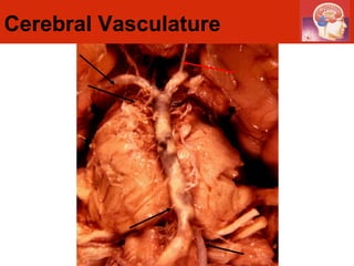
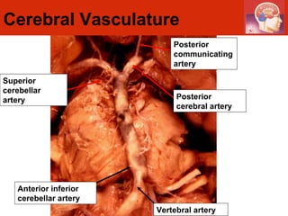
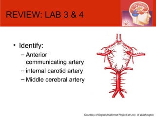
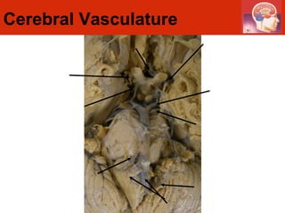
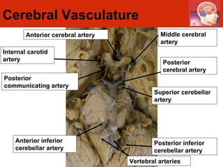
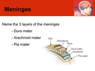
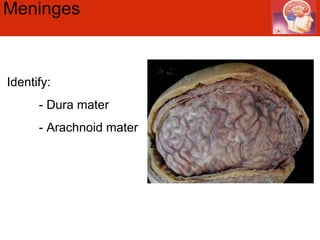
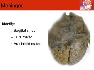
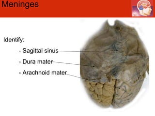
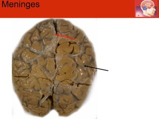
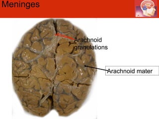
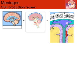
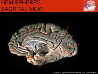
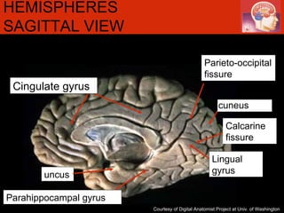
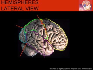
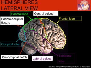
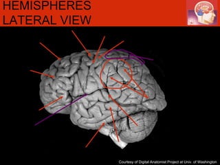
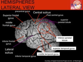
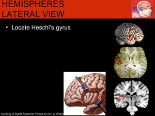
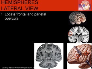
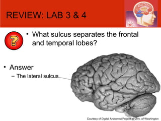
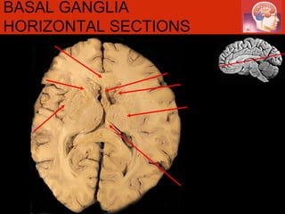
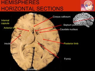
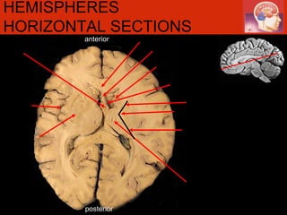
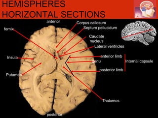
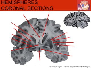
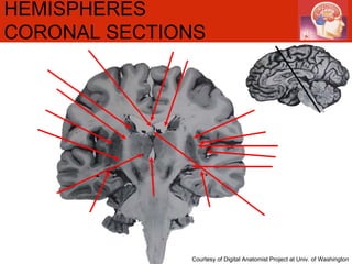
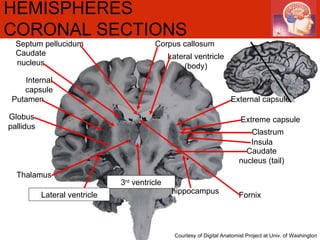
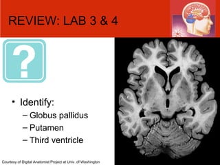
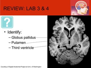
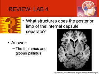
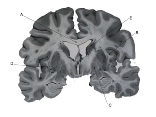
ANAT 321 REVIEW LABORATORY 3 & 4 NEUROANATOMY LAB
Cerebral Vasculature
Cerebral Vasculature Posterior communicating artery Superior cerebellar artery Anterior inferior cerebellar artery Vertebral artery Posterior cerebral artery
REVIEW: LAB 3 & 4 Identify: Anterior communicating artery  internal carotid artery Middle cerebral artery Courtesy of Digital Anatomist Project at Univ. of Washington
Cerebral Vasculature
Cerebral Vasculature Posterior communicating artery Superior cerebellar artery Anterior inferior cerebellar artery Internal carotid artery Vertebral arteries Posterior inferior cerebellar artery Middle cerebral artery Anterior cerebral artery Posterior cerebral artery
Meninges Name the 3 layers of the meninges - Dura mater - Arachnoid mater - Pia mater
Meninges  Identify: - Dura mater - Arachnoid mater
Meninges  Identify: - Sagittal sinus - Dura mater - Arachnoid mater
Meninges
Meninges  Arachnoid granulations Arachnoid mater
Meninges  CSF production review
HEMISPHERES SAGITTAL VIEW  Courtesy of Digital Anatomist Project at Univ. of Washington
HEMISPHERES SAGITTAL VIEW  Courtesy of Digital Anatomist Project at Univ. of Washington Calcarine fissure Parieto-occipital fissure cuneus Lingual gyrus Cingulate gyrus uncus Parahippocampal gyrus
HEMISPHERES LATERAL VIEW  Courtesy of Digital Anatomist Project at Univ. of Washington
HEMISPHERES LATERAL VIEW  Courtesy of Digital Anatomist Project at Univ. of Washington Lateral sulcus Temporal lobe Frontal lobe Pre-occipital notch Parieto-occipital fissure Central sulcus Occipital lobe Parietal lobe
HEMISPHERES LATERAL VIEW   Courtesy of Digital Anatomist Project at Univ. of Washington
HEMISPHERES LATERAL VIEW   Courtesy of Digital Anatomist Project at Univ. of Washington Central sulcus Inferior temporal gyrus superior temporal gyrus middle temporal gyrus Post central gyrus precentral gyrus Lateral sulcus inferior frontal gyrus middle frontal gyrus Superior frontal gyrus superior parietal lobule Inferior parietal lobule
HEMISPHERES LATERAL VIEW  Locate Heschl’s gyrus Courtesy of Digital Anatomist Project at Univ. of Washington
HEMISPHERES LATERAL VIEW  Locate frontal and parietal opercula Courtesy of Digital Anatomist Project at Univ. of Washington
REVIEW: LAB 3 & 4 What sulcus separates the frontal and temporal lobes? Courtesy of Digital Anatomist Project at Univ. of Washington Answer The lateral sulcus
REVIEW: LAB 3 & 4 What gyrus lies inferior to the calcarine sulcus? Courtesy of Digital Anatomist Project at Univ. of Washington Answer The lingual gyrus
REVIEW: LAB 3 &4 What structures comprise the basal ganglia? Caudate nucleus Putamen (lateral) Globus pallidus (medial)
anterior posterior HEMISPHERES HORIZONTAL SECTIONS Courtesy of Digital Anatomist Project at Univ. of Washington
anterior posterior Internal capsule HEMISPHERES HORIZONTAL SECTIONS Courtesy of Digital Anatomist Project at Univ. of Washington Caudate nucleus Lateral ventricles Corpus callosum Thalamus Corpus callosum fornix Septum pellucidum Extreme capsule  External capsule Claustrum insula Putamen Anterior limb genu Posterior limb
anterior posterior BASAL GANGLIA HORIZONTAL SECTIONS
anterior posterior Internal capsule HEMISPHERES HORIZONTAL SECTIONS Fornix Posterior limb insula Anterior limb Septum pellucidum Caudate nucleus Corpus callosum
anterior posterior HEMISPHERES HORIZONTAL SECTIONS
anterior posterior HEMISPHERES HORIZONTAL SECTIONS Lateral ventricles Corpus callosum Thalamus Septum pellucidum Insula anterior limb posterior limb genu Caudate nucleus Putamen Internal capsule fornix
HEMISPHERES CORONAL SECTIONS   Courtesy of Digital Anatomist Project at Univ. of Washington
HEMISPHERES CORONAL SECTIONS   Caudate nucleus putamen Internal capsule External capsule extreme capsule clastrum insula Globus pallidus Lateral ventricle Septum pellucidum fornix hypothalamus 3 rd  ventricle
HEMISPHERES CORONAL SECTIONS   Courtesy of Digital Anatomist Project at Univ. of Washington
HEMISPHERES CORONAL SECTIONS   Courtesy of Digital Anatomist Project at Univ. of Washington Caudate nucleus Putamen  Internal capsule External capsule Extreme capsule Clastrum Insula Globus pallidus Lateral ventricle (body) Septum pellucidum 3 rd  ventricle hippocampus Lateral ventricle Corpus callosum Caudate nucleus (tail) Fornix Thalamus
REVIEW: LAB 3 & 4 Identify: Globus pallidus Putamen Third ventricle  Courtesy of Digital Anatomist Project at Univ. of Washington
REVIEW: LAB 4 What structures does the posterior limb of the internal capsule separate?  Answer:  The thalamus and globus pallidus Courtesy of Digital Anatomist Project at Univ. of Washington
QUIZ
E B C D A
Answer Key A: corpus callosum B: extreme capsule C: globus pallidus D: claustrum E: caudate nucleus
A B C D E
Answer Key A: lateral ventricle B: fornix C: posterior limb of internal capsule D: caudate nucleus E: insula
A B C D E Courtesy of Digital Anatomist Project at Univ. of Washington
Answer Key A: anterior communicating artery B: posterior communicating artery C: superior cerebellar artery D: vertebral artery E: anterior cerebral artery
A B C D E Courtesy of Digital Anatomist Project at Univ. of Washington
Answer Key A: cingulate gyrus  B: fornix C: parahippocampal gyrus D: cuneus E: parieto-occipital fissure
A B C D E Courtesy of Digital Anatomist Project at Univ. of Washington
Answer Key A: internal carotid artery B: superior cerebellar artery C: posterior inferior cerebellar artery D: superior cerebellar artery  E: vertebral artery

ANAT-321 demonstrator lab: review lab 3 & 4

  • 1.
    ANAT 321 REVIEWLABORATORY 3 & 4 NEUROANATOMY LAB
  • 2.
  • 3.
    Cerebral Vasculature Posteriorcommunicating artery Superior cerebellar artery Anterior inferior cerebellar artery Vertebral artery Posterior cerebral artery
  • 4.
    REVIEW: LAB 3& 4 Identify: Anterior communicating artery internal carotid artery Middle cerebral artery Courtesy of Digital Anatomist Project at Univ. of Washington
  • 5.
    REVIEW: LAB 3& 4 Identify: Anterior communicating artery internal carotid artery Middle cerebral artery Courtesy of Digital Anatomist Project at Univ. of Washington
  • 6.
  • 7.
    Cerebral Vasculature Posteriorcommunicating artery Superior cerebellar artery Anterior inferior cerebellar artery Internal carotid artery Vertebral arteries Posterior inferior cerebellar artery Middle cerebral artery Anterior cerebral artery Posterior cerebral artery
  • 8.
    Meninges Name the3 layers of the meninges - Dura mater - Arachnoid mater - Pia mater
  • 9.
    Meninges Identify:- Dura mater - Arachnoid mater
  • 10.
    Meninges Identify:- Dura mater - Arachnoid mater
  • 11.
    Meninges Identify:- Sagittal sinus - Dura mater - Arachnoid mater
  • 12.
    Meninges Identify:- Sagittal sinus - Dura mater - Arachnoid mater
  • 13.
  • 14.
    Meninges Arachnoidgranulations Arachnoid mater
  • 15.
    Meninges CSFproduction review
  • 16.
    HEMISPHERES SAGITTAL VIEW Courtesy of Digital Anatomist Project at Univ. of Washington
  • 17.
    HEMISPHERES SAGITTAL VIEW Courtesy of Digital Anatomist Project at Univ. of Washington Calcarine fissure Parieto-occipital fissure cuneus Lingual gyrus Cingulate gyrus uncus Parahippocampal gyrus
  • 18.
    HEMISPHERES LATERAL VIEW Courtesy of Digital Anatomist Project at Univ. of Washington
  • 19.
    HEMISPHERES LATERAL VIEW Courtesy of Digital Anatomist Project at Univ. of Washington Lateral sulcus Temporal lobe Frontal lobe Pre-occipital notch Parieto-occipital fissure Central sulcus Occipital lobe Parietal lobe
  • 20.
    HEMISPHERES LATERAL VIEW Courtesy of Digital Anatomist Project at Univ. of Washington
  • 21.
    HEMISPHERES LATERAL VIEW Courtesy of Digital Anatomist Project at Univ. of Washington Central sulcus Inferior temporal gyrus superior temporal gyrus middle temporal gyrus Post central gyrus precentral gyrus Lateral sulcus inferior frontal gyrus middle frontal gyrus Superior frontal gyrus superior parietal lobule Inferior parietal lobule
  • 22.
    HEMISPHERES LATERAL VIEW Locate Heschl’s gyrus Courtesy of Digital Anatomist Project at Univ. of Washington
  • 23.
    HEMISPHERES LATERAL VIEW Locate frontal and parietal opercula Courtesy of Digital Anatomist Project at Univ. of Washington
  • 24.
    REVIEW: LAB 3& 4 What sulcus separates the frontal and temporal lobes? Courtesy of Digital Anatomist Project at Univ. of Washington Answer The lateral sulcus
  • 25.
    REVIEW: LAB 3& 4 What gyrus lies inferior to the calcarine sulcus? Courtesy of Digital Anatomist Project at Univ. of Washington Answer The lingual gyrus
  • 26.
    REVIEW: LAB 3&4 What structures comprise the basal ganglia? Caudate nucleus Putamen (lateral) Globus pallidus (medial)
  • 27.
    anterior posterior HEMISPHERESHORIZONTAL SECTIONS Courtesy of Digital Anatomist Project at Univ. of Washington
  • 28.
    anterior posterior Internalcapsule HEMISPHERES HORIZONTAL SECTIONS Courtesy of Digital Anatomist Project at Univ. of Washington Caudate nucleus Lateral ventricles Corpus callosum Thalamus Corpus callosum fornix Septum pellucidum Extreme capsule External capsule Claustrum insula Putamen Anterior limb genu Posterior limb
  • 29.
    anterior posterior BASALGANGLIA HORIZONTAL SECTIONS
  • 30.
    anterior posterior Internalcapsule HEMISPHERES HORIZONTAL SECTIONS Fornix Posterior limb insula Anterior limb Septum pellucidum Caudate nucleus Corpus callosum
  • 31.
    anterior posterior HEMISPHERESHORIZONTAL SECTIONS
  • 32.
    anterior posterior HEMISPHERESHORIZONTAL SECTIONS Lateral ventricles Corpus callosum Thalamus Septum pellucidum Insula anterior limb posterior limb genu Caudate nucleus Putamen Internal capsule fornix
  • 33.
    HEMISPHERES CORONAL SECTIONS Courtesy of Digital Anatomist Project at Univ. of Washington
  • 34.
    HEMISPHERES CORONAL SECTIONS Caudate nucleus putamen Internal capsule External capsule extreme capsule clastrum insula Globus pallidus Lateral ventricle Septum pellucidum fornix hypothalamus 3 rd ventricle
  • 35.
    HEMISPHERES CORONAL SECTIONS Courtesy of Digital Anatomist Project at Univ. of Washington
  • 36.
    HEMISPHERES CORONAL SECTIONS Courtesy of Digital Anatomist Project at Univ. of Washington Caudate nucleus Putamen Internal capsule External capsule Extreme capsule Clastrum Insula Globus pallidus Lateral ventricle (body) Septum pellucidum 3 rd ventricle hippocampus Lateral ventricle Corpus callosum Caudate nucleus (tail) Fornix Thalamus
  • 37.
    REVIEW: LAB 3& 4 Identify: Globus pallidus Putamen Third ventricle Courtesy of Digital Anatomist Project at Univ. of Washington
  • 38.
    REVIEW: LAB 3& 4 Identify: Globus pallidus Putamen Third ventricle Courtesy of Digital Anatomist Project at Univ. of Washington
  • 39.
    REVIEW: LAB 4What structures does the posterior limb of the internal capsule separate? Answer: The thalamus and globus pallidus Courtesy of Digital Anatomist Project at Univ. of Washington
  • 40.
  • 41.
    E B CD A
  • 42.
    Answer Key A:corpus callosum B: extreme capsule C: globus pallidus D: claustrum E: caudate nucleus
  • 43.
    A B CD E
  • 44.
    Answer Key A:lateral ventricle B: fornix C: posterior limb of internal capsule D: caudate nucleus E: insula
  • 45.
    A B CD E Courtesy of Digital Anatomist Project at Univ. of Washington
  • 46.
    Answer Key A:anterior communicating artery B: posterior communicating artery C: superior cerebellar artery D: vertebral artery E: anterior cerebral artery
  • 47.
    A B CD E Courtesy of Digital Anatomist Project at Univ. of Washington
  • 48.
    Answer Key A:cingulate gyrus B: fornix C: parahippocampal gyrus D: cuneus E: parieto-occipital fissure
  • 49.
    A B CD E Courtesy of Digital Anatomist Project at Univ. of Washington
  • 50.
    Answer Key A:internal carotid artery B: superior cerebellar artery C: posterior inferior cerebellar artery D: superior cerebellar artery E: vertebral artery