SlideShare a Scribd company logo
1 of 7
Download to read offline
Biji G
International Journal of Image Processing (IJIP), Volume (12) : Issue (3) : 2018 83
An Efficient Automatic Segmentation Method For Leukocytes
Biji G biji2engg@gmail.com
Assistant Professor
Dept of Electrical Engineering
Govt Engineering College Bartonhill
Thiruvananthapuram,695035,India
Abstract
Blood tests are of the most important and counting of leukocytes in peripheral blood is commonly
used in basic clinical diagnosis. A major requirement for this paper is an efficient method to
segment cell images. This work presents an accurate segmentation method for automatic count
of white blood cells. First a simple thresholding approach is applied to give initial labels to pixels
in the blood cell images. The algorithm is based on information about blood smear images, and
then the labels are adjusted with a shape detection method based on large regional context
information to produce meaningful results. This approach makes use of knowledge of blood cell
structure, the experimental result shows that this method is more powerful than traditional
methods that use only local context information. It can perform accurate segmentation of white
blood cells even if they have unsharp boundaries.
Keywords: Leukocytes, Thresholding, Pixels, Peripheral Blood, Segmentation.
1. INTRODUCTION
There are many different classes of white blood cells present in blood smear images. Differential
count of these various types of cells gives valuable information and plays an important role in the
diagnosis of differential diseases. It’s a tedious task to count class of cells manually. An automatic
counter using computer vision helps this medical test rapidly and accurately. The first step of this
automatic analysis is a segmentation of blood cells images, which differentiates meaningful
objects from the background. In these case attempting to identify white blood cells with an
accurate segmentation methods.
This step is crucial because the result is the basis of subsequent analysis. The success of
classification depends mainly on the correct segmentation of images. It’s also difficult and
challenging problem due to the complex nature of the cells and uncertainty in the microscopic
images. Cells often overlap each other and have variation of different size and shape. The
contrast between the cells boundary and the background varies according to illumination in
consistency.
Many segmentation methods of blood cell images have been proposed. Histogram thresholding,
edge detection and regional growing methods are often used. Thresholding techniques always
can’t produce meaningful result since no spatial information is used during the selection of the
segmentation threshold. They are often combined with mathematical morphology operation. Edge
detection method perform poorly on cell image because not all cells boundaries are sharp, so it’s
difficult to get all the edge information and locate the cells
Most of these mentioned methods are on short decision processes and often make wrong crisp
decisions. Relaxation methods are proposed to avoid it. A fuzzy patch label relaxation algorithm
used patches that provide more useful and meaningful context information that compact local
context to obtain better segmentation results. But sometimes the information of a few neighbour
patches still not enough to make correct decision. For example, too bright illumination often leads
Biji G
International Journal of Image Processing (IJIP), Volume (12) : Issue (3) : 2018 84
to bright gaps inside cytoplasm, and then a part of cytoplasm may be labeled as red blood cells
since it is separate from the other parts of the cytoplasm and is similar to a red blood cell in
position relationship. The neighbour patches play poorly on recovering this mistake. Hence a
shape detection method is proposed to provide fast and accurate segmentation by using large
regional context information. Since more information is used to help make decision, the approach
is more robust and efficient.
In this paper presents a white blood cell segmentation approach that consists of three steps. First
is simple thresholding approach combined with mathematical morphology operation is applied to
give initial labels to pixel in the blood cell images. The algorithm is based on priori information
about blood smear images derived from a learning process. Next the labels are adjusted with a
shape detection method based on large regional context information, which makes use of
knowledge of blood cell structure. At last the regions of white blood cells are marked and the
shapes of the regions are arranged to form rounded boundaries. The following sections give a
detailed explanation of the proposed method along with some experiments results.
2. PROPOSED METHOD
2.1 Initial Segmentation
The goal of initial segmentation is to separate four different regions roughly, background, red
cells, cytoplasm and nucleus. Information of colours, brightness and gradients are used in
thresholding to create initial labels of the pixels. The bright white or yellow regions correspond to
background. The dark regions correspond to nucleus. The regions that have intermediate
brightness and small gradients correspond to red cells. Other regions are labeled as cytoplasm
temporarily. Most thresholds are derived from priori information of blood smear images. But some
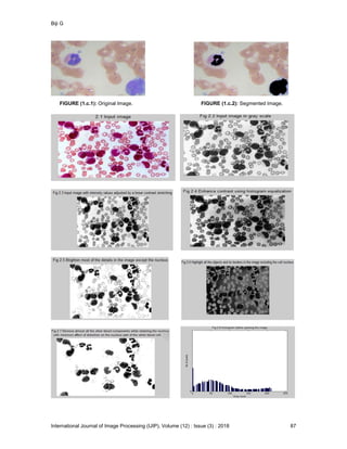
sensitive thresholds should be selected automatically to adapt to the illumination variation, which
greatly influence the segmentation result.
To select an optimal threshold, we change it from a smaller value to larger one by a small step
and segment the image with it. If the segmentation is correct with this threshold, the edges of the
segmented image should have relatively large gradients, which correspond the real edges of
different regions. So we calculate the number of the edge points that have relatively large
gradients while the threshold changes and select the threshold that leads to the largest number of
edge points to serve as the optimal threshold.
The regions labeled cytoplasm after thresholding are not real cytoplasm regions. They are
repartitioned to three different parts according to their connection status with other components.
Regions connected with only red cell regions are labeled as red cell. Regions connected only with
nucleus and regions connected with both are relabeled separately.
Every time the labels of pixels change, the different types of regions are smoothened with
mathematical morphology operation to get connected regions and eliminate single points and
lines caused by noises.
After initial labeling with thresholding method and morphology operation, the blood smear images
are segmented roughly into four regions. Pixels that are sure enough to belong to background or
nucleus are determined and other labels of pixels will be adjusted in the following steps.
2.2 Label Connection with Shape Detection
Now the initial nucleus regions along with cytoplasm regions around them make up the initial
leukocyte regions. They are not real leukocyte regions since there are many other regions in
them such as red cell and stain regions which may be connected with real leukocyte regions.
Some of these false regions can be eliminated by a scan method.
Real leukocyte regions are connected regions with convex shapes. False regions may be
connected with them, but there still will have some relatively large gaps between them. So when
we scan the images in a certain direction, back ground regions will be detected between false
Biji G
International Journal of Image Processing (IJIP), Volume (12) : Issue (3) : 2018 85
regions and real regions. Separate regions in this direction can be eliminated. That is although
the false regions are connected with real leukocyte regions somewhere; they are separate in
certain directions and can be detected by scanning in these directions.Fuzzy C-Means is a
clustering method which allows single data belong to more than one clusters. This method (Dunn,
1973) is a pattern recognition based on minimization of the following objective function given as
Equation (2) below:
The FCM algorithm attempts to partition a finite collection of elements X={X1,X2, ….. Xn} into a
collection of c fuzzy clusters with respect to some given criterion. Given a finite set of data, the
algorithm returns a list of cluster centres C={C1,C2, ….. Cn} and a partition matrix
Like the K-means clustering, the FCM aims to minimize an objective function:
2
1 1
ji
N
i
c
j
m
ijm cxuJ   
----------------(1)
where:













c
k
m
ki
ji
ij
cx
cx
u
1
1
2
1
-------------------(2)




 N
i
m
ij
N
i
i
m
ij
j
u
xu
c
1
1
..
----------------------(3)
where each element uij tells the degree to which element xi belongs to cluster Cj .
This differs from the k-means objective function by the addition of the membership values uij and
the fuzzifier m, with m≥1. The fuzzifier m determines the level of cluster fuzziness. A
large m results in smaller memberships ||*|| is any norm expressing the similarity between any
measured data and the center.
The algorithm is composed of the following steps:
1. Initialize U=[uij] matrix, U(0)
2. At k-step: calculate the centers vectors C(k)=[cj] with U(k)
3. Update U(k) , U(k+1)
4. If || U(k+1) - U(k)||< then STOP; otherwise return to step 2.
After the scan process a large region containing several white blood cells has been segmented
out. Then we’ll give accurate segmentation in smaller regions each containing a single white
blood cell.Nucleus regions are always in the centre of the leukocyte. There are many “cytoplasm’’
regions around them, which may be labeled incorrectly. The real cytoplasm regions can be picked
out with a regularity detection method. The distance from a boundary point of the cell to center
Biji G
International Journal of Image Processing (IJIP), Volume (12) : Issue (3) : 2018 86
point of the nucleus is calculated to serve as the radius. If a real leukocyte region is missed, a
sharp decrease can be detected while we check the radiuses of boundary points in turn. If a false
region is added into the leukocyte region, a sharp increase will be detected. These two cases
both lead to great variation in radius. So the cytoplasm regions that together form a region with
the least variation of the radius should be labeled real leukocyte regions.
2.3 Shape Arrangement
Most pixels have been correctly labeled after the previous processes. But the unsharp boundaries
between the cells and the background often lead to small regions around the white blood cells,
which may be edge regions of the cells and influence the shapes of the cells. The labels of these
small regions can be determined according to their positions.
Hough transformation is applied to find a circle in the image of edge points, which corresponds to
the rough region of the cell. Small regions over this circle are added to the cell region and the
small gaps inside cell regions are filled to form an orbicular shape of the cell.
Although we adjust labels of points at every step to avoid making false segmentation, there is no
guarantee that the segmentation results are correct all the time. So we identify the regularity of
the final boundary of cells by calculating the variation of the radius. Great variation corresponding
to an irregular shape means possible incorrect segmentation. The irregular edges are marked to
remind users that these edges should be checked manually to avoid mistakes
3. EXPERIMENTAL RESULTS
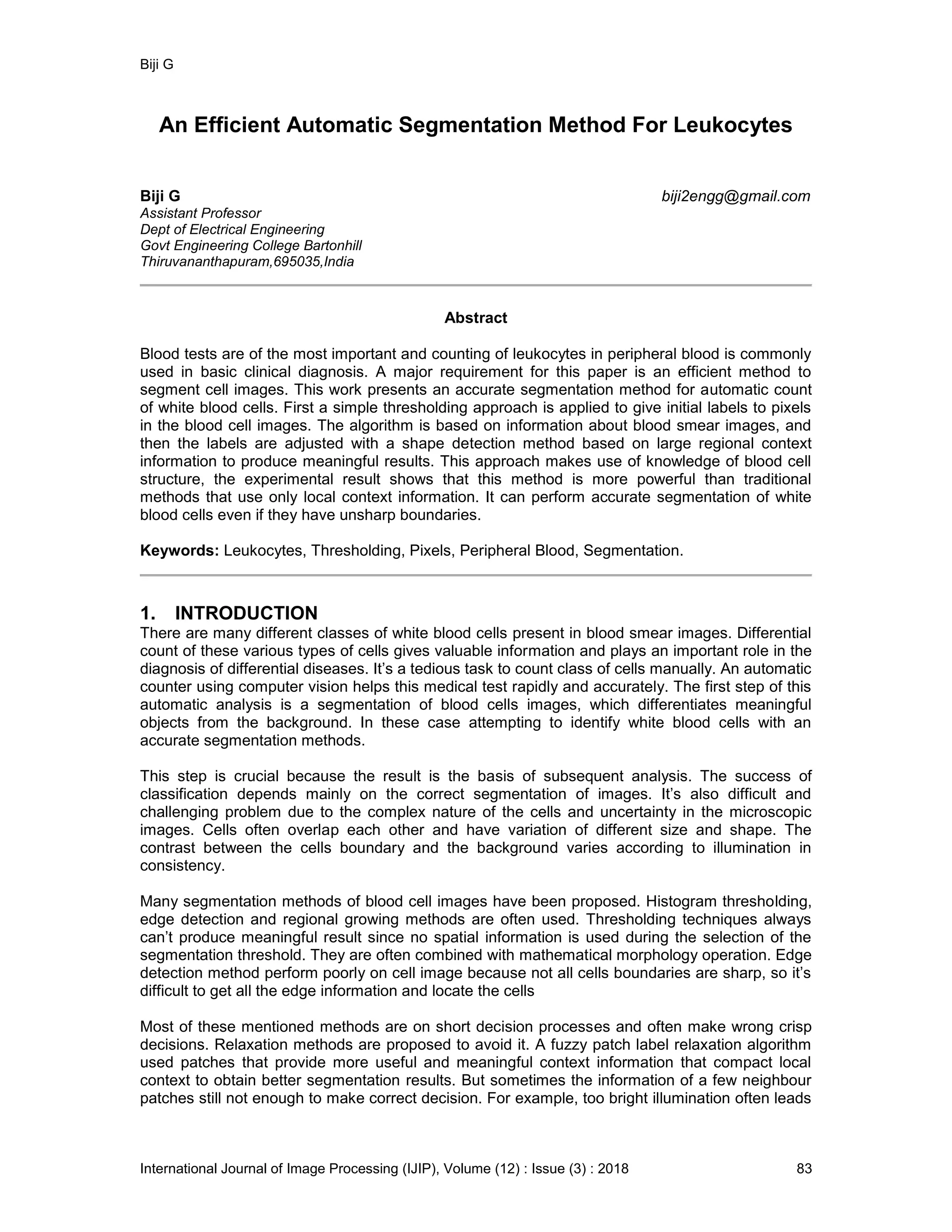
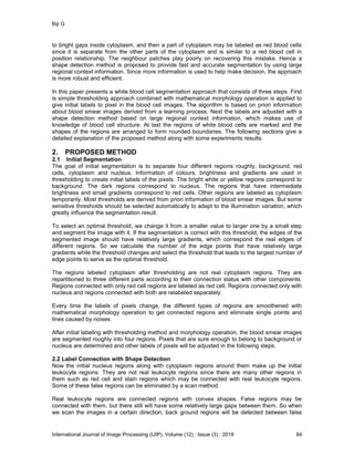
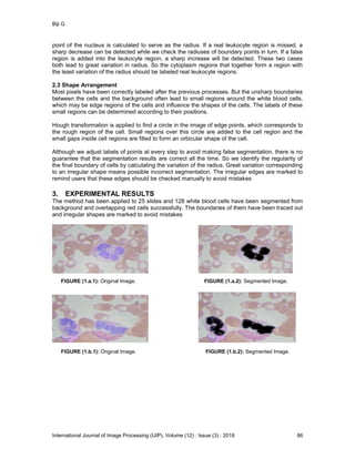
The method has been applied to 25 slides and 128 white blood cells have been segmented from
background and overlapping red cells successfully. The boundaries of them have been traced out
and irregular shapes are marked to avoid mistakes
FIGURE (1.a.1): Original Image. FIGURE (1.a.2): Segmented Image.
FIGURE (1.b.1): Original Image. FIGURE (1.b.2): Segmented Image.
Biji G
International Journal of Image Processing (IJIP), Volume (12) : Issue (3) : 2018 87
FIGURE (1.c.1): Original Image. FIGURE (1.c.2): Segmented Image.
Biji G
International Journal of Image Processing (IJIP), Volume (12) : Issue (3) : 2018 88
4. CONCLUSION
The proposed shape detection method uses information from a large region that contains a whole
white blood cell rather than information from a small local region or a few neighbour patches.
Therefore it is powerful to obtain reliable and meaningful segmentation results and trace out
accurate boundaries of white blood cells. Although the labels of the pixels are adjusted step by
step to avoid wrong crisp decisions that are difficult to be reversed, it is not an iterative approach
which makes it faster than most relaxation methods.
5. FUTURE SCOPE OF WORK
The shape detection and segmentation of white blood cell can be approximated by a quasi
circular form or a circular form. The automatic segmentation of WBC’s in complicated and
cluttered images can be approached by circle detection optimization methods.
6. REFERENCES
[1] D.Anorganingrum,”Cell segmentation with median filter and mathematical morphology
operation,” Proceedings on International Conference on Image Analysis and Processing,
pp.1043-1046, 1999.
[2] C.Di Rubeto, A.Dempster, S.Khan, B.JArra, ”SEgmentation of blood images using
morphological operators,”, Proceedings of 15th International conference on Pattern
recognition,” Vol.3,pp.397-400 , 2000
[3] M.B.Jeacocke, B.C.Lovell,”A multi-resolution algorithm for cytological image segmentation,”
Proceedings of International conference on second Australian and New Zealand if Intelligent
Information Systems”, pp.322-326, 1994 .
[4] J.Theerapattanakul, J.Plodpai and C.Pintavirooj,”An efficient method for segmentation step
of automated white blood cell classifications,” in TENCON, Bangkok 2004
[5] T. Z. Muda, R. A. Salam, “Blood cell image segmentation using hybrid K-Means and
median-cut algorithms” IEEE International Conference on Control System, Computing
and Engineering (ICCSCE), 2011.
[6] G.Cao, C.Zhong, L.Li and J.Dong,” Detection of Red blood cell in Urine micrograph,” in
ICBBE, 2009.
[7] F. H . A. Jabar, W. Ismail, K. A Rahim, and R. Hassan,"K-Means Clustering For Acute
Leukemia Blood Cells Image", Proceedings of the International Conference on Soft
Computing and Computational Mathematics (ICSCCM), 2013.
[8] H.Ramoser, V.Laurian, H.Bischort and R.Ecker,”Leukocyte segmentation and classification
in blood smear images”, Proceedings in IEEE Engineering in Medicine and Biology,
Shanghai 2005.
Biji G
International Journal of Image Processing (IJIP), Volume (12) : Issue (3) : 2018 89
[9] H. Zhang, J. E. Fritts, S.A. Goldman, “Image Segmentation Evaluation: A survey of
unsupervised methods”, IEEE proceedings on Computer Vision and Image Understanding,
Volume 110, Issue 2, Pages 260- 280, May 2008.
[10] H.T.Madhloom, S.A.Kareem, H.Ariffin, A.A.Zaidan, H.O Alanazi and B.B.Zaidan, “An
automated white Blood cell Nucleus Localization and Segmentation using Image Arithmetic
and Automatic threshold”, Journal of Applied Sciences, Vol.10, No.11, pp.959-966, 2010
[11] P. Filipczuk, M. Kowal, A. Obuchowicz, “Automatic Breast Cancer Diagnosis Based on K-
Means Clustering and Adaptive Thresholding Hybrid Segmentation”, Image Processing and
Communications Challenges. 3, 295-302, 2011.
[12] A. S. Samma and R. A. Salam, “Adaptation of K-Means Algorithm for Image Segmentation”,
International Journal of Information and Communication Engineering , 2009.
[13] N. H. Harun, M. Y. Mashor, and R. Hassan, “Automated Blasts Segmentation Techniques
Based on Clustering Algorithm for Acute Leukaemia Blood Samples”, Journal of Advanced
Computer Science and Technology Research 1. 96-109, 2011.
[14] J. Freixenet, X. Munoz, D. Raba, J. Marti, X. Cufi, “Yet another survey on image
segmentation: Region and boundary information integration.” ,Notes in Computer Science
Volume 2352, pp 408-422, 2002.
[15] D. Comaniciu and P. Meer, " Mean shift: A robust approach toward feature space analysis”,
IEEE Transactions on Pattern Analysis and Machine Intelligence,24:603–619, 2002.
[16] M. Kunt, M. Benard, and R. Leonardi, “ Recent results in high compression image coding”
IEEE Transactions Circuits System, vol.34, 1987, pp.1306–1336.

More Related Content

What's hot

REVIEW OF MACHINE LEARNING APPLICATIONS AND DATASETS IN CLASSIFICATION OF ACU...
REVIEW OF MACHINE LEARNING APPLICATIONS AND DATASETS IN CLASSIFICATION OF ACU...REVIEW OF MACHINE LEARNING APPLICATIONS AND DATASETS IN CLASSIFICATION OF ACU...
REVIEW OF MACHINE LEARNING APPLICATIONS AND DATASETS IN CLASSIFICATION OF ACU...IJDKP
 
Mammogram image segmentation using rough clustering
Mammogram image segmentation using rough clusteringMammogram image segmentation using rough clustering
Mammogram image segmentation using rough clusteringeSAT Journals
 
Automatic Detection of Malaria Parasites for Estimating Parasitemia
Automatic Detection of Malaria Parasites for Estimating ParasitemiaAutomatic Detection of Malaria Parasites for Estimating Parasitemia
Automatic Detection of Malaria Parasites for Estimating ParasitemiaCSCJournals
 
Mammogram image segmentation using rough
Mammogram image segmentation using roughMammogram image segmentation using rough
Mammogram image segmentation using rougheSAT Publishing House
 
A comparative study of clustering and biclustering of microarray data
A comparative study of clustering and biclustering of microarray dataA comparative study of clustering and biclustering of microarray data
A comparative study of clustering and biclustering of microarray dataijcsit
 
Fractals for complexity analysis of diabetic retinopathy in retinal vasculatu...
Fractals for complexity analysis of diabetic retinopathy in retinal vasculatu...Fractals for complexity analysis of diabetic retinopathy in retinal vasculatu...
Fractals for complexity analysis of diabetic retinopathy in retinal vasculatu...eSAT Publishing House
 
Fractals for complexity analysis of diabetic retinopathy in retinal vasculatu...
Fractals for complexity analysis of diabetic retinopathy in retinal vasculatu...Fractals for complexity analysis of diabetic retinopathy in retinal vasculatu...
Fractals for complexity analysis of diabetic retinopathy in retinal vasculatu...eSAT Journals
 
International Journal of Engineering Research and Development (IJERD)
International Journal of Engineering Research and Development (IJERD)International Journal of Engineering Research and Development (IJERD)
International Journal of Engineering Research and Development (IJERD)IJERD Editor
 
Survey on chromosome image analysis for abnormality detection in leukemias
Survey on chromosome image analysis for abnormality detection in leukemiasSurvey on chromosome image analysis for abnormality detection in leukemias
Survey on chromosome image analysis for abnormality detection in leukemiaseSAT Journals
 
A SIMPLE APPROACH FOR RELATIVELY AUTOMATED HIPPOCAMPUS SEGMENTATION FROM SAGI...
A SIMPLE APPROACH FOR RELATIVELY AUTOMATED HIPPOCAMPUS SEGMENTATION FROM SAGI...A SIMPLE APPROACH FOR RELATIVELY AUTOMATED HIPPOCAMPUS SEGMENTATION FROM SAGI...
A SIMPLE APPROACH FOR RELATIVELY AUTOMATED HIPPOCAMPUS SEGMENTATION FROM SAGI...ijbbjournal
 
new image processing techniques using elitism immigrants multiple objective ...
 new image processing techniques using elitism immigrants multiple objective ... new image processing techniques using elitism immigrants multiple objective ...
new image processing techniques using elitism immigrants multiple objective ...khalil IBRAHIM
 
M-FISH KARYOTYPING - A NEW APPROACH BASED ON WATERSHED TRANSFORM
M-FISH KARYOTYPING - A NEW APPROACH BASED ON WATERSHED TRANSFORMM-FISH KARYOTYPING - A NEW APPROACH BASED ON WATERSHED TRANSFORM
M-FISH KARYOTYPING - A NEW APPROACH BASED ON WATERSHED TRANSFORMIJCSEIT Journal
 
Classification of Abnormalities in Brain MRI Images Using PCA and SVM
Classification of Abnormalities in Brain MRI Images Using PCA and SVMClassification of Abnormalities in Brain MRI Images Using PCA and SVM
Classification of Abnormalities in Brain MRI Images Using PCA and SVMIJERA Editor
 
Application of genetic algorithm for liver cancer detection
Application of genetic algorithm for liver cancer detectionApplication of genetic algorithm for liver cancer detection
Application of genetic algorithm for liver cancer detectioneSAT Publishing House
 
Intensity Non-uniformity Correction for Image Segmentation
Intensity Non-uniformity Correction for Image SegmentationIntensity Non-uniformity Correction for Image Segmentation
Intensity Non-uniformity Correction for Image SegmentationIOSR Journals
 
Brain Tumor Segmentation and Volume Estimation from T1-Contrasted and T2 MRIs
Brain Tumor Segmentation and Volume Estimation from T1-Contrasted and T2 MRIsBrain Tumor Segmentation and Volume Estimation from T1-Contrasted and T2 MRIs
Brain Tumor Segmentation and Volume Estimation from T1-Contrasted and T2 MRIsCSCJournals
 
Brain Tumor Segmentation and Extraction of MR Images Based on Improved Waters...
Brain Tumor Segmentation and Extraction of MR Images Based on Improved Waters...Brain Tumor Segmentation and Extraction of MR Images Based on Improved Waters...
Brain Tumor Segmentation and Extraction of MR Images Based on Improved Waters...IOSR Journals
 

What's hot (20)

REVIEW OF MACHINE LEARNING APPLICATIONS AND DATASETS IN CLASSIFICATION OF ACU...
REVIEW OF MACHINE LEARNING APPLICATIONS AND DATASETS IN CLASSIFICATION OF ACU...REVIEW OF MACHINE LEARNING APPLICATIONS AND DATASETS IN CLASSIFICATION OF ACU...
REVIEW OF MACHINE LEARNING APPLICATIONS AND DATASETS IN CLASSIFICATION OF ACU...
 
Mammogram image segmentation using rough clustering
Mammogram image segmentation using rough clusteringMammogram image segmentation using rough clustering
Mammogram image segmentation using rough clustering
 
Automatic Detection of Malaria Parasites for Estimating Parasitemia
Automatic Detection of Malaria Parasites for Estimating ParasitemiaAutomatic Detection of Malaria Parasites for Estimating Parasitemia
Automatic Detection of Malaria Parasites for Estimating Parasitemia
 
Mammogram image segmentation using rough
Mammogram image segmentation using roughMammogram image segmentation using rough
Mammogram image segmentation using rough
 
A comparative study of clustering and biclustering of microarray data
A comparative study of clustering and biclustering of microarray dataA comparative study of clustering and biclustering of microarray data
A comparative study of clustering and biclustering of microarray data
 
Fractals for complexity analysis of diabetic retinopathy in retinal vasculatu...
Fractals for complexity analysis of diabetic retinopathy in retinal vasculatu...Fractals for complexity analysis of diabetic retinopathy in retinal vasculatu...
Fractals for complexity analysis of diabetic retinopathy in retinal vasculatu...
 
Fractals for complexity analysis of diabetic retinopathy in retinal vasculatu...
Fractals for complexity analysis of diabetic retinopathy in retinal vasculatu...Fractals for complexity analysis of diabetic retinopathy in retinal vasculatu...
Fractals for complexity analysis of diabetic retinopathy in retinal vasculatu...
 
International Journal of Engineering Research and Development (IJERD)
International Journal of Engineering Research and Development (IJERD)International Journal of Engineering Research and Development (IJERD)
International Journal of Engineering Research and Development (IJERD)
 
A02010106
A02010106A02010106
A02010106
 
Survey on chromosome image analysis for abnormality detection in leukemias
Survey on chromosome image analysis for abnormality detection in leukemiasSurvey on chromosome image analysis for abnormality detection in leukemias
Survey on chromosome image analysis for abnormality detection in leukemias
 
A SIMPLE APPROACH FOR RELATIVELY AUTOMATED HIPPOCAMPUS SEGMENTATION FROM SAGI...
A SIMPLE APPROACH FOR RELATIVELY AUTOMATED HIPPOCAMPUS SEGMENTATION FROM SAGI...A SIMPLE APPROACH FOR RELATIVELY AUTOMATED HIPPOCAMPUS SEGMENTATION FROM SAGI...
A SIMPLE APPROACH FOR RELATIVELY AUTOMATED HIPPOCAMPUS SEGMENTATION FROM SAGI...
 
new image processing techniques using elitism immigrants multiple objective ...
 new image processing techniques using elitism immigrants multiple objective ... new image processing techniques using elitism immigrants multiple objective ...
new image processing techniques using elitism immigrants multiple objective ...
 
MR Brain Volume Analysis Using BrainAssist
MR Brain Volume Analysis Using BrainAssistMR Brain Volume Analysis Using BrainAssist
MR Brain Volume Analysis Using BrainAssist
 
M-FISH KARYOTYPING - A NEW APPROACH BASED ON WATERSHED TRANSFORM
M-FISH KARYOTYPING - A NEW APPROACH BASED ON WATERSHED TRANSFORMM-FISH KARYOTYPING - A NEW APPROACH BASED ON WATERSHED TRANSFORM
M-FISH KARYOTYPING - A NEW APPROACH BASED ON WATERSHED TRANSFORM
 
Classification of Abnormalities in Brain MRI Images Using PCA and SVM
Classification of Abnormalities in Brain MRI Images Using PCA and SVMClassification of Abnormalities in Brain MRI Images Using PCA and SVM
Classification of Abnormalities in Brain MRI Images Using PCA and SVM
 
Application of genetic algorithm for liver cancer detection
Application of genetic algorithm for liver cancer detectionApplication of genetic algorithm for liver cancer detection
Application of genetic algorithm for liver cancer detection
 
Intensity Non-uniformity Correction for Image Segmentation
Intensity Non-uniformity Correction for Image SegmentationIntensity Non-uniformity Correction for Image Segmentation
Intensity Non-uniformity Correction for Image Segmentation
 
Brain Tumor Segmentation and Volume Estimation from T1-Contrasted and T2 MRIs
Brain Tumor Segmentation and Volume Estimation from T1-Contrasted and T2 MRIsBrain Tumor Segmentation and Volume Estimation from T1-Contrasted and T2 MRIs
Brain Tumor Segmentation and Volume Estimation from T1-Contrasted and T2 MRIs
 
Brain Tumor Segmentation and Extraction of MR Images Based on Improved Waters...
Brain Tumor Segmentation and Extraction of MR Images Based on Improved Waters...Brain Tumor Segmentation and Extraction of MR Images Based on Improved Waters...
Brain Tumor Segmentation and Extraction of MR Images Based on Improved Waters...
 
A01110107
A01110107A01110107
A01110107
 

Similar to An Efficient Automatic Segmentation Method For Leukocytes

Cancer cell segmentation and detection using nc ratio
Cancer cell segmentation and detection using nc ratioCancer cell segmentation and detection using nc ratio
Cancer cell segmentation and detection using nc ratioeSAT Publishing House
 
Tracking of Fluorescent Cells Based on the Wavelet Otsu Model
Tracking of Fluorescent Cells Based on the Wavelet Otsu ModelTracking of Fluorescent Cells Based on the Wavelet Otsu Model
Tracking of Fluorescent Cells Based on the Wavelet Otsu Modelrahulmonikasharma
 
Automatic Diagnosis of Abnormal Tumor Region from Brain Computed Tomography I...
Automatic Diagnosis of Abnormal Tumor Region from Brain Computed Tomography I...Automatic Diagnosis of Abnormal Tumor Region from Brain Computed Tomography I...
Automatic Diagnosis of Abnormal Tumor Region from Brain Computed Tomography I...ijcseit
 
Diagnosis of Rectal Cancer through Images
Diagnosis of Rectal Cancer through ImagesDiagnosis of Rectal Cancer through Images
Diagnosis of Rectal Cancer through ImagesEswar Publications
 
Comparative performance analysis of segmentation techniques
Comparative performance analysis of segmentation techniquesComparative performance analysis of segmentation techniques
Comparative performance analysis of segmentation techniquesIAEME Publication
 
Comparison of Image Segmentation Algorithms for Brain Tumor Detection
Comparison of Image Segmentation Algorithms for Brain Tumor DetectionComparison of Image Segmentation Algorithms for Brain Tumor Detection
Comparison of Image Segmentation Algorithms for Brain Tumor DetectionIJMTST Journal
 
CONVOLUTIONAL NEURAL NETWORK BASED RETINAL VESSEL SEGMENTATION
CONVOLUTIONAL NEURAL NETWORK BASED RETINAL VESSEL SEGMENTATIONCONVOLUTIONAL NEURAL NETWORK BASED RETINAL VESSEL SEGMENTATION
CONVOLUTIONAL NEURAL NETWORK BASED RETINAL VESSEL SEGMENTATIONCSEIJJournal
 
Convolutional Neural Network based Retinal Vessel Segmentation
Convolutional Neural Network based Retinal Vessel SegmentationConvolutional Neural Network based Retinal Vessel Segmentation
Convolutional Neural Network based Retinal Vessel SegmentationCSEIJJournal
 
An enhanced liver stages classification in 3 d ct and 3d-us images using glrl...
An enhanced liver stages classification in 3 d ct and 3d-us images using glrl...An enhanced liver stages classification in 3 d ct and 3d-us images using glrl...
An enhanced liver stages classification in 3 d ct and 3d-us images using glrl...Bathshebaparimala
 
Geometric Correction for Braille Document Images
Geometric Correction for Braille Document Images  Geometric Correction for Braille Document Images
Geometric Correction for Braille Document Images csandit
 
GAUSSIAN KERNEL BASED FUZZY C-MEANS CLUSTERING ALGORITHM FOR IMAGE SEGMENTATION
GAUSSIAN KERNEL BASED FUZZY C-MEANS CLUSTERING ALGORITHM FOR IMAGE SEGMENTATIONGAUSSIAN KERNEL BASED FUZZY C-MEANS CLUSTERING ALGORITHM FOR IMAGE SEGMENTATION
GAUSSIAN KERNEL BASED FUZZY C-MEANS CLUSTERING ALGORITHM FOR IMAGE SEGMENTATIONcscpconf
 
GAUSSIAN KERNEL BASED FUZZY C-MEANS CLUSTERING ALGORITHM FOR IMAGE SEGMENTATION
GAUSSIAN KERNEL BASED FUZZY C-MEANS CLUSTERING ALGORITHM FOR IMAGE SEGMENTATIONGAUSSIAN KERNEL BASED FUZZY C-MEANS CLUSTERING ALGORITHM FOR IMAGE SEGMENTATION
GAUSSIAN KERNEL BASED FUZZY C-MEANS CLUSTERING ALGORITHM FOR IMAGE SEGMENTATIONcsandit
 
Performance Analysis of SVM Classifier for Classification of MRI Image
Performance Analysis of SVM Classifier for Classification of MRI ImagePerformance Analysis of SVM Classifier for Classification of MRI Image
Performance Analysis of SVM Classifier for Classification of MRI ImageIRJET Journal
 
ENHANCING SEGMENTATION APPROACHES FROM FUZZY K-C-MEANS TO FUZZY-MPSO BASED LI...
ENHANCING SEGMENTATION APPROACHES FROM FUZZY K-C-MEANS TO FUZZY-MPSO BASED LI...ENHANCING SEGMENTATION APPROACHES FROM FUZZY K-C-MEANS TO FUZZY-MPSO BASED LI...
ENHANCING SEGMENTATION APPROACHES FROM FUZZY K-C-MEANS TO FUZZY-MPSO BASED LI...Christo Ananth
 
Melanoma Cell Detection in Lymph Nodes Histopathological Images using Deep Le...
Melanoma Cell Detection in Lymph Nodes Histopathological Images using Deep Le...Melanoma Cell Detection in Lymph Nodes Histopathological Images using Deep Le...
Melanoma Cell Detection in Lymph Nodes Histopathological Images using Deep Le...sipij
 
Articles -Signal & Image Processing: An International Journal (SIPIJ)
Articles -Signal & Image Processing: An International Journal (SIPIJ)Articles -Signal & Image Processing: An International Journal (SIPIJ)
Articles -Signal & Image Processing: An International Journal (SIPIJ)sipij
 

Similar to An Efficient Automatic Segmentation Method For Leukocytes (20)

Cancer cell segmentation and detection using nc ratio
Cancer cell segmentation and detection using nc ratioCancer cell segmentation and detection using nc ratio
Cancer cell segmentation and detection using nc ratio
 
Tracking of Fluorescent Cells Based on the Wavelet Otsu Model
Tracking of Fluorescent Cells Based on the Wavelet Otsu ModelTracking of Fluorescent Cells Based on the Wavelet Otsu Model
Tracking of Fluorescent Cells Based on the Wavelet Otsu Model
 
RBC
RBCRBC
RBC
 
A0150106
A0150106A0150106
A0150106
 
poster
posterposter
poster
 
Automatic Diagnosis of Abnormal Tumor Region from Brain Computed Tomography I...
Automatic Diagnosis of Abnormal Tumor Region from Brain Computed Tomography I...Automatic Diagnosis of Abnormal Tumor Region from Brain Computed Tomography I...
Automatic Diagnosis of Abnormal Tumor Region from Brain Computed Tomography I...
 
wbc_rbc
wbc_rbcwbc_rbc
wbc_rbc
 
Diagnosis of Rectal Cancer through Images
Diagnosis of Rectal Cancer through ImagesDiagnosis of Rectal Cancer through Images
Diagnosis of Rectal Cancer through Images
 
Comparative performance analysis of segmentation techniques
Comparative performance analysis of segmentation techniquesComparative performance analysis of segmentation techniques
Comparative performance analysis of segmentation techniques
 
Comparison of Image Segmentation Algorithms for Brain Tumor Detection
Comparison of Image Segmentation Algorithms for Brain Tumor DetectionComparison of Image Segmentation Algorithms for Brain Tumor Detection
Comparison of Image Segmentation Algorithms for Brain Tumor Detection
 
CONVOLUTIONAL NEURAL NETWORK BASED RETINAL VESSEL SEGMENTATION
CONVOLUTIONAL NEURAL NETWORK BASED RETINAL VESSEL SEGMENTATIONCONVOLUTIONAL NEURAL NETWORK BASED RETINAL VESSEL SEGMENTATION
CONVOLUTIONAL NEURAL NETWORK BASED RETINAL VESSEL SEGMENTATION
 
Convolutional Neural Network based Retinal Vessel Segmentation
Convolutional Neural Network based Retinal Vessel SegmentationConvolutional Neural Network based Retinal Vessel Segmentation
Convolutional Neural Network based Retinal Vessel Segmentation
 
An enhanced liver stages classification in 3 d ct and 3d-us images using glrl...
An enhanced liver stages classification in 3 d ct and 3d-us images using glrl...An enhanced liver stages classification in 3 d ct and 3d-us images using glrl...
An enhanced liver stages classification in 3 d ct and 3d-us images using glrl...
 
Geometric Correction for Braille Document Images
Geometric Correction for Braille Document Images  Geometric Correction for Braille Document Images
Geometric Correction for Braille Document Images
 
GAUSSIAN KERNEL BASED FUZZY C-MEANS CLUSTERING ALGORITHM FOR IMAGE SEGMENTATION
GAUSSIAN KERNEL BASED FUZZY C-MEANS CLUSTERING ALGORITHM FOR IMAGE SEGMENTATIONGAUSSIAN KERNEL BASED FUZZY C-MEANS CLUSTERING ALGORITHM FOR IMAGE SEGMENTATION
GAUSSIAN KERNEL BASED FUZZY C-MEANS CLUSTERING ALGORITHM FOR IMAGE SEGMENTATION
 
GAUSSIAN KERNEL BASED FUZZY C-MEANS CLUSTERING ALGORITHM FOR IMAGE SEGMENTATION
GAUSSIAN KERNEL BASED FUZZY C-MEANS CLUSTERING ALGORITHM FOR IMAGE SEGMENTATIONGAUSSIAN KERNEL BASED FUZZY C-MEANS CLUSTERING ALGORITHM FOR IMAGE SEGMENTATION
GAUSSIAN KERNEL BASED FUZZY C-MEANS CLUSTERING ALGORITHM FOR IMAGE SEGMENTATION
 
Performance Analysis of SVM Classifier for Classification of MRI Image
Performance Analysis of SVM Classifier for Classification of MRI ImagePerformance Analysis of SVM Classifier for Classification of MRI Image
Performance Analysis of SVM Classifier for Classification of MRI Image
 
ENHANCING SEGMENTATION APPROACHES FROM FUZZY K-C-MEANS TO FUZZY-MPSO BASED LI...
ENHANCING SEGMENTATION APPROACHES FROM FUZZY K-C-MEANS TO FUZZY-MPSO BASED LI...ENHANCING SEGMENTATION APPROACHES FROM FUZZY K-C-MEANS TO FUZZY-MPSO BASED LI...
ENHANCING SEGMENTATION APPROACHES FROM FUZZY K-C-MEANS TO FUZZY-MPSO BASED LI...
 
Melanoma Cell Detection in Lymph Nodes Histopathological Images using Deep Le...
Melanoma Cell Detection in Lymph Nodes Histopathological Images using Deep Le...Melanoma Cell Detection in Lymph Nodes Histopathological Images using Deep Le...
Melanoma Cell Detection in Lymph Nodes Histopathological Images using Deep Le...
 
Articles -Signal & Image Processing: An International Journal (SIPIJ)
Articles -Signal & Image Processing: An International Journal (SIPIJ)Articles -Signal & Image Processing: An International Journal (SIPIJ)
Articles -Signal & Image Processing: An International Journal (SIPIJ)
 

Recently uploaded

Physiochemical properties of nanomaterials and its nanotoxicity.pptx
Physiochemical properties of nanomaterials and its nanotoxicity.pptxPhysiochemical properties of nanomaterials and its nanotoxicity.pptx
Physiochemical properties of nanomaterials and its nanotoxicity.pptxAArockiyaNisha
 
Biological Classification BioHack (3).pdf
Biological Classification BioHack (3).pdfBiological Classification BioHack (3).pdf
Biological Classification BioHack (3).pdfmuntazimhurra
 
All-domain Anomaly Resolution Office U.S. Department of Defense (U) Case: “Eg...
All-domain Anomaly Resolution Office U.S. Department of Defense (U) Case: “Eg...All-domain Anomaly Resolution Office U.S. Department of Defense (U) Case: “Eg...
All-domain Anomaly Resolution Office U.S. Department of Defense (U) Case: “Eg...Sérgio Sacani
 
❤Jammu Kashmir Call Girls 8617697112 Personal Whatsapp Number 💦✅.
❤Jammu Kashmir Call Girls 8617697112 Personal Whatsapp Number 💦✅.❤Jammu Kashmir Call Girls 8617697112 Personal Whatsapp Number 💦✅.
❤Jammu Kashmir Call Girls 8617697112 Personal Whatsapp Number 💦✅.Nitya salvi
 
Lucknow 💋 Russian Call Girls Lucknow Finest Escorts Service 8923113531 Availa...
Lucknow 💋 Russian Call Girls Lucknow Finest Escorts Service 8923113531 Availa...Lucknow 💋 Russian Call Girls Lucknow Finest Escorts Service 8923113531 Availa...
Lucknow 💋 Russian Call Girls Lucknow Finest Escorts Service 8923113531 Availa...anilsa9823
 
GBSN - Microbiology (Unit 2)
GBSN - Microbiology (Unit 2)GBSN - Microbiology (Unit 2)
GBSN - Microbiology (Unit 2)Areesha Ahmad
 
Natural Polymer Based Nanomaterials
Natural Polymer Based NanomaterialsNatural Polymer Based Nanomaterials
Natural Polymer Based NanomaterialsAArockiyaNisha
 
Pulmonary drug delivery system M.pharm -2nd sem P'ceutics
Pulmonary drug delivery system M.pharm -2nd sem P'ceuticsPulmonary drug delivery system M.pharm -2nd sem P'ceutics
Pulmonary drug delivery system M.pharm -2nd sem P'ceuticssakshisoni2385
 
Pests of cotton_Borer_Pests_Binomics_Dr.UPR.pdf
Pests of cotton_Borer_Pests_Binomics_Dr.UPR.pdfPests of cotton_Borer_Pests_Binomics_Dr.UPR.pdf
Pests of cotton_Borer_Pests_Binomics_Dr.UPR.pdfPirithiRaju
 
DIFFERENCE IN BACK CROSS AND TEST CROSS
DIFFERENCE IN  BACK CROSS AND TEST CROSSDIFFERENCE IN  BACK CROSS AND TEST CROSS
DIFFERENCE IN BACK CROSS AND TEST CROSSLeenakshiTyagi
 
Isotopic evidence of long-lived volcanism on Io
Isotopic evidence of long-lived volcanism on IoIsotopic evidence of long-lived volcanism on Io
Isotopic evidence of long-lived volcanism on IoSérgio Sacani
 
Discovery of an Accretion Streamer and a Slow Wide-angle Outflow around FUOri...
Discovery of an Accretion Streamer and a Slow Wide-angle Outflow around FUOri...Discovery of an Accretion Streamer and a Slow Wide-angle Outflow around FUOri...
Discovery of an Accretion Streamer and a Slow Wide-angle Outflow around FUOri...Sérgio Sacani
 
Chromatin Structure | EUCHROMATIN | HETEROCHROMATIN
Chromatin Structure | EUCHROMATIN | HETEROCHROMATINChromatin Structure | EUCHROMATIN | HETEROCHROMATIN
Chromatin Structure | EUCHROMATIN | HETEROCHROMATINsankalpkumarsahoo174
 
fundamental of entomology all in one topics of entomology
fundamental of entomology all in one topics of entomologyfundamental of entomology all in one topics of entomology
fundamental of entomology all in one topics of entomologyDrAnita Sharma
 
Biopesticide (2).pptx .This slides helps to know the different types of biop...
Biopesticide (2).pptx  .This slides helps to know the different types of biop...Biopesticide (2).pptx  .This slides helps to know the different types of biop...
Biopesticide (2).pptx .This slides helps to know the different types of biop...RohitNehra6
 
Nightside clouds and disequilibrium chemistry on the hot Jupiter WASP-43b
Nightside clouds and disequilibrium chemistry on the hot Jupiter WASP-43bNightside clouds and disequilibrium chemistry on the hot Jupiter WASP-43b
Nightside clouds and disequilibrium chemistry on the hot Jupiter WASP-43bSérgio Sacani
 
Raman spectroscopy.pptx M Pharm, M Sc, Advanced Spectral Analysis
Raman spectroscopy.pptx M Pharm, M Sc, Advanced Spectral AnalysisRaman spectroscopy.pptx M Pharm, M Sc, Advanced Spectral Analysis
Raman spectroscopy.pptx M Pharm, M Sc, Advanced Spectral AnalysisDiwakar Mishra
 
Unlocking the Potential: Deep dive into ocean of Ceramic Magnets.pptx
Unlocking  the Potential: Deep dive into ocean of Ceramic Magnets.pptxUnlocking  the Potential: Deep dive into ocean of Ceramic Magnets.pptx
Unlocking the Potential: Deep dive into ocean of Ceramic Magnets.pptxanandsmhk
 

Recently uploaded (20)

Physiochemical properties of nanomaterials and its nanotoxicity.pptx
Physiochemical properties of nanomaterials and its nanotoxicity.pptxPhysiochemical properties of nanomaterials and its nanotoxicity.pptx
Physiochemical properties of nanomaterials and its nanotoxicity.pptx
 
Biological Classification BioHack (3).pdf
Biological Classification BioHack (3).pdfBiological Classification BioHack (3).pdf
Biological Classification BioHack (3).pdf
 
All-domain Anomaly Resolution Office U.S. Department of Defense (U) Case: “Eg...
All-domain Anomaly Resolution Office U.S. Department of Defense (U) Case: “Eg...All-domain Anomaly Resolution Office U.S. Department of Defense (U) Case: “Eg...
All-domain Anomaly Resolution Office U.S. Department of Defense (U) Case: “Eg...
 
❤Jammu Kashmir Call Girls 8617697112 Personal Whatsapp Number 💦✅.
❤Jammu Kashmir Call Girls 8617697112 Personal Whatsapp Number 💦✅.❤Jammu Kashmir Call Girls 8617697112 Personal Whatsapp Number 💦✅.
❤Jammu Kashmir Call Girls 8617697112 Personal Whatsapp Number 💦✅.
 
Lucknow 💋 Russian Call Girls Lucknow Finest Escorts Service 8923113531 Availa...
Lucknow 💋 Russian Call Girls Lucknow Finest Escorts Service 8923113531 Availa...Lucknow 💋 Russian Call Girls Lucknow Finest Escorts Service 8923113531 Availa...
Lucknow 💋 Russian Call Girls Lucknow Finest Escorts Service 8923113531 Availa...
 
GBSN - Microbiology (Unit 2)
GBSN - Microbiology (Unit 2)GBSN - Microbiology (Unit 2)
GBSN - Microbiology (Unit 2)
 
Natural Polymer Based Nanomaterials
Natural Polymer Based NanomaterialsNatural Polymer Based Nanomaterials
Natural Polymer Based Nanomaterials
 
CELL -Structural and Functional unit of life.pdf
CELL -Structural and Functional unit of life.pdfCELL -Structural and Functional unit of life.pdf
CELL -Structural and Functional unit of life.pdf
 
Pulmonary drug delivery system M.pharm -2nd sem P'ceutics
Pulmonary drug delivery system M.pharm -2nd sem P'ceuticsPulmonary drug delivery system M.pharm -2nd sem P'ceutics
Pulmonary drug delivery system M.pharm -2nd sem P'ceutics
 
Pests of cotton_Borer_Pests_Binomics_Dr.UPR.pdf
Pests of cotton_Borer_Pests_Binomics_Dr.UPR.pdfPests of cotton_Borer_Pests_Binomics_Dr.UPR.pdf
Pests of cotton_Borer_Pests_Binomics_Dr.UPR.pdf
 
DIFFERENCE IN BACK CROSS AND TEST CROSS
DIFFERENCE IN  BACK CROSS AND TEST CROSSDIFFERENCE IN  BACK CROSS AND TEST CROSS
DIFFERENCE IN BACK CROSS AND TEST CROSS
 
Isotopic evidence of long-lived volcanism on Io
Isotopic evidence of long-lived volcanism on IoIsotopic evidence of long-lived volcanism on Io
Isotopic evidence of long-lived volcanism on Io
 
Discovery of an Accretion Streamer and a Slow Wide-angle Outflow around FUOri...
Discovery of an Accretion Streamer and a Slow Wide-angle Outflow around FUOri...Discovery of an Accretion Streamer and a Slow Wide-angle Outflow around FUOri...
Discovery of an Accretion Streamer and a Slow Wide-angle Outflow around FUOri...
 
Chromatin Structure | EUCHROMATIN | HETEROCHROMATIN
Chromatin Structure | EUCHROMATIN | HETEROCHROMATINChromatin Structure | EUCHROMATIN | HETEROCHROMATIN
Chromatin Structure | EUCHROMATIN | HETEROCHROMATIN
 
The Philosophy of Science
The Philosophy of ScienceThe Philosophy of Science
The Philosophy of Science
 
fundamental of entomology all in one topics of entomology
fundamental of entomology all in one topics of entomologyfundamental of entomology all in one topics of entomology
fundamental of entomology all in one topics of entomology
 
Biopesticide (2).pptx .This slides helps to know the different types of biop...
Biopesticide (2).pptx  .This slides helps to know the different types of biop...Biopesticide (2).pptx  .This slides helps to know the different types of biop...
Biopesticide (2).pptx .This slides helps to know the different types of biop...
 
Nightside clouds and disequilibrium chemistry on the hot Jupiter WASP-43b
Nightside clouds and disequilibrium chemistry on the hot Jupiter WASP-43bNightside clouds and disequilibrium chemistry on the hot Jupiter WASP-43b
Nightside clouds and disequilibrium chemistry on the hot Jupiter WASP-43b
 
Raman spectroscopy.pptx M Pharm, M Sc, Advanced Spectral Analysis
Raman spectroscopy.pptx M Pharm, M Sc, Advanced Spectral AnalysisRaman spectroscopy.pptx M Pharm, M Sc, Advanced Spectral Analysis
Raman spectroscopy.pptx M Pharm, M Sc, Advanced Spectral Analysis
 
Unlocking the Potential: Deep dive into ocean of Ceramic Magnets.pptx
Unlocking  the Potential: Deep dive into ocean of Ceramic Magnets.pptxUnlocking  the Potential: Deep dive into ocean of Ceramic Magnets.pptx
Unlocking the Potential: Deep dive into ocean of Ceramic Magnets.pptx
 

An Efficient Automatic Segmentation Method For Leukocytes

  • 1. Biji G International Journal of Image Processing (IJIP), Volume (12) : Issue (3) : 2018 83 An Efficient Automatic Segmentation Method For Leukocytes Biji G biji2engg@gmail.com Assistant Professor Dept of Electrical Engineering Govt Engineering College Bartonhill Thiruvananthapuram,695035,India Abstract Blood tests are of the most important and counting of leukocytes in peripheral blood is commonly used in basic clinical diagnosis. A major requirement for this paper is an efficient method to segment cell images. This work presents an accurate segmentation method for automatic count of white blood cells. First a simple thresholding approach is applied to give initial labels to pixels in the blood cell images. The algorithm is based on information about blood smear images, and then the labels are adjusted with a shape detection method based on large regional context information to produce meaningful results. This approach makes use of knowledge of blood cell structure, the experimental result shows that this method is more powerful than traditional methods that use only local context information. It can perform accurate segmentation of white blood cells even if they have unsharp boundaries. Keywords: Leukocytes, Thresholding, Pixels, Peripheral Blood, Segmentation. 1. INTRODUCTION There are many different classes of white blood cells present in blood smear images. Differential count of these various types of cells gives valuable information and plays an important role in the diagnosis of differential diseases. It’s a tedious task to count class of cells manually. An automatic counter using computer vision helps this medical test rapidly and accurately. The first step of this automatic analysis is a segmentation of blood cells images, which differentiates meaningful objects from the background. In these case attempting to identify white blood cells with an accurate segmentation methods. This step is crucial because the result is the basis of subsequent analysis. The success of classification depends mainly on the correct segmentation of images. It’s also difficult and challenging problem due to the complex nature of the cells and uncertainty in the microscopic images. Cells often overlap each other and have variation of different size and shape. The contrast between the cells boundary and the background varies according to illumination in consistency. Many segmentation methods of blood cell images have been proposed. Histogram thresholding, edge detection and regional growing methods are often used. Thresholding techniques always can’t produce meaningful result since no spatial information is used during the selection of the segmentation threshold. They are often combined with mathematical morphology operation. Edge detection method perform poorly on cell image because not all cells boundaries are sharp, so it’s difficult to get all the edge information and locate the cells Most of these mentioned methods are on short decision processes and often make wrong crisp decisions. Relaxation methods are proposed to avoid it. A fuzzy patch label relaxation algorithm used patches that provide more useful and meaningful context information that compact local context to obtain better segmentation results. But sometimes the information of a few neighbour patches still not enough to make correct decision. For example, too bright illumination often leads
  • 2. Biji G International Journal of Image Processing (IJIP), Volume (12) : Issue (3) : 2018 84 to bright gaps inside cytoplasm, and then a part of cytoplasm may be labeled as red blood cells since it is separate from the other parts of the cytoplasm and is similar to a red blood cell in position relationship. The neighbour patches play poorly on recovering this mistake. Hence a shape detection method is proposed to provide fast and accurate segmentation by using large regional context information. Since more information is used to help make decision, the approach is more robust and efficient. In this paper presents a white blood cell segmentation approach that consists of three steps. First is simple thresholding approach combined with mathematical morphology operation is applied to give initial labels to pixel in the blood cell images. The algorithm is based on priori information about blood smear images derived from a learning process. Next the labels are adjusted with a shape detection method based on large regional context information, which makes use of knowledge of blood cell structure. At last the regions of white blood cells are marked and the shapes of the regions are arranged to form rounded boundaries. The following sections give a detailed explanation of the proposed method along with some experiments results. 2. PROPOSED METHOD 2.1 Initial Segmentation The goal of initial segmentation is to separate four different regions roughly, background, red cells, cytoplasm and nucleus. Information of colours, brightness and gradients are used in thresholding to create initial labels of the pixels. The bright white or yellow regions correspond to background. The dark regions correspond to nucleus. The regions that have intermediate brightness and small gradients correspond to red cells. Other regions are labeled as cytoplasm temporarily. Most thresholds are derived from priori information of blood smear images. But some sensitive thresholds should be selected automatically to adapt to the illumination variation, which greatly influence the segmentation result. To select an optimal threshold, we change it from a smaller value to larger one by a small step and segment the image with it. If the segmentation is correct with this threshold, the edges of the segmented image should have relatively large gradients, which correspond the real edges of different regions. So we calculate the number of the edge points that have relatively large gradients while the threshold changes and select the threshold that leads to the largest number of edge points to serve as the optimal threshold. The regions labeled cytoplasm after thresholding are not real cytoplasm regions. They are repartitioned to three different parts according to their connection status with other components. Regions connected with only red cell regions are labeled as red cell. Regions connected only with nucleus and regions connected with both are relabeled separately. Every time the labels of pixels change, the different types of regions are smoothened with mathematical morphology operation to get connected regions and eliminate single points and lines caused by noises. After initial labeling with thresholding method and morphology operation, the blood smear images are segmented roughly into four regions. Pixels that are sure enough to belong to background or nucleus are determined and other labels of pixels will be adjusted in the following steps. 2.2 Label Connection with Shape Detection Now the initial nucleus regions along with cytoplasm regions around them make up the initial leukocyte regions. They are not real leukocyte regions since there are many other regions in them such as red cell and stain regions which may be connected with real leukocyte regions. Some of these false regions can be eliminated by a scan method. Real leukocyte regions are connected regions with convex shapes. False regions may be connected with them, but there still will have some relatively large gaps between them. So when we scan the images in a certain direction, back ground regions will be detected between false
  • 3. Biji G International Journal of Image Processing (IJIP), Volume (12) : Issue (3) : 2018 85 regions and real regions. Separate regions in this direction can be eliminated. That is although the false regions are connected with real leukocyte regions somewhere; they are separate in certain directions and can be detected by scanning in these directions.Fuzzy C-Means is a clustering method which allows single data belong to more than one clusters. This method (Dunn, 1973) is a pattern recognition based on minimization of the following objective function given as Equation (2) below: The FCM algorithm attempts to partition a finite collection of elements X={X1,X2, ….. Xn} into a collection of c fuzzy clusters with respect to some given criterion. Given a finite set of data, the algorithm returns a list of cluster centres C={C1,C2, ….. Cn} and a partition matrix Like the K-means clustering, the FCM aims to minimize an objective function: 2 1 1 ji N i c j m ijm cxuJ    ----------------(1) where:              c k m ki ji ij cx cx u 1 1 2 1 -------------------(2)      N i m ij N i i m ij j u xu c 1 1 .. ----------------------(3) where each element uij tells the degree to which element xi belongs to cluster Cj . This differs from the k-means objective function by the addition of the membership values uij and the fuzzifier m, with m≥1. The fuzzifier m determines the level of cluster fuzziness. A large m results in smaller memberships ||*|| is any norm expressing the similarity between any measured data and the center. The algorithm is composed of the following steps: 1. Initialize U=[uij] matrix, U(0) 2. At k-step: calculate the centers vectors C(k)=[cj] with U(k) 3. Update U(k) , U(k+1) 4. If || U(k+1) - U(k)||< then STOP; otherwise return to step 2. After the scan process a large region containing several white blood cells has been segmented out. Then we’ll give accurate segmentation in smaller regions each containing a single white blood cell.Nucleus regions are always in the centre of the leukocyte. There are many “cytoplasm’’ regions around them, which may be labeled incorrectly. The real cytoplasm regions can be picked out with a regularity detection method. The distance from a boundary point of the cell to center
  • 4. Biji G International Journal of Image Processing (IJIP), Volume (12) : Issue (3) : 2018 86 point of the nucleus is calculated to serve as the radius. If a real leukocyte region is missed, a sharp decrease can be detected while we check the radiuses of boundary points in turn. If a false region is added into the leukocyte region, a sharp increase will be detected. These two cases both lead to great variation in radius. So the cytoplasm regions that together form a region with the least variation of the radius should be labeled real leukocyte regions. 2.3 Shape Arrangement Most pixels have been correctly labeled after the previous processes. But the unsharp boundaries between the cells and the background often lead to small regions around the white blood cells, which may be edge regions of the cells and influence the shapes of the cells. The labels of these small regions can be determined according to their positions. Hough transformation is applied to find a circle in the image of edge points, which corresponds to the rough region of the cell. Small regions over this circle are added to the cell region and the small gaps inside cell regions are filled to form an orbicular shape of the cell. Although we adjust labels of points at every step to avoid making false segmentation, there is no guarantee that the segmentation results are correct all the time. So we identify the regularity of the final boundary of cells by calculating the variation of the radius. Great variation corresponding to an irregular shape means possible incorrect segmentation. The irregular edges are marked to remind users that these edges should be checked manually to avoid mistakes 3. EXPERIMENTAL RESULTS The method has been applied to 25 slides and 128 white blood cells have been segmented from background and overlapping red cells successfully. The boundaries of them have been traced out and irregular shapes are marked to avoid mistakes FIGURE (1.a.1): Original Image. FIGURE (1.a.2): Segmented Image. FIGURE (1.b.1): Original Image. FIGURE (1.b.2): Segmented Image.
  • 5. Biji G International Journal of Image Processing (IJIP), Volume (12) : Issue (3) : 2018 87 FIGURE (1.c.1): Original Image. FIGURE (1.c.2): Segmented Image.
  • 6. Biji G International Journal of Image Processing (IJIP), Volume (12) : Issue (3) : 2018 88 4. CONCLUSION The proposed shape detection method uses information from a large region that contains a whole white blood cell rather than information from a small local region or a few neighbour patches. Therefore it is powerful to obtain reliable and meaningful segmentation results and trace out accurate boundaries of white blood cells. Although the labels of the pixels are adjusted step by step to avoid wrong crisp decisions that are difficult to be reversed, it is not an iterative approach which makes it faster than most relaxation methods. 5. FUTURE SCOPE OF WORK The shape detection and segmentation of white blood cell can be approximated by a quasi circular form or a circular form. The automatic segmentation of WBC’s in complicated and cluttered images can be approached by circle detection optimization methods. 6. REFERENCES [1] D.Anorganingrum,”Cell segmentation with median filter and mathematical morphology operation,” Proceedings on International Conference on Image Analysis and Processing, pp.1043-1046, 1999. [2] C.Di Rubeto, A.Dempster, S.Khan, B.JArra, ”SEgmentation of blood images using morphological operators,”, Proceedings of 15th International conference on Pattern recognition,” Vol.3,pp.397-400 , 2000 [3] M.B.Jeacocke, B.C.Lovell,”A multi-resolution algorithm for cytological image segmentation,” Proceedings of International conference on second Australian and New Zealand if Intelligent Information Systems”, pp.322-326, 1994 . [4] J.Theerapattanakul, J.Plodpai and C.Pintavirooj,”An efficient method for segmentation step of automated white blood cell classifications,” in TENCON, Bangkok 2004 [5] T. Z. Muda, R. A. Salam, “Blood cell image segmentation using hybrid K-Means and median-cut algorithms” IEEE International Conference on Control System, Computing and Engineering (ICCSCE), 2011. [6] G.Cao, C.Zhong, L.Li and J.Dong,” Detection of Red blood cell in Urine micrograph,” in ICBBE, 2009. [7] F. H . A. Jabar, W. Ismail, K. A Rahim, and R. Hassan,"K-Means Clustering For Acute Leukemia Blood Cells Image", Proceedings of the International Conference on Soft Computing and Computational Mathematics (ICSCCM), 2013. [8] H.Ramoser, V.Laurian, H.Bischort and R.Ecker,”Leukocyte segmentation and classification in blood smear images”, Proceedings in IEEE Engineering in Medicine and Biology, Shanghai 2005.
  • 7. Biji G International Journal of Image Processing (IJIP), Volume (12) : Issue (3) : 2018 89 [9] H. Zhang, J. E. Fritts, S.A. Goldman, “Image Segmentation Evaluation: A survey of unsupervised methods”, IEEE proceedings on Computer Vision and Image Understanding, Volume 110, Issue 2, Pages 260- 280, May 2008. [10] H.T.Madhloom, S.A.Kareem, H.Ariffin, A.A.Zaidan, H.O Alanazi and B.B.Zaidan, “An automated white Blood cell Nucleus Localization and Segmentation using Image Arithmetic and Automatic threshold”, Journal of Applied Sciences, Vol.10, No.11, pp.959-966, 2010 [11] P. Filipczuk, M. Kowal, A. Obuchowicz, “Automatic Breast Cancer Diagnosis Based on K- Means Clustering and Adaptive Thresholding Hybrid Segmentation”, Image Processing and Communications Challenges. 3, 295-302, 2011. [12] A. S. Samma and R. A. Salam, “Adaptation of K-Means Algorithm for Image Segmentation”, International Journal of Information and Communication Engineering , 2009. [13] N. H. Harun, M. Y. Mashor, and R. Hassan, “Automated Blasts Segmentation Techniques Based on Clustering Algorithm for Acute Leukaemia Blood Samples”, Journal of Advanced Computer Science and Technology Research 1. 96-109, 2011. [14] J. Freixenet, X. Munoz, D. Raba, J. Marti, X. Cufi, “Yet another survey on image segmentation: Region and boundary information integration.” ,Notes in Computer Science Volume 2352, pp 408-422, 2002. [15] D. Comaniciu and P. Meer, " Mean shift: A robust approach toward feature space analysis”, IEEE Transactions on Pattern Analysis and Machine Intelligence,24:603–619, 2002. [16] M. Kunt, M. Benard, and R. Leonardi, “ Recent results in high compression image coding” IEEE Transactions Circuits System, vol.34, 1987, pp.1306–1336.