Downloaded 16 times


![April 15, 2006 Volume 73, Number 8 www.aafp.org/afp American Family Physician 1377
Amenorrhea
Gonadotropin levels can further help determine the
source of the abnormality. Elevated follicle-stimulat-
ing hormone (FSH) or luteinizing hormone (LH) levels
suggest an ovarian abnormality (hypergonadotropic
hypogonadism). Normal or low FSH or LH levels suggest
a pituitary or hypothalamic abnormality (hypogonado-
tropic hypogonadism). Magnetic resonance imaging
(MRI) of the sella turcica can rule out a pituitary
tumor. Normal MRI indicates a hypothalamic cause of
amenorrhea.3
Differential Diagnosis of Primary Amenorrhea
Causes of primary amenorrhea should be evaluated in the
context of the presence or absence of secondary sexual
characteristics. Table 43,6,15
includes the differential diag-
nosis of primary amenorrhea.
PRESENCE OF SECONDARY SEXUAL CHARACTERISTICS
If a patient with amenorrhea has breast development and
minimal or no pubic hair, the usual diagnosis is androgen
insensitivity syndrome (i.e., patient is phenotypically female
but genetically male with undescended testes). A karyotype
analysis is needed to determine proper treatment. If testes
are present, they should be removed because of the high
risk of malignant transformation after puberty.1
If a patient has normal secondary sexual characteris-
tics, including pubic hair, the physician should perform
MRI or ultrasonography to determine if a uterus is
present. Müllerian agenesis (the congenital absence of
a vagina and abnormal uterine development [usually
rudimentary]) causes approximately 15 percent of pri-
mary amenorrhea.16
The etiology is thought to involve
embryonic activation of the antimüllerian hormone,
causing malformation of the female genital tract.7,17
Patients may have cyclic abdominal pain if there is endo-
metrial tissue in the rudimentary uterus, mittelschmerz,
or breast tenderness. An absent or truncated vagina and
an abnormal adult uterus confirm müllerian agenesis.
Karyotype analysis should be performed to determine if
the patient is genetically female.8
If the patient has a normal uterus, outflow tract
obstructionshouldbeconsidered.Animperforatehymen
or a transverse vaginal septum can cause congenital out-
flow tract obstruction, which typically is associated with
cyclic abdominal pain from blood accumulation in the
uterus and vagina.1
If the outflow tract is patent, the
Evaluation of Primary Amenorrhea
Figure 1. Algorithm for the evaluation of primary amenorrhea. (FSH = follicle-stimulating hormone; LH = luteinizing
hormone.)
Information from references 1, 7, 9, and 10.
History and physical examination completed
for a patient with primary amenorrhea
Secondary sexual characteristics present
Perform ultrasonography of uterus.Measure FSH and LH levels.
Hypogonadotropic
hypogonadism (Table 4)
No Yes
Uterus present
or normal
Uterus absent
or abnormal
FSH >20 IU per L and
LH >40 IU per L
FSH and LH <5 IU per L
Hypergonadotropic
hypogonadism
Karyotype analysis
45,XO
Turner’s
syndrome
46,XX
Premature
ovarian failure
Müllerian
agenesis
Androgen
insensitivity
syndrome
Karyotype analysis
46,XX46,XY
Outflow obstruction
Imperforate hymen
or transverse
vaginal septum
Evaluate for secondary
amenorrhea (Figure 2).
No Yes](https://image.slidesharecdn.com/amenorrhea-150622020458-lva1-app6891/85/Amenorrhea-4-320.jpg)


![April 15, 2006 Volume 73, Number 8 www.aafp.org/afp American Family Physician 1381
Amenorrhea
ovarian failure is characterized by amenorrhea, hypoes-
trogenism, and increased gonadotropin levels occurring
before 40 years of age and is not always irreversible27
(0.1 percent of women are affected by 30 years of age
and one percent by 40 years of age).28
Approximately
50 percent of women with premature ovarian failure have
intermittent ovarian functioning29
with a 5 to 10 percent
chance of achieving natural conception.
Women with premature ovarian failure have an
increased risk of osteoporosis and heart disease.29-31
The condition also can be associated with autoim-
mune endocrine disorders such as hypothyroidism,
Addison’s disease, and diabetes mellitus.27,29
Therefore,
fasting glucose, thyroid-stimulating hormone (TSH),
and, if clinically appropriate, morning cortisol levels
should be measured. Other laboratory testing should be
determined based on the individual patient.32
Approxi-
mately 20 to 40 percent of women with premature ovar-
ian failure will develop another autoimmune disorder;
therefore, if initial laboratory tests are normal, periodic
screening should be considered. Patients younger than
30 years should receive a karyotype analysis to rule
out the presence of a Y chromosome and the need for
removal of gonadal tissue.29
Ovarian biopsy and anti-
ovarian antibody testing have not been shown to have
clinical benefit.27,29
HYPOGONADOTROPIC HYPOGONADISM
Hypothalamic amenorrhea is associated with abnor-
malities in gonadotropin-releasing hormone (GnRH)
secretion and disruption of the hypothalamic-pituitary-
ovarian axis. The condition often is caused by excessive
weight loss, exercise, or stress. Other causes are listed
in Table 4.3,6,15
The mechanism of how stress or weight
loss affects GnRH secretion is unknown.33-35
Treatment
of hypothalamic amenorrhea depends on the etiology.
Women with excessive weight loss should be screened
for eating disorders and treated if anorexia nervosa or
bulimia nervosa is diagnosed. Menses usually will return
after a healthy body weight is acheived.35
Young athletes may develop a combination of health
conditions called the female athlete triad that includes
an eating disorder, amenorrhea, and osteoporosis. Men-
ses may return after a modest increase in caloric intake
or a decrease in athletic training. Similar to patients
with eating disorders, athletes with continued amenor-
rhea are at risk of bone loss. In adolescent athletes, the
bone loss occurs during peak bone mass development
and may not be reversible.36,37
Weight-bearing exercise
may partially protect against bone loss.38
In patients with amenorrhea caused by eating disor-
ders or excessive exercise, the use of oral contraceptive
pills or menopausal hormone therapy may decrease
bone turnover and partially reverse bone loss; however,
neither therapy has been shown to significantly increase
bone mass.38
Bisphosphonates, traditionally used to
treat postmenopausal osteoporosis, are possible terato-
gens and have not been studied as a therapy in women
of reproductive age. Adequate calcium and vitamin D
intake are recommended for these patients.
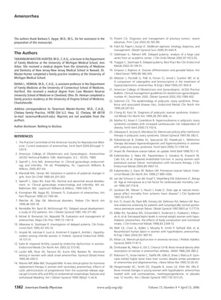
TABLE 5
Laboratory Evaluation of Hyperandrogenism
Findings Indications
Serum testosterone (normal: 20 to 80 ng per dL [0.7 to 2.8 nmol per L])
200 ng per dL (6.9 nmol per L) Consider hyperandrogenic chronic anovulation*
>200 ng per dL Evaluate for androgen-secreting tumor
Serum dehydroepiandrosterone sulfate (normal: 250 to 300 ng per dL [0.7 to 0.8 μmol per L])
700 ng per dL (1.9 μmol per L) Consider hyperandrogenic chronic anovulation*
>700 ng per dL Evaluate for adrenal or ovarian tumor
Serum 17-hydroxyprogesterone (normal: <2 ng per mL (6.1 nmol per L])†
>4 ng per mL (12.1 nmol per L) Consider adrenocorticotropic stimulation test to diagnose
congenital adrenal hyperplasia
Dexamethasone suppression test (if clinically indicated)††
Morning cortisol level > 5 μg per dL (138 nmol per L)§ Evaluate for Cushing’s disease
*— These values are not specific for diagnosis of hyperandrogenic chronic anovulation.
†—Morning level during follicular phase of menstrual cycle.
††—For an overnight dexamethasone suppression test, the physician should administer a 1-mg dose of dexamethasone orally between 11 p.m. and
midnight and draw a single blood sample for serum cortisol testing at 8 a.m. the following day.
§—Morning cortisol level in a healthy patient with an intact hypothalamic-pituitary axis. There is some variability in the cutoff values that can affect
sensitivity and specificity of the test. Patients should receive further testing to confirm Cushing’s disease.
Information from references 6 and 21.](https://image.slidesharecdn.com/amenorrhea-150622020458-lva1-app6891/85/Amenorrhea-8-320.jpg)

A thorough history and physical examination as well as laboratory testing can help narrow the differential diagnosis of amenorrhea. In patients with primary amenorrhea, the presence or absence of sexual development should direct the evaluation. Constitutional delay of growth and puberty commonly causes primary amenorrhea in patients with no sexual development. If the patient has normal pubertal development and a uterus, the most common etiology is congenital outflow tract obstruction with a transverse vaginal septum or imperforate hymen. If laboratory tests are normal, challenge tests can help determine if the cause is an outflow tract abnormality or inadequate estrogenization. The treatment of primary and secondary amenorrhea is based on the causative factor and aims to