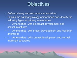
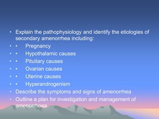
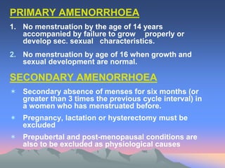
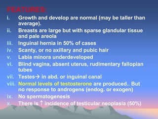
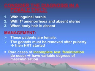
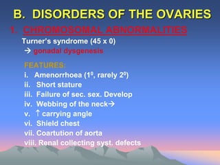
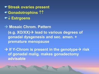
AMENORRHOEA
Primary & Secondary
King Khalid University Hospital
Department of Obstetrics &
Gynecology
Course 482
Objectives
• Define primary and secondary amenorrhea
• Explain the pathophysiology amnorrhoea and identify the
following types of primary amenorroea.
• • Amenorrhea with no breast development and
sexual infantilism
• • Amenorrhea with breast Development and mullerian
anomalies
• • Amenorrhea With breast development and normal
mullerian structures
• Explain the pathophysiology and identify the etiologies of
secondary amenorrhea including:
• • Pregnancy
• • Hypothalamic causes
• • Pituitary causes
• • Ovarian causes
• • Uterine causes
• • Hyperandrogenism
• Describe the symptoms and signs of amenorrhea
• Outline a plan for investigation and management of
amenorrhoea
PRIMARY AMENORRHOEA
1. No menstruation by the age of 14 years
accompanied by failure to grow properly or
develop sec. sexual characteristics.
2. No menstruation by age of 16 when growth and
sexual development are normal.
SECONDARY AMENORRHOEA
 Secondary absence of menses for six months (or
greater than 3 times the previous cycle interval) in
a women who has menstruated before.
 Pregnancy, lactation or hysterectomy must be
excluded
 Prepubertal and post-menopausal conditions are
also to be excluded as physiological causes
CLINICAL APPROACH
There is a difference of opinion about the age
at which Primary Amenorrhoea should be
investigated  18 yrs. often suggested.
Provided the patient has developed normal
sec. sex. Characteristics and
cryptomenorrhoea has been excluded.
While those patient with Primary amenorrhoea
and sexual infantilism should be investigated
at  age of 15 years or 16 years (may be
earlier).
• Accurate, adequate history is essential to
reach a firm diagnosis
• Specific questioning is necessary to
establish diagnosis of Primary or Secondary
amenorrhoea
• Is the amenorrhoea is truly secondary (e.g.
prev. menses were actucally steroid –
induced)
• Careful physical examination aids in
reaching a fairly firm provisional diagnosis
• In minority, there is a need to go beyond
simple out-patient investigation.
CAUSES OF AMENORRHOEA
A. Disorder of outflow tract and or uterus
B. Disorders of ovary
C. Disorders of Ant. Pituitary
D. Disorders of Hypothalamus
A. DISORDERS OF OUTFLOW TRACT &
OR UTERUS
1. CRYPTOMENORRHOEA
Vaginal atresia or imperforate hymen prevent menstrual loss
from escaping.
FEATURES:
 Prim. Amenorrhoea in a teenage girl with normal
sexual development present
Complaining of:
i. Intermittent lower abd. pain
ii. Possible difficulty of mict.
iii. Palpable lower abd. swelling (Haematometra)
iv. Bulging, bluish membrane at lower end of vagina
(Haematocolpus).
MANAGEMENT: INCISE MEMBRANE
2. ABSENCE OR HYPOPLASIA OF
VAGINA:
FEATURES:
 Growth, develop, and ovarian function are
usually normal.
 Uterus may be normal or rudimentary
 Renal anomalies (in 30%) or skeletal
defects (in 10%) may be present.
MANAGEMENT:
Create a functional vagina by surgery or dilators
3. TESTICULAR FEMINIZATION:
(Androgen Insensitivity)
 Phenotype is woman. Genotype is
man (xy)  testes are present.
 Inherited by an X-linked recessive
gene… (familial)
 Resulting in absence of cytosol
androgen receptor
FEATURES:
i. Growth and develop are normal (may be taller than
average).
ii. Breasts are large but with sparse glandular tissue
and pale areola
iii. Inguinal hernia in 50% of cases
iv. Scanty, or no axillary and pubic hair
v. Labia minora underdeveloped
vi. Blind vagina, absent uterus, rudimentary fallopian
tubes
vii. Testes in abd. or inguinal canal
viii. Normal levels of testosterone are produced.. But
no response to androgens (endog. or exogen)
ix. No spermatogenesis
x. There is  incidence of testicular neoplasia (50%)
CONSIDER THE DIAGNOSIS IN A
FEMALE CHILD:
1. With inguinal hernia
2. With 10 amenorrhoea and absent uterus
3. When body hair is absent
MANAGEMENT:
 These patients are female.
 The gonads must be removed after puberty
 then HRT started
 Rare cases of incomplete test. feminization
do occur  have variable degress of
masculinization
4. ASHERMAN’S SYNDROME:
Sec. amenorrhoea following distruction of
the endomet. by overzealous curettage
multip. Synechiae show up on
“Hysterography”.
MANAGEMENT:
Under G.A. breakdown intraut. Adhesions
through hysteroscopeinsert an IUCD to
deter reformation hormone therapy (E2 + P)
5. INFECTION
e.g. Tuberculosis. Ut. Schistosomiasis
B. DISORDERS OF THE OVARIES
1. CHROMOSOMAL ABNORMALITIES
Turner’s syndrome (45 x 0)
 gonadal dysgenesis
FEATURES:
i. Amenorrhoea (10, rarely 20)
ii. Short stature
iii. Failure of sec. sex. Develop
iv. Webbing of the neck
v.  carrying angle
vi. Shield chest
vii. Coartution of aorta
viii. Renal collecting syst. defects
Streak ovaries present
Gonadotrophins 
 Estrgoens
 Mosaic Chrom. Pattern
(e.g. XO/XX) lead to various degrees of
gonadal dysgenesis and sec. amen. +
premature menopause
 If Y-Chrom is present in the genotype risk
of gonadal malig. makes gonadectomy
advisable
2. GONADAL AGENESIS:
(Failure of gonadal develop): no
other cong. abn.
3. RESISTANT OVARY SYNDROME
 A rare condition
 Normal ovarian develop and
potential
 FSH 
 It may resolve spontaneously
 If hot flushes  Rx. With estrogen
4. PREMATURE MENOPAUSE:
Ovarian failure….due to
i. Auto-immune dis. (associated with Addison’s
dis. ??)
ii. Viral infection (e.g. mumps)
iii. Cytotoxic drugs
5. PCOs:
 Mostly present with classical Stein-Leventhal
syndrome (of oligomenorrhoea, obesity,
hirsuitism, and infertility)
 However a substantial group will have sec.
amenorrhoea with no obesity or hirsuitism
 Diagnosis is made by finding  LH/FSH ratio
 Confirmation is made by laparoscopy.
 USS +
C. DISORDERS OF PITUITARY
1. Pituitary Tumor causing
“Hyperprolactinemia”
 40% of women with hyperprolactinemia
will have a pituitary adenoma
Pit. Fossa XR is necessary in all cases of
amenorrhoea – particular 20.
FEATURES: In coned view:
 Erosion of clinoid process
 Enlarge of pituitary fossa
 Double flooring of fossa
 If any of above features seen
 CT san or MRI + Assessment of visual fields
MANAGEMENT:
 Bromocriptine (Dopamine agonist)
 Suppres prolactin sec.
 Correct estrogen deficiency
 Permits ovulation
  Size of most prolactinomas
 Surgical removal of tumor
 if extracellar manifestation (e.g.
press. on optic chiasma) or if patient
cannot tolerate or respond to
medical Rx.
2. OTHER CAUSE OF  PROLACT.
♣ Drugs: e.g. phenothiazines, methyl-dopa,
metclopramide, anti-histamines,
oestrogens and morphine.
3. CRANIOPHARYNGIOMA
♣ Other intracranial tumor
4. SHEEHAN’S SYNDROME
♣ Necrosis of ant. pituitary due to severe
PPH
 Pan – or partial hypopituitarism
♣ It is rare problem today due to better
obstetric care and adequate blood
transfusion
D. DISORDERS OF HYPOTHALAMUS
♣ Commonest reason for
hypogonadotrophic sec. amenorrhoea
♣ Often associated with stress e.g. in
migrants, young women when leave
home, university students
♣ Diagnosis by exclusion of pituitary
lesions.
♣ Hormone therapy or ovulation
induction is not indicated unless
patient wishes to become pregnant
1. WEIGHT – LOSS ASSOCIATED
AMENORRHOEA
A loss of > 10 kg is frequently associated with
amenorrhoea
i. In young women and teen ages girls
become obsessed with their body image and
starve themselves.
ii. Jogger’s amenorrhoea:
 This is seen frequently in women training
for marathon racing, in ballet dancers and
other form of athletes.
CAUSES:
 + redistribution between proportion of
body fat mass and body muscle mass.
 May be also mediated by exercise related
changes in -endorphins
iii. ANOREXIA NERVOSA
Associated with sec. amenorrhoea
(misnomer no loss of appetite)
2. AMENORRHOEA AND ANOSMIA:
rare cause of amenorrhoea of hypogonadotrophic –
hypo-gonadism.
(Counterpart in males is Kallman’s syndrome)
POST-PILL AMENORRHOEA:
♣ There is no evidence that Est. prog. Contraceptive pills
predispose to amenorrhoea.. once pill taking is
ceased.
♣ An irregular men. cycle frequently precedes pill taking
♣ If this assumption of amenorrhoea being merely an
after-effect of pill taking  many cases of
hyperprolactinemia will be missed (1:5)
♣ And Premat. ovarian failure will be missed in 1:10
cases
♣ Once other causes are excluded, this type of ameno.
Responds well to ovulation induction with Clomiphene
citrate if preg. is desired.
INVESTIGATION OF AMENORRHOEA
1. S. Prolactin level and TFT
2. Karyotyping…if chrom. anomaly is
suspected on clinical grounds
3. Progesterone withdrawal test….to
check endog. estrogen.
e.g. Provera (medroxy-prog)  if
bleeding PV=reactive endom. and
patent outflow tract.
• If PRL is norm. + no galactorrhoea ---no need for further
investigation for pituitary tumor
• If GALACTOR is present further evaluation of pit. gland is
necessary .. regardless of level of PRL and menstrual pattern
• If PRL is signific. elevated (excluding stress)  Radiology
exam of pituitary to exclude tum.
• Visual fields assessment – if X-Ray abnormal
• FSH & LH level… especially if no withdrawal bleeding following
prog. Challenge.
•  LH (<5 IU/ml) hypogonafotrophic- hypogonadism
•  FSH (>40 IU/ml) on successive readings  ovarian failure
If women < 35 years = premet. ovar. failure (menopause)
check karyotype. (if Y-Chrom +  high risk of gonadal
malignancy
4. USS:
Of uterus and ovaries  can be useful to investigating and
monitor Rx. Of these women
FLOW CHAR FOR INVESTIGATING OF SEC. AMENORRHOEA
Complete History
Full Ph. exam.,tubal patency +
sperm count
Amen.
Traumatica
Proof by FSH, LH, TSH, P RL, X-RAY
Hysterogram of Pit. Fossa
Hyperprolac. Abn.Fossa
FSH, Low or
LH N LH, FSH
Tomograms
TSH 
? Premat Clomid Thyroxin Tumor
Menopause
Biopsy Failed No Tum MPS
?”Resist. Response
Ovary
Srug. & OR
ext. radioth.
HPG Repeat
or MPS
HMG
Bromocriptine

Amenorrhoea Causes and management .ppt

  • 1.
    AMENORRHOEA Primary & Secondary KingKhalid University Hospital Department of Obstetrics & Gynecology Course 482
  • 2.
    Objectives • Define primaryand secondary amenorrhea • Explain the pathophysiology amnorrhoea and identify the following types of primary amenorroea. • • Amenorrhea with no breast development and sexual infantilism • • Amenorrhea with breast Development and mullerian anomalies • • Amenorrhea With breast development and normal mullerian structures
  • 3.
    • Explain thepathophysiology and identify the etiologies of secondary amenorrhea including: • • Pregnancy • • Hypothalamic causes • • Pituitary causes • • Ovarian causes • • Uterine causes • • Hyperandrogenism • Describe the symptoms and signs of amenorrhea • Outline a plan for investigation and management of amenorrhoea
  • 4.
    PRIMARY AMENORRHOEA 1. Nomenstruation by the age of 14 years accompanied by failure to grow properly or develop sec. sexual characteristics. 2. No menstruation by age of 16 when growth and sexual development are normal. SECONDARY AMENORRHOEA  Secondary absence of menses for six months (or greater than 3 times the previous cycle interval) in a women who has menstruated before.  Pregnancy, lactation or hysterectomy must be excluded  Prepubertal and post-menopausal conditions are also to be excluded as physiological causes
  • 5.
    CLINICAL APPROACH There isa difference of opinion about the age at which Primary Amenorrhoea should be investigated  18 yrs. often suggested. Provided the patient has developed normal sec. sex. Characteristics and cryptomenorrhoea has been excluded. While those patient with Primary amenorrhoea and sexual infantilism should be investigated at  age of 15 years or 16 years (may be earlier).
  • 6.
    • Accurate, adequatehistory is essential to reach a firm diagnosis • Specific questioning is necessary to establish diagnosis of Primary or Secondary amenorrhoea • Is the amenorrhoea is truly secondary (e.g. prev. menses were actucally steroid – induced) • Careful physical examination aids in reaching a fairly firm provisional diagnosis • In minority, there is a need to go beyond simple out-patient investigation.
  • 7.
    CAUSES OF AMENORRHOEA A.Disorder of outflow tract and or uterus B. Disorders of ovary C. Disorders of Ant. Pituitary D. Disorders of Hypothalamus
  • 8.
    A. DISORDERS OFOUTFLOW TRACT & OR UTERUS 1. CRYPTOMENORRHOEA Vaginal atresia or imperforate hymen prevent menstrual loss from escaping. FEATURES:  Prim. Amenorrhoea in a teenage girl with normal sexual development present Complaining of: i. Intermittent lower abd. pain ii. Possible difficulty of mict. iii. Palpable lower abd. swelling (Haematometra) iv. Bulging, bluish membrane at lower end of vagina (Haematocolpus). MANAGEMENT: INCISE MEMBRANE
  • 9.
    2. ABSENCE ORHYPOPLASIA OF VAGINA: FEATURES:  Growth, develop, and ovarian function are usually normal.  Uterus may be normal or rudimentary  Renal anomalies (in 30%) or skeletal defects (in 10%) may be present. MANAGEMENT: Create a functional vagina by surgery or dilators
  • 10.
    3. TESTICULAR FEMINIZATION: (AndrogenInsensitivity)  Phenotype is woman. Genotype is man (xy)  testes are present.  Inherited by an X-linked recessive gene… (familial)  Resulting in absence of cytosol androgen receptor
  • 11.
    FEATURES: i. Growth anddevelop are normal (may be taller than average). ii. Breasts are large but with sparse glandular tissue and pale areola iii. Inguinal hernia in 50% of cases iv. Scanty, or no axillary and pubic hair v. Labia minora underdeveloped vi. Blind vagina, absent uterus, rudimentary fallopian tubes vii. Testes in abd. or inguinal canal viii. Normal levels of testosterone are produced.. But no response to androgens (endog. or exogen) ix. No spermatogenesis x. There is  incidence of testicular neoplasia (50%)
  • 12.
    CONSIDER THE DIAGNOSISIN A FEMALE CHILD: 1. With inguinal hernia 2. With 10 amenorrhoea and absent uterus 3. When body hair is absent MANAGEMENT:  These patients are female.  The gonads must be removed after puberty  then HRT started  Rare cases of incomplete test. feminization do occur  have variable degress of masculinization
  • 13.
    4. ASHERMAN’S SYNDROME: Sec.amenorrhoea following distruction of the endomet. by overzealous curettage multip. Synechiae show up on “Hysterography”. MANAGEMENT: Under G.A. breakdown intraut. Adhesions through hysteroscopeinsert an IUCD to deter reformation hormone therapy (E2 + P) 5. INFECTION e.g. Tuberculosis. Ut. Schistosomiasis
  • 14.
    B. DISORDERS OFTHE OVARIES 1. CHROMOSOMAL ABNORMALITIES Turner’s syndrome (45 x 0)  gonadal dysgenesis FEATURES: i. Amenorrhoea (10, rarely 20) ii. Short stature iii. Failure of sec. sex. Develop iv. Webbing of the neck v.  carrying angle vi. Shield chest vii. Coartution of aorta viii. Renal collecting syst. defects
  • 16.
    Streak ovaries present Gonadotrophins  Estrgoens  Mosaic Chrom. Pattern (e.g. XO/XX) lead to various degrees of gonadal dysgenesis and sec. amen. + premature menopause  If Y-Chrom is present in the genotype risk of gonadal malig. makes gonadectomy advisable
  • 17.
    2. GONADAL AGENESIS: (Failureof gonadal develop): no other cong. abn. 3. RESISTANT OVARY SYNDROME  A rare condition  Normal ovarian develop and potential  FSH   It may resolve spontaneously  If hot flushes  Rx. With estrogen
  • 18.
    4. PREMATURE MENOPAUSE: Ovarianfailure….due to i. Auto-immune dis. (associated with Addison’s dis. ??) ii. Viral infection (e.g. mumps) iii. Cytotoxic drugs 5. PCOs:  Mostly present with classical Stein-Leventhal syndrome (of oligomenorrhoea, obesity, hirsuitism, and infertility)  However a substantial group will have sec. amenorrhoea with no obesity or hirsuitism  Diagnosis is made by finding  LH/FSH ratio  Confirmation is made by laparoscopy.  USS +
  • 19.
    C. DISORDERS OFPITUITARY 1. Pituitary Tumor causing “Hyperprolactinemia”  40% of women with hyperprolactinemia will have a pituitary adenoma Pit. Fossa XR is necessary in all cases of amenorrhoea – particular 20. FEATURES: In coned view:  Erosion of clinoid process  Enlarge of pituitary fossa  Double flooring of fossa  If any of above features seen  CT san or MRI + Assessment of visual fields
  • 20.
    MANAGEMENT:  Bromocriptine (Dopamineagonist)  Suppres prolactin sec.  Correct estrogen deficiency  Permits ovulation   Size of most prolactinomas  Surgical removal of tumor  if extracellar manifestation (e.g. press. on optic chiasma) or if patient cannot tolerate or respond to medical Rx.
  • 21.
    2. OTHER CAUSEOF  PROLACT. ♣ Drugs: e.g. phenothiazines, methyl-dopa, metclopramide, anti-histamines, oestrogens and morphine. 3. CRANIOPHARYNGIOMA ♣ Other intracranial tumor 4. SHEEHAN’S SYNDROME ♣ Necrosis of ant. pituitary due to severe PPH  Pan – or partial hypopituitarism ♣ It is rare problem today due to better obstetric care and adequate blood transfusion
  • 22.
    D. DISORDERS OFHYPOTHALAMUS ♣ Commonest reason for hypogonadotrophic sec. amenorrhoea ♣ Often associated with stress e.g. in migrants, young women when leave home, university students ♣ Diagnosis by exclusion of pituitary lesions. ♣ Hormone therapy or ovulation induction is not indicated unless patient wishes to become pregnant
  • 23.
    1. WEIGHT –LOSS ASSOCIATED AMENORRHOEA A loss of > 10 kg is frequently associated with amenorrhoea i. In young women and teen ages girls become obsessed with their body image and starve themselves. ii. Jogger’s amenorrhoea:  This is seen frequently in women training for marathon racing, in ballet dancers and other form of athletes. CAUSES:  + redistribution between proportion of body fat mass and body muscle mass.  May be also mediated by exercise related changes in -endorphins iii. ANOREXIA NERVOSA Associated with sec. amenorrhoea (misnomer no loss of appetite)
  • 24.
    2. AMENORRHOEA ANDANOSMIA: rare cause of amenorrhoea of hypogonadotrophic – hypo-gonadism. (Counterpart in males is Kallman’s syndrome) POST-PILL AMENORRHOEA: ♣ There is no evidence that Est. prog. Contraceptive pills predispose to amenorrhoea.. once pill taking is ceased. ♣ An irregular men. cycle frequently precedes pill taking ♣ If this assumption of amenorrhoea being merely an after-effect of pill taking  many cases of hyperprolactinemia will be missed (1:5) ♣ And Premat. ovarian failure will be missed in 1:10 cases ♣ Once other causes are excluded, this type of ameno. Responds well to ovulation induction with Clomiphene citrate if preg. is desired.
  • 25.
    INVESTIGATION OF AMENORRHOEA 1.S. Prolactin level and TFT 2. Karyotyping…if chrom. anomaly is suspected on clinical grounds 3. Progesterone withdrawal test….to check endog. estrogen. e.g. Provera (medroxy-prog)  if bleeding PV=reactive endom. and patent outflow tract.
  • 26.
    • If PRLis norm. + no galactorrhoea ---no need for further investigation for pituitary tumor • If GALACTOR is present further evaluation of pit. gland is necessary .. regardless of level of PRL and menstrual pattern • If PRL is signific. elevated (excluding stress)  Radiology exam of pituitary to exclude tum. • Visual fields assessment – if X-Ray abnormal • FSH & LH level… especially if no withdrawal bleeding following prog. Challenge. •  LH (<5 IU/ml) hypogonafotrophic- hypogonadism •  FSH (>40 IU/ml) on successive readings  ovarian failure If women < 35 years = premet. ovar. failure (menopause) check karyotype. (if Y-Chrom +  high risk of gonadal malignancy 4. USS: Of uterus and ovaries  can be useful to investigating and monitor Rx. Of these women
  • 27.
    FLOW CHAR FORINVESTIGATING OF SEC. AMENORRHOEA Complete History Full Ph. exam.,tubal patency + sperm count Amen. Traumatica Proof by FSH, LH, TSH, P RL, X-RAY Hysterogram of Pit. Fossa Hyperprolac. Abn.Fossa FSH, Low or LH N LH, FSH Tomograms TSH  ? Premat Clomid Thyroxin Tumor Menopause Biopsy Failed No Tum MPS ?”Resist. Response Ovary Srug. & OR ext. radioth. HPG Repeat or MPS HMG Bromocriptine