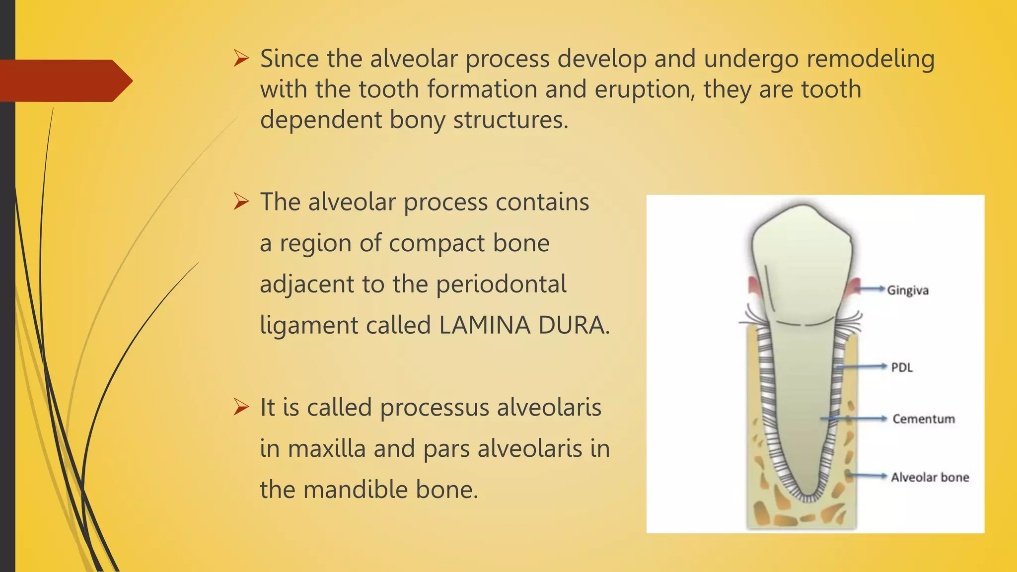
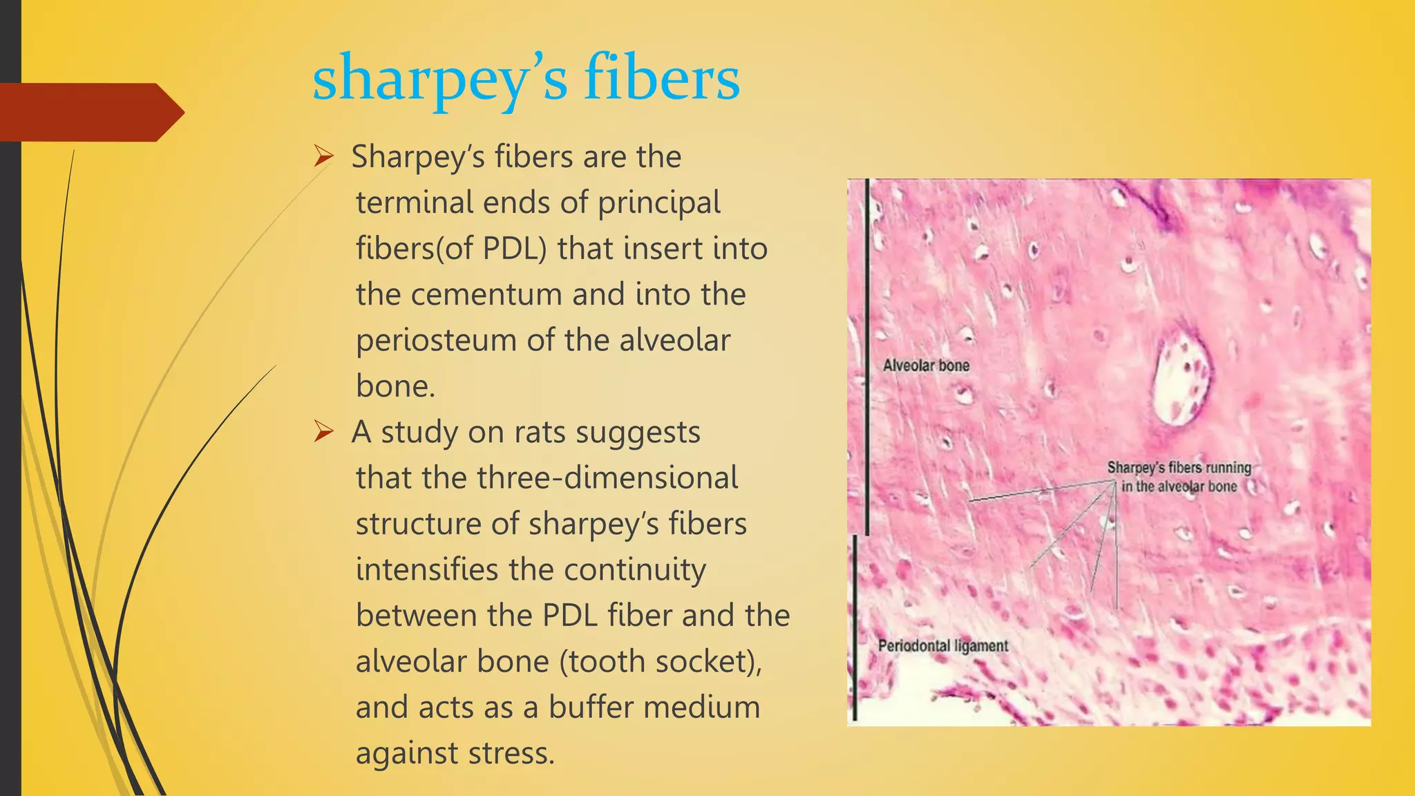
ALVEOLAR
BONE
DEPT. OF PERIODONTOLOGY
KULBHUSHAN GARG
ROLL NO. 30
BATCH “d”
MAY JUNE
CONTENT
 ALVEOLAR BONE AND ALVEOLAR PROCESS
 FUNCTIONS OF ALVEOLAR BONE
 STRUCTURAL ELEMENTS OF ALVEOLAR BONE
-BONE CELLS
• OSTEOPROGENITOR CELLS – STEM CELLS
• OSTEOBLASTS – FORMS BONE
• OSTEOCYTES – MAINTAINS BONE
• OSTEOCLASTS – RESORBS BONE
 SHARPEY’S FIBERS
 VASCULAR SUPPLY AND LYMPHATIC VESSELS
 GROSS MORPHOLOGY OF ALVEOLAR BONE
 STRUCTURES OF ALVEOLAR BONE
ALVEOLAR BONE
 Alveolar bone or alveolar process is that portion of maxilla
and mandible that supports the roots of the tooth.
 If the teeth is lost the alveolar process disappears.
 It is composed of 2 parts the alveolar bone proper and the
supporting bone.
 Since the alveolar process develop and undergo remodeling
with the tooth formation and eruption, they are tooth
dependent bony structures.
 The alveolar process contains
a region of compact bone
adjacent to the periodontal
ligament called LAMINA DURA.
 It is called processus alveolaris
in maxilla and pars alveolaris in
the mandible bone.
Functions of alveolar process
 It supports the tooth roots on the facial and on the
palatal/lingual sides.
 It is the one responsible for the separation of teeth from
mesial and distal.
 Also contributes to absorption and distribution of occlusal
pressure produced in tooth to tooth contact.
Structural elements of bone
 Bone cells
• Osteoprogenitor cells – stem cells
• Osteoblasts – forms bone
• Osteocytes – maintains bone
• Osteoclasts – resorbs bone
 Bone matrix
 Sharpey’s fibres
 Vascular supply and lymphatic drainage
Osteoprogenitor cells
 Stem cells derived from the mesenchyme.
 Possess mitotic potential and the ability to differentiate into
mature bone cells.
 Resemble mesenchymal cells and are spindle – shaped, with
pale – staining elongated nuclei and sparse cytoplasm(small
amount of ER and poorly developed golgi complex).
 Mostly found in the inner portion of the periosteum, in the
endosteum, and within vascular canals of compact bone.
osteoblasts
 Uninucleated cells.
 Synthesize both collagenous and non collagenous bone
proteins.
 Osteoblasts also synthesize the enzyme alkaline phosphatase,
which is needed locally for the mineralization of osteoid.
 The precursor cell of the osteoblast is the preosteoblast.
 Osteoblasts have all the characteristics of hard tissue -forming
cells.
 When the bone is no longer forming, the surfaces of the
osteoblasts become inactive and are called lining cells.
osteocytes
 These are osteoblasts secreted in the bone matrixes that are
entrapped in lacunae.
 An osteocyte lies in its own lacuna and contacts its
neighboring osteocytes cytoplasmically through canaliculi.
 The most important function of osteoblast – osteocyte
complex is to prevent hypomineralization of bone by
continually pumping calcium back to the bloodstream.
osteoclasts
 Derived from a monocytic – macrophage system, which are
responsible for bone resorbtion.
 They are multinucleated cells with fine, fingerlike cytoplasmic
processes and are rich in lysosomes that contain tartrate –
resistant acid phosphatase(TRAP).
 Osteoclasts lie in the resorbtion craters known as howship’s
lacunae on the bone surface or in deep resorption cavities
called cutting cones.
 These bone cells can only resorb mineralized bone matrix.
Bone matrix
 Bone matrix consists of organic and inorganic components.
 The association of these substances gives bone its hardness
and resistance.
 The organic component is composed of collagen fibers with
predominately type 1 collagen(95%) and amorphous material,
including glycosaminoglycans that are associated with
proteins.
 Osteoid is uncalcified organic matrix.
 Inorganic matter represents about 50% of the dry weight of
bone matrix.
 Composed of abundant calcium and phosphorous as well as
smaller amounts of bicarbonate, citrate, magnesium,
potassium, and sodium.
 Calcium forms hydroxyapatite crystals with phosphorous but
is also present in an amorphous form.
 Concentrically arranged bony lamellae
 Types of layering (lamella)
• Circumferential lamella – external, internal
• Concentric/ haversian lamella
• Interstitial or intermediate lamella
sharpey’s fibers
 Sharpey’s fibers are the
terminal ends of principal
fibers(of PDL) that insert into
the cementum and into the
periosteum of the alveolar
bone.
 A study on rats suggests
that the three-dimensional
structure of sharpey’s fibers
intensifies the continuity
between the PDL fiber and the
alveolar bone (tooth socket),
and acts as a buffer medium
against stress.
Vascular supply of alveolar process
 Alveolar process of the maxilla
• Anterior and posterior alveolar arteries (branch from the maxilla and infraorbital
arteries).
 Alveolar process of the mandible
• Inferior alveolar arteries (internal).
• Periosteal branches of submental and buccal arteries (external).
Lymphatic drainage
 Smallest lymph vessel – lymph capillaries.
 All third molars – jugulodigastric lymph nodes.
 Mandibular incisors – submental lymph nodes.
 Rest – submandibular lymph nodes.
Gross morphology
Alveolar socket
 Also called dental
alveolus.
 Are sockets in the jaws in
which the roots of teeth
are held in the alveolar
process with the
periodontal ligament.
 Interdental septa
• “septa” – in latin, it means
“fence” or “wall”.
• Are plates of bone that separate
each individual sockets from one
another.
 Inter – radicular septa
• Are thin plates of bone that separate the roots of multi – rooted teeth.
Cribriform plate
 Also called as bundle bone.
 A type of alveolar bone so called because of the ‘bundle’ pattern
caused by continuation of principal (sharpey’s) fibers into it.
 Is the compact layer of bone lining the tooth socket (alveolar
socket).
 Reflects the sieve – like appearance produced by numerous
Volkmann’s canals passing from the alveolar bone to the PDL.
 Numerous sharpey’s fiber pass through it.
Structures of alveolar bone
cortical plate
 Outer bony plate of varying thickness,
which is the outside wall of the maxilla
and mandible, covered with periosteum.
 Continuous with the lamina cribriformis at
the orifice of the alveoli – alveolar crest.
 Consists of haversian systems (osteons) and
interstitial lamellae.
 Thicker in the mandible than maxilla.
 Generally greater on the lingual than on the
buccal/facial aspect.
spongiosa
 Are spongy (or cancellous/trabecullar) bone between the 2
bony plates and between the lamina cribriformis of adjascent
teeth or roots.
 Consists of delicate trabeculae, between which are narrow
spaces, filled mostly with fatty marrow.
 Regions of maxillary tuberosity and the angle of mandible.
Periosteum and endosteum
 Layers of differentiated osteogenic connective tissue covers all
bone surfaces.
 Tissue covering outer surface of bone is termed as
periosteum.
 Tissue lining the internal bone cavities is called endosteum.
THANK YOU

alveolar bone ppt.pptx

  • 1.
    ALVEOLAR BONE DEPT. OF PERIODONTOLOGY KULBHUSHANGARG ROLL NO. 30 BATCH “d” MAY JUNE
  • 2.
    CONTENT  ALVEOLAR BONEAND ALVEOLAR PROCESS  FUNCTIONS OF ALVEOLAR BONE  STRUCTURAL ELEMENTS OF ALVEOLAR BONE -BONE CELLS • OSTEOPROGENITOR CELLS – STEM CELLS • OSTEOBLASTS – FORMS BONE • OSTEOCYTES – MAINTAINS BONE • OSTEOCLASTS – RESORBS BONE  SHARPEY’S FIBERS  VASCULAR SUPPLY AND LYMPHATIC VESSELS  GROSS MORPHOLOGY OF ALVEOLAR BONE  STRUCTURES OF ALVEOLAR BONE
  • 3.
    ALVEOLAR BONE  Alveolarbone or alveolar process is that portion of maxilla and mandible that supports the roots of the tooth.  If the teeth is lost the alveolar process disappears.  It is composed of 2 parts the alveolar bone proper and the supporting bone.
  • 4.
     Since thealveolar process develop and undergo remodeling with the tooth formation and eruption, they are tooth dependent bony structures.  The alveolar process contains a region of compact bone adjacent to the periodontal ligament called LAMINA DURA.  It is called processus alveolaris in maxilla and pars alveolaris in the mandible bone.
  • 5.
    Functions of alveolarprocess  It supports the tooth roots on the facial and on the palatal/lingual sides.  It is the one responsible for the separation of teeth from mesial and distal.  Also contributes to absorption and distribution of occlusal pressure produced in tooth to tooth contact.
  • 6.
    Structural elements ofbone  Bone cells • Osteoprogenitor cells – stem cells • Osteoblasts – forms bone • Osteocytes – maintains bone • Osteoclasts – resorbs bone  Bone matrix  Sharpey’s fibres  Vascular supply and lymphatic drainage
  • 7.
    Osteoprogenitor cells  Stemcells derived from the mesenchyme.  Possess mitotic potential and the ability to differentiate into mature bone cells.  Resemble mesenchymal cells and are spindle – shaped, with pale – staining elongated nuclei and sparse cytoplasm(small amount of ER and poorly developed golgi complex).  Mostly found in the inner portion of the periosteum, in the endosteum, and within vascular canals of compact bone.
  • 8.
    osteoblasts  Uninucleated cells. Synthesize both collagenous and non collagenous bone proteins.  Osteoblasts also synthesize the enzyme alkaline phosphatase, which is needed locally for the mineralization of osteoid.  The precursor cell of the osteoblast is the preosteoblast.  Osteoblasts have all the characteristics of hard tissue -forming cells.  When the bone is no longer forming, the surfaces of the osteoblasts become inactive and are called lining cells.
  • 9.
    osteocytes  These areosteoblasts secreted in the bone matrixes that are entrapped in lacunae.  An osteocyte lies in its own lacuna and contacts its neighboring osteocytes cytoplasmically through canaliculi.  The most important function of osteoblast – osteocyte complex is to prevent hypomineralization of bone by continually pumping calcium back to the bloodstream.
  • 10.
    osteoclasts  Derived froma monocytic – macrophage system, which are responsible for bone resorbtion.  They are multinucleated cells with fine, fingerlike cytoplasmic processes and are rich in lysosomes that contain tartrate – resistant acid phosphatase(TRAP).  Osteoclasts lie in the resorbtion craters known as howship’s lacunae on the bone surface or in deep resorption cavities called cutting cones.  These bone cells can only resorb mineralized bone matrix.
  • 11.
    Bone matrix  Bonematrix consists of organic and inorganic components.  The association of these substances gives bone its hardness and resistance.  The organic component is composed of collagen fibers with predominately type 1 collagen(95%) and amorphous material, including glycosaminoglycans that are associated with proteins.
  • 12.
     Osteoid isuncalcified organic matrix.  Inorganic matter represents about 50% of the dry weight of bone matrix.  Composed of abundant calcium and phosphorous as well as smaller amounts of bicarbonate, citrate, magnesium, potassium, and sodium.  Calcium forms hydroxyapatite crystals with phosphorous but is also present in an amorphous form.
  • 13.
     Concentrically arrangedbony lamellae  Types of layering (lamella) • Circumferential lamella – external, internal • Concentric/ haversian lamella • Interstitial or intermediate lamella
  • 15.
    sharpey’s fibers  Sharpey’sfibers are the terminal ends of principal fibers(of PDL) that insert into the cementum and into the periosteum of the alveolar bone.  A study on rats suggests that the three-dimensional structure of sharpey’s fibers intensifies the continuity between the PDL fiber and the alveolar bone (tooth socket), and acts as a buffer medium against stress.
  • 16.
    Vascular supply ofalveolar process  Alveolar process of the maxilla • Anterior and posterior alveolar arteries (branch from the maxilla and infraorbital arteries).  Alveolar process of the mandible • Inferior alveolar arteries (internal). • Periosteal branches of submental and buccal arteries (external).
  • 17.
    Lymphatic drainage  Smallestlymph vessel – lymph capillaries.  All third molars – jugulodigastric lymph nodes.  Mandibular incisors – submental lymph nodes.  Rest – submandibular lymph nodes.
  • 18.
    Gross morphology Alveolar socket Also called dental alveolus.  Are sockets in the jaws in which the roots of teeth are held in the alveolar process with the periodontal ligament.
  • 19.
     Interdental septa •“septa” – in latin, it means “fence” or “wall”. • Are plates of bone that separate each individual sockets from one another.  Inter – radicular septa • Are thin plates of bone that separate the roots of multi – rooted teeth.
  • 20.
    Cribriform plate  Alsocalled as bundle bone.  A type of alveolar bone so called because of the ‘bundle’ pattern caused by continuation of principal (sharpey’s) fibers into it.  Is the compact layer of bone lining the tooth socket (alveolar socket).  Reflects the sieve – like appearance produced by numerous Volkmann’s canals passing from the alveolar bone to the PDL.  Numerous sharpey’s fiber pass through it.
  • 21.
    Structures of alveolarbone cortical plate  Outer bony plate of varying thickness, which is the outside wall of the maxilla and mandible, covered with periosteum.  Continuous with the lamina cribriformis at the orifice of the alveoli – alveolar crest.  Consists of haversian systems (osteons) and interstitial lamellae.  Thicker in the mandible than maxilla.  Generally greater on the lingual than on the buccal/facial aspect.
  • 22.
    spongiosa  Are spongy(or cancellous/trabecullar) bone between the 2 bony plates and between the lamina cribriformis of adjascent teeth or roots.  Consists of delicate trabeculae, between which are narrow spaces, filled mostly with fatty marrow.  Regions of maxillary tuberosity and the angle of mandible.
  • 23.
    Periosteum and endosteum Layers of differentiated osteogenic connective tissue covers all bone surfaces.  Tissue covering outer surface of bone is termed as periosteum.  Tissue lining the internal bone cavities is called endosteum.
  • 24.