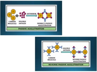
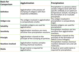
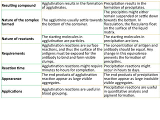
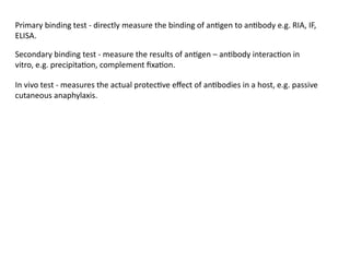
Serological techniques
Antigen and Antibodies
• An antigen is any substance that causes your immune system to produce antibodies
against it.
• Antigens are large molecules of proteins, present on the surface of the pathogen-
such as bacteria, fungi, viruses, and other foreign particles.
• When these harmful agents enter the body, it induces an immune response in the
body for the production of antibodies.
• For example: When a common cold virus enters the body, it causes the body to
produce antibodies to prevent from getting sick.
• Special part of antigen that binds with the antibody is called epitope.
• Antibody (Ab) is also known as an immunoglobulin(Ig).
• These are large, Y-shaped blood proteins produced by plasma cells.
• They bind to foreign particles and invade them.
• These particles are foreign bodies that get attacked by Antibody.
• B cells get activated immediately releasing antibodies into the bloodstream.
• Special part of antibody that binds with the antigen is called paratope.
ANTIGEN
• Immunogenicity is ability to generate immune response.
• Antigenicity is the ability to bind with specific antibody.
• Hapten is non-protein, foreign substances that require a carrier molecule to induce an
immune response.
• Hapten + carrier = Immunogenic.
• Any foreign substances that when entering our body sometimes self-elicit a series of
immune responses and are precisely called immunogens. Whereas some of them don’t
directly elicit an immune response but require the help of some other molecules (carrier
proteins) to do so and are called hapten. The immunogens and hapten are collectively
called antigens.
• Polyclonal antibodies
• Polyclonal antibodies are produced by different B cells in a host animal and recognize
multiple epitopes of a single antigen. The most common choices of antigens are protein
or synthetic peptide
Feature Monoclonal Antibodies Polyclonal Antibodies
Origin
Derived from a single clone of
B cells or hybridoma cells.
Derived from multiple clones
of B cells.
Specificity
Highly specific, recognizing a
single epitope on an antigen.
Less specific, recognizing
multiple epitopes on an
antigen.
Production Method
Produced from immortalized
hybridoma cells generated by
fusing a specific antibody-
producing B cell with a
myeloma cell.
Produced by injecting an
antigen into an animal (e.g.,
rabbit, mouse), triggering
multiple B cell clones to
produce antibodies.
Uniformity
Uniform in structure and
specificity, as they are clones
from a single parent cell.
Heterogeneous mixture with
antibodies from different B cell
clones targeting various
epitopes of the antigen.
Applications
Commonly used in targeted
therapies (e.g., cancer
treatment), diagnostics (e.g.,
pregnancy tests), and research
(e.g., studying protein
interactions).
Used in various research and
diagnostic applications,
particularly where broad
antigen recognition is
advantageous (e.g., western
blotting, ELISA assays).
Antigen-Antibody Reaction
• The antigen and the antibody are combine specifically with each other this
interaction is called antigen and antibody reaction.
• The abbreviation is Ag-Ab reaction.
• These serve as the building blocks of humoral or antibody-mediated immunity.
• These reactions serve as the foundation for the detection of both specific and non-
specific Ags, such as enzymes that cause non-specific diseases. Serological reactions
are referred to as Ag-Ab reactions when they occur in vitro.
• In Vitro (Done in artificial conditions): It includes a series of serological tests
performed in laboratories to detect antigens or antibodies in case of many diseases.
Stages of Antigen-Antibody Reaction
There are three stages to the interactions between Ag and Ab.
The first stage of the reaction entails the formation of the Ag-Ab complex.
The second stage results in visible phenomena like agglutination, precipitation, etc.
The third stage involves the destruction of Ag or neutralization of Ag
.
Properties of Antigen-Antibody Reaction
• Significantly specific reaction
• Occurs in a noticeable manner
• Non-covalent reactions (Ionic bonds, Van der Waals forces,
Hydrophobic interactions, Hydrogen bonds)
• Antibodies and antigens are not denatured
• Reversible
• Affinity: This refers to how strongly an antigen binds to a certain
antigen-binding site on an antibody.
• Avidity: It is a more general concept than affinity. It represents the
Ag-Ab complex’s total strength. It depends on:
• The antibody’s affinity
• Antibody and antigen valencies (the number of binding sites)
• How epitopes and paratopes are structurally arranged.
• Cross-Reactivity: This term describes an antibody’s capacity to
bind to similar epitopes on other antigens.
Types of Antigen-Antibody Reaction
The types of antigen-antibody reactions are as follows:
• Precipitation Reaction
• Agglutination Reaction
• Complement Fixation
• Immunofluorescence
• ELISA – Enzyme-Linked ImmunoSorbent Assay
• Precipitation Reaction:
 An insoluble precipitate of Ag-Ab complex is produced when a soluble Ag and its Ab
combine in the presence of an electrolyte (NaCl) at a specific pH and temperature.
 precipitin: Any antibody which reacts with an antigen to form a precipitate.
 At specific pH and Temperature.
 In the presence of electrolytes
 The proportion of Ag and Ab in the reaction must be equivalent for the reaction to
occur.
 On either side of the equivalence zone, precipitation is actually prevented because of
an excess of either antigen or antibody.
 The zone of antibody excess is known as the pro zone phenomenon.
 the zone of antigen excess is known as the post zone phenomenon.
 Antigen access(post zone): Too much antigen prevents efficient crosslinking/lattice
formation. antigens are larger in number than antibodies
 Antibody access(pro zone): Too much antibody prevents efficient crosslinking/lattice
formation. antigens are lesser than antibodies
 Equivalent Antigen and Antibody(zone of equivalent): Maximum amount of lattice
(Precipitate) is formed
This phenomenon in terms of
lattice formation was
explained in 1934 by Marrack.
 Antigens are soluble molecules and are larger in size in precipitation reactions.
 There are several precipitation methods applied in the clinical laboratory for the
diagnosis of disease.
 These can be performed in semi-solid media such as agar or agarose, or in non-gel
support media such as cellulose acetate.
 Precipitation or immunoprecipitation is the non-covalent interaction between
soluble (small) antigens and soluble antibodies that bind to form an insoluble
precipitate.
 It can occur both in vivo and in vitro.
Amount
of
precipitation
Increase in antigen concentration
Post zone
Pro zone
Zone of equivalent
Agglutination Reaction
 Agglutination is an antigen-antibody reaction in which a particulate antigen
combines with its antibody in the presence of electrolytes at a specified temperature
and pH resulting in the formation of visible clumping of particles.
 Antibodies that produce such reactions are called agglutinins.
 The agglutination process involving red blood cells is termed hemagglutination, and
the process with white blood cells is termed leukoagglutination.
 Agglutination has been used for the detection of antigens in bacteria which ultimately
helps in the identification of those bacteria.
 The endpoint of the test is the observation of clumps resulting from that antigen-
antibody complex formation.
 In agglutination reactions, serial dilutions of the antibody solution are made and a
constant amount of particulate antigen is added to serially diluted antibody solutions
 They are very sensitive and the result of the test can be read visually with ease.
 The condition of excess antibody, however, is called a prozone phenomenon. At a high
concentration of antibody, the number of epitopes are outnumbered by antigen-binding
sites.
Types of Agglutination Reactions
Agglutination reactions can be broadly divided into three groups:
Active/Direct agglutination
Passive agglutination
Hemagglutination
 Agglutination reactions where the antigens are found naturally on a particle are known
as direct agglutination.
 In active agglutination, direct agglutination of particulate antigen with specific antibody
occurs.
 Direct bacterial agglutination uses whole pathogens as a source of antigen.
 The binding of antibodies to surface antigens on the bacteria results in visible clumps
Active agglutination
 Slide Agglutination: This is a fast and convenient way to identify the presence of
agglutinating antibodies. Determining species of bacteria
 Tube agglutination: It is agglutination test performed in tube and standard quantitative
technique for determination of antibody titre. In this method serum is diluted in a series
of tubes and standard antigen suspensions (specific for the suspected disease) are
added to it. After incubation, antigen-antibody reaction is indicated visible clumps of
agglutination. Used for widal test, brucellosis.
 Heterphile agglutination: Detect the presence of heterophilic antibodies in the patient’s
serum.
 Passive agglutination employs carrier particles that are coated with soluble antigens.
 In this either antibody or antigen is attached to certain inert carrier thereby, particles or
cells gets agglutinated when corresponding antigen or antibody reacts.
 Latex particles, Carbon particles, Bantonite etc. are used as inert carriers.
 When the antibody instead of antigens is adsorbed on the carrier particle for detection
of antigens, it is called reverse passive agglutination.
 RBCs are used as carrier particles in hemagglutination tests. RBCs of sheep, human,
chick, etc. are commonly used in the test. When RBCs are coated with antigen to detect
antibodies in the serum, the test is called indirect hemagglutination (IHA) test.
 Hemagglutination uses erythrocytes as the biological carriers of bacterial antigens, and
purified polysaccharides or proteins for determining the presence of corresponding
antibodies in a specimen.
 When antibodies are attached to the RBCs to detect microbial antigen, it is known
as reverse passive hemagglutination (RPHA).
 Viral hemagglutination: Many viruses including influenza, mumps, and measles have the
ability to agglutinate RBCs without antigen–antibody reactions. This process is called
viral hemagglutination.
 This hemagglutination can be inhibited by antibody specifically directed against the
virus, and this phenomenon is called hemagglutination inhibition.
Basis for
Comparison
Agglutination Precipitation
Definition
Agglutination is the process of
clumping of antigens with their
respective antibodies.
Precipitation is a process where
soluble antigens bind with their
specific antibody at an optimum
temperature and pH, resulting in
the formation of an insoluble
precipitate.
Antigen size
The antigen involved in agglutination
is comparatively smaller.
The antigen involved in
precipitation is comparatively
larger.
Solubility Insoluble antigens are used for
agglutination.
Soluble antigens are used for
precipitation.
Sensitivity Agglutination reactions are more
sensitive than precipitation reactions.
Precipitation reactions are less
sensitive than agglutination
reactions.
Principle
Agglutination is based on the
principle of the clumping of particles.
Precipitation is based on the
principle of the formation of
lattices (cross-linkages).
Reactions involved
Agglutination involves complex-
forming chemical reactions.
Precipitation involves chemical
reactions between ions and salt
molecules.
Media
No gel matrix is required for
agglutination.
A liquid or semi-solid matrix is
required for precipitation.
Resulting compound
Agglutination results in the formation
of agglutinates.
Precipitation results in the
formation of precipitates.
Nature of the complex
formed
The agglutinins usually settle towards
the bottom of the container.
The precipitins might either
remain suspended or settle down
towards the bottom. In
flocculation, the flocculants float
on the surface of the liquid
matrix.
Nature of reactants
The starting molecules in
agglutination are particles.
The starting molecules in
precipitation are ions.
Requirements
Agglutination reactions are surface
reactions, and thus the surface of the
antigens must be exposed for the
antibody to bind and form visible
clumps.
The concentration of antigen and
antibody should be equal. Any
change in this equivalence
prevents the formation of
precipitins.
Reaction time
Agglutination reactions might require
minutes to hours for completion.
Precipitation reactions might
occur in hours to days.
Appearance
The end products of agglutination
reaction appear as large visible
aggregates.
The end products of precipitation
reaction appear as large insoluble
visible aggregates.
Applications
Agglutination reactions are useful in
blood grouping.
Precipitation reactions are useful
in quantitative analysis and
pigment formation.
Primary binding test - directly measure the binding of antigen to antibody e.g. RIA, IF,
ELISA.
Secondary binding test - measure the results of antigen – antibody interaction in
vitro, e.g. precipitation, complement fixation.
In vivo test - measures the actual protective effect of antibodies in a host, e.g. passive
cutaneous anaphylaxis.
ENZYME LINKED IMMUNOSORBENT ASSAY
(ELISA)
An immunological technique using an enzyme as a label to detect (assay) presence of a
target protein (antigen or antibody).
• A conjugate in ELISA is an enzyme (label) bound to an antibody which bind to target
protein
• Can be qualitative or quantitative.
• Very sensitive.
• Commonly used in medicine and scientific research.
The enzyme-linked immunosorbent assay (ELISA) is an immunological assay commonly
used to measure antibodies, antigens, proteins and glycoproteins in biological samples.
Enzyme-Linked Immunosorbent Assay (ELISA) is a modern molecular technique for the
detection of antigen-antibody interaction with the help of an enzyme.
An ELISA test uses components of the immune system (such as IgG or IgM antibodies)
and chemicals for the detection of immune responses in the body.
Following the antigen-antibody reaction, chromogenic substrate specific to the enzyme
(o- phenyldiamine dihydrochloride for peroxidase, p-nitrophenyl phosphate for alkaline
phosphatase, etc.) is added. The substrate is acted upon (usually hydrolyzed) by
an enzyme attached to the antigen-antibody complex to give color change. The color in
the reaction can be read visually or The reaction is detected by reading the optical
density (estimated colorimetrically) using a microassay plate reader i.e. ELISA reader.
Usually, a standard curve based on known concentrations of antigen or antibody is
prepared from which the unknown quantities are calculated.
The antigen or antibody is coated on a solid surface such as in a plastic tube or well of a
microtiter plate. Thus, after the antigen and antibody have combined (Antigen-antibody
complex formed) they remain firmly attached to a solid surface during subsequent
washing stages.
ELISA Requirements
Coated plates (Microtitre plates): Commonly used ones are 96 well polystyrene plates.
Coated with antigens or antibodies at the bottom of the well.
Sample diluents: To dilute the sample before application in some cases of ELISA test.
Wash Buffers: Help to wash away the unrequired contaminants and unbound antigens or
antibodies. Eg. Triphosphate buffer (ph 7.40) and detergents such as Tween-20.
Enzyme-linked Antibodies: For this, most common enzymes used are
AP(Alkaline Phosphatase)
HRP(Horseradish Peroxidase)
Substrates: Specific chromogenic substrates are used for the respective enzymes used.
Commonly used substrates are o-phenylene diamine for HRP enzyme and p-nitrophenyl
phosphate for AP enzyme.
Stop solution: It stops the enzyme and substrate reaction. It can be acids such as sulphuric
acid.
ELISA Principle
Antigens and antibodies react specifically to form the Ag-Ab complex. Antibodies can be
linked or attached to enzymes. The enzyme-linked antibodies can modify the specific
substrates used to produce a color change within the preparation. The enzyme activity is
measured with a colorimeter in a specific wavelength of light to determine the magnitude
of the infection in the patient.
1. Direct ELISA
It is the simplest and quickest of all other types of ELISA
A single enzyme-linked antibody is used which directly interacts with the antigen
present in the sample
Direct ELISA Procedure
Addition of sample(containing antigen) to the well of the microtitre plate
Antigen gets adsorbed to the surface of the well.
Washing to remove unbound antigens
The addition of an enzyme-linked antibody that with the antigens if present.
Washing to remove unbound antibodies
Addition of chromogenic substrate
Visualization of color change and result interpretation
Direct ELISA Applications
Used in the identification of biomolecules.
Also for the diagnosis of infections of Mycoplasma bovis
Indirect ELISA Procedure
Addition of known antigens specific to the antibody of interest
Careful washing to remove unadsorbed antigens or some adsorbed contaminants.
Addition of serum samples to their respective wells
If there is the presence of specific antibodies, they will bind to the antigens.
Wash again.
Addition of enzyme-linked secondary antibodies to the well
Wash again
Addition of substrate
Visualization of color change and result interpretation
Indirect ELISA Applications
Detection of HIV, Rubella, Dengue viruses, etc.
To detect certain drugs in serum.
. Indirect ELISA
Developed in 1978.
It is the most popular type of ELISA in use.
Antibody detection is carried out in indirect ELISA. A secondary antibody linked with the
enzyme is used.
3. Sandwich ELISA
Developed in 1977.
Detection of antigens.
Two different antibodies i.e. capture antibodies(for attachment sample antigens) and
enzyme-linked secondary antibodies are required.
The antigen of interest first binds with the capture antibodies and then the secondary
antibodies bind with the epitope antigen forming a sandwich-like structure with
antigen in between antibodies.
Sandwich ELISA Procedure
Addition of capture antibodies in the microtitre well
Washing
Addition of serum sample(containing antigen) in the well
Washing
Addition of enzyme-linked secondary antibodies in the well
Washing
Addition of substrate
Visualization of color development and result interpretation
Sandwich ELISA Applications
Mainly used for detection of Rotavirus and enterotoxins of fecal E. coli.
Pregnancy tests kids are based on this technique.
4. Competitive ELISA
Developed in 1976.
Detection of antibodies.
The basic concept of this type is that there occurs competition of binding between the
sample antibodies(if present) and enzyme-linked secondary antibodies to the antigens. If
the sample contains specific antibodies of interest, they will bind to the antigens and
won’t allow enzyme-linked antibodies to bind with antigens which are then washed away.
Hence after the addition of substrate, there won’t be a color change indicating a positive
test and vice-versa.
Competitive ELISA Procedure
Addition of HIV antigens in the microtitre well
Washing
Addition of serum sample
Washing
Addition of enzyme-linked HIV specific antibodies
Washing
Addition of substrate
Visualization of color change and result interpretation
Competitive ELISA Application
Mainly used for the detection of HIV
ELISA Result Interpretation
Qualitative interpretation i.e. presence or absence of antigen is done by visualizing the
color change in the solution.
If the Ag-Ab reaction takes place and bound enzyme linked with antibodies remain in the
solution, they will modify the substrate, and color change can be seen.
Quantitative interpretation i.e. concentration of antigen or antibody can be identified by
measuring the optical density of the solution with the help of an ELISA reader
spectrophotometer.
Then optical density vs concentration curve is prepared for both the standard sample and
unknown sample and compared to determine the concentration in the unknown sample.
ELISA Applications
It is one of the most sensitive and effective methods for the detection of different viral,
bacterial, and fungal infections.
Screening test for HIV infection.
Different EISA test kits are available for the detection of dengue fever, TB, and Hepatitis B
infections.
Pregnancy test kits based on ELISA are also available.
Qualitative and quantitative estimation of various proteins, hormones, toxins, etc.
Also used in the detection of different food allergens.
Advantages of ELISA
Protocols are simple.
Highly specific and sensitive
Highly efficient
Low-cost reagents
No requirement of any unsafe materials such as radioactive substances as in Radio
Immunoassay
Limitations of ELISA
Work-Intensive
Preparation of enzyme-linked antibodies can be difficult and expensive.
Since the antibodies are unstable so for transport and storage proper refrigeration is
required.
The antigen and antibody reaction in which the antigen-antibody complex formed is visible
in the form of clumps is called agglutination.
It occurs on the surface of the cells or components involved as antigens are expressed on
their surface. It occurs between insoluble antigens and soluble antibodies.
Agglutination reaction is of two types:
Direct agglutination: It includes slide agglutination, tube agglutination, coombs’ test, and
heterophile agglutination test.
Passive agglutination: It includes latex agglutination, hemagglutination test and,
cooaglutination test.
Hemagglutination is a type of passive agglutination reaction.
Hemagglutination assay (HA) is a type of immunoassay in which erythrocytes are used as
carrier particles and are commonly preferred for serological diagnosis of various infections.
George Hirst was the person to discover hemagglutination tests. He was an American
virologist.
HEMAAGGLUTINATION
Requirements
RBC suspension: It is coated with antigens specific to the antibody to be detected or
antibodies specific to antigens to be detected; hence called a carrier particle. RBCs can be
of humans, sheep, chicks, etc.
Serum or blood sample
Microtitre plates such as 96 well-microtitre (V-bottomed) plate
Diluent: Phosphate Buffered Saline (PBS)
Negative and Positive Control Samples
RDT kits (generally with slides, reagents, and control) are available in case of Rapid
Hemagglutination assay
Principle of Hemagglutination Assay
The primary theme of the hemagglutination test is that when any antigens present on the
surface of Red Blood Cells come in contact with any complementary antibody and vice-
versa, they combine to agglutinate and form noticeable clumps, which can be observed
clearly distinguishing the positive test from the negative one.
Types of Hemagglutination Assay
It is of mainly two types based on the methodology:
A. Rapid Hemagglutination Assay
As the name suggests, in around one minute, this test can determine the presence of a
haemagglutinating agent. Hence, it can also be called Rapid Diagnosis Test (RDT). The
negative and positive control samples must be tested only once when testing multiple
samples.
Whenever a haemagglutination test is performed, the settling pattern of the red blood cell
suspension must be tested. This is done by combining diluent with red blood cells and
allowing them to settle.
i. The diluent should be dispensed.
ii. Add red blood cells and gently shake them to combine.
iii. Allow the red blood cells to settle before examining the pattern
iv. Examine the cells to see if they are setting in a normal pattern and have no auto-
agglutination. In the micro-agglutination assay, there will be a distinct button of cells and
an even suspension with no signs of clumping in the rapid assay.
Procedure of Rapid Hemagglutination Assay
Place four separate drops of 10% chicken red blood cells on a glass slide or the provided one
in the kit.
Add one drop of each control and test sample to each drop of blood along with PBS. To
dispense each sample, use separate tips, pipettes, or a flamed loop.
At first, PBS is dropped, followed by control and unidentified samples.
It should be mixed for one minute by rotating the slide or tile.
Observe the result and compare it with the positive and negative control provided in the kit
to analyze the result.
B. Micro-hemagglutination Assay
This method is useful for testing the presence or absence of haemagglutinin in allantoic
fluid from many embryonated eggs. It is a more time-consuming method than RDT. Red
blood cells are dissolved in a 1% solution. Cells settle faster in V-bottom plates, and the
slight difference between positive and negative results is greater than in U-bottom plates.
Procedure of Micro-hemagglutination Assay
Fill out a recording sheet with information about the samples being tested. The samples
and controls will be placed in the wells indicated on this sheet.
Take the sample of about 50 ml with a micropipette and dispense it into a well of the
microwell plate. Use a different tip for each sample to prevent contamination of samples.
Place negative and positive controls on one of the plates.
Pour 50 mL of PBS into each well. These wells will serve as auto-agglutination controls for
red blood cells.
Fill each well with 25 mL of 1% red blood cells.
Gently tap the plate’s sides to mix. Cover the plate with a plate cover.
Let the plate stand for about 40 minutes, and observe/record the data.
Complement Fixation
Complements are some special chemicals or protein components that are a part of our
humoral immunity and aid in establishing antigen-antibody complexes, engulfing,
degrading, and washing them away. They are found in the blood or attached near
membranes. Eg. C1, C2, C3, C4 proteins, etc.
Definition- Complement fixation is one of the most important and one of the classical
techniques for determining antigen-antibody complexes present in the testing
sample.
In complement fixation, the antigen-antibody complex formed within the solution gets
fixed with the complement proteins and the further process takes place, and hence it is
named so.
Note: Only antigen alone or antibody alone cannot fix the complement. Fixing
generally represents that the complement protein is in use.
It occurs in both conditions i.e. in vivo (in the human body) and in vitro (in artificial
conditions like lab tests).
Complement Fixation Test Requirements
Samples such as serum or CSF (may or may not contain the specific antigens or antibodies
of interest)
Known complementary antigens based on the component desired to be detected.
Complement Proteins: The native complement present in the sample is inactivated.
Complement obtained from the serum of other organisms such as Guinea pig is added to
the sample during the test.
Indicator System: Sheep erythrocytes or RBCs coated with antibodies (mainly derived from
Rabbit serum) on the surface. These RBCs can also be called sensitized RBCs.
Complement Fixation Test Principle
When antigen and antibody interact with each other, they form a complex called antigen-
antibody (Ag-Ab ) complex. The complex then interacts with complement protein and gets
fixed with it. After fixing, the complement degrades or gets cleaved into two fragments i.e.
smaller and larger fragments.
For eg. C2 gets fragmented into C2a and C2b. The larger fragments or active sites remain
attached to the Ag-Ab complex whereas smaller fragments separate and act as the
signaling molecule. The signaling molecule provides the signal to macrophages for
engulfment of complex and destruction of the antigen. It is a biological or in vivo
mechanism.
The complement fixation test is based on the principle that the Ag-Ab complex can only
fix the complement and its effect on the hemolysis of RBC used in the indicator system.
If the sample contains desired antibodies or antigens, the Ag-Ab complex will be formed in
the sample after the addition of a complementary reactant(antigen or antibody, based on
the component being detected), and the indicator system will not be able to react to the
added complement(as it already gets fixed with Ag-Ab complex) which results in no change
in the indicator system.
No change in the indicator system refers to no lysis of RBC or no hemolysis.
Positive test
Antibody in sample + Antigen (added) + Complement → Ag-Ab Complex Fixed with
Complement
Complement fixed Ag-Ab + Indicator System → No change (No hemolysis)
Negative test
Sample with no antibody + Antigen (added) + Complement → Free Complement
Antigen (added) + Antibody in indicator system (On RBC) → Ag-Ab complex
Ag-Ab complex + Complement → Fixed Complement System → Hemolysis
Immunochromatography
Immunochromatography is a simple, rapid test that detects the presence of a specific
antigen or antibody in a sample
Immuno-diffusion
Immuno-diffusion is a technique for the detection or measurement of antibodies and
antigens by their precipitation which involves diffusion through a substance such as agar or
gel agarose. Simply, it denotes precipitation in gel.
It refers to any of the several techniques for obtaining a precipitate between an antibody and
its specific antigen.
This can be achieved by:
a) Suspending antigen/antibody in a gel and letting the other migrate through it from a well
or,
b) Letting both antibody and antigen migrate through the gel from separate wells such that
they form an area of precipitation.
Based on the method employed, immuno-diffusion may be:
Radial immunodiffusion
Ouchterlony Double Diffusion
Radial immunodiffusion (RID) or Mancini method is also known as Mancini
immunodiffusion or single radial immunodiffusion assay. It is a single diffusion technique
whereby a solution containing the antigen is placed into wells in a gel or agar surface
evenly impregnated with antibody. The diameter of the ring that precipitates around the
well as a result of antigen antibody reaction corresponds to the amount of antigen in the
solution.
Objectives of Radial Immunodiffusion
The Mancini immunodiffusion test may be carried out with one or more of the following
objectives:
To detect antigen-antibody complexes.
Describe the circumstances under which antigen-antibody complexes precipitate out.
Determine relative concentration of antigens.
Principle of Radial Immunodiffusion
Radial immuno-diffusion is a type of precipitation reaction. It is thus based on the
principles of the precipitin curve which states that antigen-antibody interact forming
visible cross-linked precipitate when the proper ratio of antigen to antibody is present.
In the test, antibody is incorporated into agar and poured into a glass plate to form a
uniform layer. Circular wells are cut into the agar and antigen is introduced into the
wells. Specific antigens to the impregnated antibodies diffuse through the agar in all
directions from the well and react with the antibody present forming visible precipitate
or a precipitin ring. Ring shaped bands of precipitates from concentrically around the
well indicating reaction. The diameter of the precipitate ring formed, corresponds to
the amount of antigen in the solution
Procedure of Radial Immunodiffusion
An agar containing an appropriate antiserum (antibody) is poured in plates.
Carefully circular wells are cut and removed from the plates.
A single or series of standards containing known concentration of antigen are placed in
separate wells, while control and “unknown” samples are placed in other remaining
wells.
As the antigen diffuses radially, a ring of precipitate will form in the area of optimal
antigen – antibody concentration.
The ring diameters are measured and noted.
A standard curve is prepared using the ring diameters of the standards versus their
concentrations. This curve is then used to determine the concentration of the control
and unknown samples.
The presence of a precipitin ring around the antigen wells indicate specific antigen-
antibody interaction.
Absence of precipitin ring suggest absence of reaction.
The greater the amount of antigen in the well, the farther the ring will form from the
well
Result Interpretation of Radial Immunodiffusion
Applications of Radial Immunodiffusion
Immuno-diffusion techniques are mostly used in immunology to determine the quantity
or concentration of an antigen in a sample.
Estimation of the immunoglobulin classes in sera.
Estimation of IgG, IgM antibodies in sera to influenza viruses.
Advantages of Radial Immunodiffusion
Precipitation in gels is believed to provide more specific and sensitive results than other
methods available.
The reaction is in the form of bands of precipitation and can be stained for better viewing
as well as preservation.
If a large number of antigens are present, each antigen-antibody reaction will give rise to
a separate line of precipitation.
This technique also indicates identity, cross reaction and non identity between different
antigens.
Limitations of Radial Immunodiffusion
Long reaction time (18-48 hours)
It has also been proposed that the results of Mancini’s test is influenced by the presence
bound metal cations in the test samples (protein).
Single diffusion menthod of precipitation is considered relatively wasteful than other
methods.
The test has been recently replaced by more sensitive and automated methods, such as
nephelometry and enzyme-linked immunosorbent assays.
Immuno-diffusion is a technique for the detection or measurement of antibodies and
antigens by their precipitation which involves diffusion through a substance such as agar
or gel agarose. Simply, it denotes precipitation in gel.
It refers to one of the several techniques for obtaining a precipitate between an antibody
and its specific antigen.
Immunodiffusion reactions are classified based on the:
Number of reactants diffusing (Single diffusion/Double diffusion)
Direction of diffusion (One dimension/Two dimension)
They thus may be of the following types:
Single diffusion in one dimension
Single diffusion in two dimensions
Double diffusion in one dimension
Double diffusion in two dimensions
Double Immuno-diffusion
Double immunodiffusion is an agar gel immunodiffusion.
It is a special precipitation reaction on gels where antibodies react with specific antigens
forming large antigen-antibody complexes which can be observed as a line of the
precipitate.
In double immunodiffusion, both the antibody and antigen are allowed to diffuse into the
gel.
After application of the reactants in their respective compartments, the antigen and the
antibody diffuse toward each other in the common gel and a precipitate is formed at the
place of equivalence.
Double diffusion in one dimension
The method also called Oakley–Fulthrope procedure involves the incorporation of the
antibody in agar gel in a test tube, above which a layer of plain agar is placed. The
antigen is then layered on top of this plain agar. During incubation, the antigen and
antibody move toward each other through the intervening layer of plain agar. In this zone
of plain agar, both antigen and antibody react with each other to form a band of
precipitation at their optimum concentration.
Double diffusion in two dimensions
It is more commonly known as Ouchterlony double diffusion or passive double
immunodiffusion. In this method, both the antigen and antibody diffuse independently
through agar gel in two dimensions, horizontally and vertically.
Objectives
The Ouchterlony double immunodiffusion test may be carried out with one or more of
the following objectives:
To detect antigen-antibody complexes.
Describe the circumstances under which antigen-antibody complexes precipitate out.
Detect the presence of an antigen-specific antibody.
To test the similarity between antigens.
Principle
In the test, an antigen solution or a sample extract of interest is placed in wells bore on
gel plates while sera or purified antibodies are placed in other remaining wells (Mostly,
an antibody well is placed centrally). On incubation, both the antigens in the solution
and the antibodies each diffuse out of their respective wells. In case of the antibodies
recognizing the antigens, they interact together to form visible immune complexes
which precipitate in the gel to give a thin white line (precipitin line) indicating a reaction.
In case multiple wells are filled with different antigen mixtures and antibodies, the
precipitate developed between two specific wells indicate the corresponding pair of
antigen-antibodies.
Materials Required
Glass plate or Petri plate, Agarose, Gel borer, Buffer, Antiserum, Antigen solutions
Procedure
Dissolve 100 mg of agarose in 10 ml of the buffer by boiling to completely dissolve the
agarose.
Cool solution to 55 °C and pour agarose solution to a depth of 1 – 2 mm on a clean glass
plate (petri dish or rectangular plate) placed on a horizontal surface.
Allow the gel to set for 30 minutes.
Wells are punched into the gel using a gel borer corresponding to the marks on the
template if used.
Fill wells with solutions of antigen and antiserum (of same or different dilutions) until the
meniscus just disappears. Antiserum is usually placed in the central well and different
antigens are added to the wells surrounding the center well.
Incubate the glass plate in a moist chamber overnight at 37 °C.
Results
The presence of an opaque precipitant line between the antiserum and antigen wells
indicates antigen-antibody interaction.
Absence of precipitant line suggests the absence of reaction.
When more than one well is used there are many possible outcomes based on the
reactivity of the antigen and antibody selected.
The results may be either of the following:
A full identity (i.e. a continuous line): Line of precipitation at their junction forming an arc
represents serologic identity or the presence of a common epitope in antigens.
Non-identity (i.e. the two lines cross completely): A pattern of crossed lines demonstrates
two separate reactions and indicates that the compared antigens are unrelated and share
no common epitopes.
Partial identity (i.e. a continuous line with a spur at one end): The two antigens share a
common epitope, but some antibody molecules are not captured by the antigen and
traverse through the initial precipitin line to combine with additional epitopes found in the
more complex antigen.
The pattern of the lines that form can determine whether the antigens are the same.
Applications
It is useful for the analysis of antigens and antibodies.
It is used in the detection, identification, and quantification of antibodies and antigens,
such as immunoglobulins and extractable nuclear antigens.
Agar gel immunodiffusions are used as serologic tests that historically have been reported
to identify antibodies to various pathogenic organisms such as Blastomyces.
Demonstration of antibodies in serodiagnosis of smallpox.
Identification of fungal antigens.
Elek’s precipitation test in the gel is a special test used for demonstration of toxigenicity of
Corynebacterium diphtheriae.
Immunoelectrophoresis
Immunoelectrophoresis refers to precipitation in agar under an electric field.
It is a process of a combination of immuno-diffusion and electrophoresis.
An antigen mixture is first separated into its component parts by electrophoresis and
then tested by double immuno-diffusion.
Antigens are placed into wells cut in a gel (without antibody) and electrophoresed. A
trough is then cut in the gel into which antibodies are placed.
The antibodies diffuse laterally to meet diffusing antigens, and lattice formation and
precipitation occur permitting determination of the nature of the antigens.
The term “immunoelectrophoresis” was first coined by Grabar and Williams in 1953.
Principle of Immunoelectrophoresis
When an electric current is applied to a slide layered with gel, the antigen mixture
placed in wells is separated into individual antigen components according to their
charge and size. Following electrophoresis, the separated antigens are reacted
with specific antisera placed in troughs parallel to the electrophoretic migration
and diffusion is allowed to occur. Antiserum present in the trough moves toward
the antigen components resulting in the formation of separate precipitin lines in
18-24 hrs, each indicating reaction between individual proteins with its antibody.
Procedure of Immunoelectrophoresis
Agarose gel is prepared on a glass slide put in a horizontal position.
Using the sample template, wells are borne on the application zone carefully.
The sample is diluted 2:3 with protein diluent solution (20μl antigen solution +10 μl
diluent).
Using a 5 μl pipette, 5 μl of control and sample is applied across each corresponding slit
(Control slit and Sample slit).
The gel is placed into the electrophoresis chamber with the samples on the cathodic side,
and electrophoresis runs for 20 mins/ 100 volts.
After electrophoresis completes, 20 μl of the corresponding antiserum is added to
troughs in a moist chamber and incubated for 18- 20 hours at room temperature in a
horizontal position.
The agarose gel is placed on a horizontal position and dried with blotter sheets.
The gel in saline solution is soaked for 10 minutes and the drying and washing repeated
twice again.
The gel is dried at a temperature less than 70°C and may be stained with protein staining
solution for about 3 minutes followed by decolorizing the gel for 5 minutes in distaining
solution baths.
The gel is dried and results evaluated.
Results of Immunoelectrophoresis
The presence of elliptical precipitin arcs represents antigen-antibody interaction.
The absence of the formation of precipitate suggests no reaction.
Different antigens (proteins) can be identified based on the intensity, shape, and position
of the precipitation lines.
Applications of Immunoelectrophoresis
The test helps in the identification and approximate quantization of various proteins
present in the serum. Immunoelectrophoresis created a breakthrough in protein
identification and in immunology.
Immunoelectrophoresis is used in patients with suspected monoclonal and polyclonal
gammopathies.
The method is used to detect normal as well as abnormal proteins, such as myeloma
proteins in human serum.
Used to analyze complex protein mixtures containing different antigens.
The medical diagnostic use is of value where certain proteins are suspected of being absent
(e.g., hypogammaglobulinemia) or overproduced (e.g., multiple myeloma).
This method is useful to monitor antigen and antigen-antibody purity and to identify a
single antigen in a mixture of antigens.
Immunoelectrophoresis is an older method for qualitative analysis of M-proteins in serum
and urine.
Immunoelectrophoresis aids in the diagnosis and evaluation of the therapeutic response in
many disease states affecting the immune system.
Advantages of Immunoelectrophoresis
Immunoelectrophoresis is a powerful analytical technique with high resolving power as it
combines the separation of antigens by electrophoresis with immunodiffusion against an
antiserum.
The main advantage of immunoelectrophoresis is that a number of antigens can be
identified in serum.
Limitations of Immunoelectrophoresis
Immunoelectrophoresis is slower, less sensitive, and more difficult to interpret than
Immunofixation electrophoresis.
IEP fails to detect some small monoclonal M-proteins because the most rapidly migrating
immunoglobulins present in the highest concentrations may obscure the presence of
small M-proteins.
The use of immunoelectrophoresis in food analysis is limited by the availability of specific
antibodies.
Counter Current Immunoelectrophoresis is a modification of immunoelectrophoresis in
which antigen and antibody move in opposite directions and form precipitates in the
area where they meet in concentrations of optimal proportions.
It is also referred to as countercurrent or crossed-over immunoelectrophoresis.
The technique is similar to the Ouchterlony method, the only difference being that the
antigen movement is facilitated by electrophoresis. It is thus also called ‘voltage
facilitated double immunodiffusion’.
Objectives
Countercurrent immunoelectrophoresis is mostly carried out with one or more of the
following objectives:
To rapidly check any antisera for the presence and specificity of antibodies for a
particular antigen.
To detect antigens and/or antibodies in serum for diagnosis of a particular disease
Materials
Agarose, Antigen, Test antiserum, Positive antiserum, Assay Buffer, Electrophoresis
apparatus, Glass Slides
Principle
Counter-current immunoelectrophoresis depends on the movement of antigen towards
the anode and of antibody towards the cathode through the agar under the electric
field. The test is performed on a glass slide in agarose gel of high electro-endosmotic
flow. A pair of wells is punched out where one well is filled with antigen and the other
with the antibody. Electric current is then passed through the gel. The migration of
antigen and antibody is greatly facilitated under the electric field, and the line of
precipitation as precipitin arcs is made visible in 30–60 minutes, which indicates a
positive reaction.
Results
Precipitin line between the antigen and antisera wells indicate positive reaction or
specific antigen-antibody reaction due to the presence of antibody specific to the
antigen.
The absence of the precipitin line indicates no reaction or the absence of any
corresponding antibody-antigen.
The presence of more than one precipitin line indicates the heterogeneity of the
antibody for the antigen.
Applications
The counter-current immuno-electrophoresis has many uses:
It is a rapid and a highly specific method for detection of both antigen and antibodies in
the serum, cerebrospinal fluid, and other body fluids in the diagnosis of many infectious
diseases including bacterial, viral, fungal, and parasitic.
The test was very popular in the past for detecting various antigens such as alpha-
fetoprotein in serum and capsular antigens of Cryptococcus and Meningococcus in
cerebrospinal fluid.
Still today, it is commonly used for Hepatitis B surface antigen (HBsAg), fetoprotein,
hydatid and amoebic antigens in the serum, and cryptococcal antigen in the CSF.
It is a rapid sensitive method for detecting pneumococcal capsular antigens in sputum
Advantages
A fast method of antigen-antibody detection (takes 30 minutes).
More sensitive than electro-immunodiffusion (EID) because it involves simultaneous
electrophoresis of the antigen and the antibody in gel in opposite directions resulting in
band formation.
Much faster and more sensitive than the double diffusion technique.
Limitations
It is more expensive than agglutination based tests.
It is believed to have decreased sensitivity, speed, and simplicity, then latex agglutination
tests.
Procedure
10 ml of 1.0% Agarose (0.1 g/10 ml) in 1X Assay Buffer is prepared by heating slowly until
agarose dissolves completely.
The ends of a glass slide are marked as +ve and -ve so that when placed in the
electrophoresis apparatus, the +ve mark is faced towards the anode and the negative mark
faced towards the cathode.
The glass plate or slide is placed on a horizontal surface. 5 ml of agarose is pipetted and
spread onto the glass slide. It is allowed to solidify for 15 minutes.
Wells are cut in the gel according to the template using gel puncher. The distance between
the two wells is not kept more than 0.5 cm.
The slide is placed in the electrophoresis tank and the tank filled with 1X electrophoresis
buffer till the buffer just covers the gel surface.
10µl of antigen is added in each of the two wells towards the cathode (Negative electrode)
and 10µl of positive control antiserum and test antisera in wells towards the anode
(Positive Electrode).
The power cord is connected to the electrophoretic power supply according to the
convention.
50 V is applied and the electrophoresis is allowed to continue for about 45 minutes after
the completion of which results are interpreted.
Rocket Immunoelectrophoresis is an adaptation of radial immunodiffusion developed by
Laurell. It is also known as electroimmunoassay or electroimmunodiffusion.
It is called as “rocket electrophoresis” due to the appearance of the precipitin bands in
the shape of cone-like structures (rocket appearance) at the end of the reaction.
In rocket immunoelectrophoresis, antigen migrates in an electric field in a layer of
agarose containing an appropriate antibody.
The migration of the antigen toward the anode gives rise to rocket-shaped patterns of
precipitation. The area under the rocket is proportional to antigen concentration.
Objectives of Rocket Immunoelectrophoresis
To detect antigen-antibody complexes.
Determine the concentration of antigen in an unknown sample
Principle of Rocket Immunoelectrophoresis
Rocket immunoelectrophoresis is a quantitative one-dimensional single electro-
immunodiffusion technique. In this method antibody is incorporated in the gel at a pH value
at which the antibodies remain essentially immobile. Antigen is placed in wells cut in the gel.
Electric current is then passed through the gel, which facilitates the migration of negatively
charged antigens into the agar. As the antigen moves out of the well and enters the agarose
gel, it combines with the antibody to form immune complex which becomes visible. During
the initial phase there is considerable antigen excess over antibody and no visible
precipitation occurs. However, as the antigen sample migrates further through the agarose
gel, more antibody molecules are encountered that interact with the antigen to form
immune complex. This results in formation of a precipitin line that is conical in shape,
resembling a rocket.
The greater the amount of antigen loaded in a well, the further the antigen will have to
travel through the gel before it can interact with sufficient antibody to form a precipitate.
Thus, the height of the rocket, measured from the well to the apex and area are directly
proportional to the amount of antigen in the sample.
Materials Required for Rocket Immunoelectrophoresis
Agarose, Antigen, Antiserum, Assay Buffer, Electrophoresis apparatus, Glass slides
Procedure of Rocket Immunoelectrophoresis
About 15 ml of 1 % agarose gel is prepared.
The solution is cooled to 55-60oC and 250 µl of antiserum added to 13 ml of agarose
solution. It is well mixed for uniform distribution of antibodies.
Agarose solution containing the antiserum is poured onto to grease-free glass plate
placed on a horizontal surface and the gel is allowed to set for 30 minutes.
The glass plate is on the template and wells punched with the help of a gel puncher.
10 µl of the standard antigen and test antigen samples are added to the wells.
1X TBE buffer is poured into the electrophoresis tank such that it just covers the gel.
Electrophoresis is carried out at 80-120 volts and 60-70 mA until the antigen travels 3-4
cms from the well.
The glass plate is incubated in a moist chamber overnight at 37o C and the results
interpreted.
In case positive for reaction, the tips of the precipitin peaks are marked and the peak
height measured from the upper edge of the well to the tip of the peak.
A graph is plotted of the rocket height (on Y-axis) versus the concentration of antigen (on
X-axis) on a semi-log graph sheet. The concentration of the unknown is determined from
the graph by finding the concentration against the rocket height.
Result
A precipitation ‘rocket’ spreading out from the loading well indicate positive reaction or
specific antigen-antibody reaction due to the presence of antibody specific to the antigen.
The absence of the precipitation indicates no reaction or the absence of any corresponding
antibody – antigen.
The height of the rocket, and its area are directly proportional to the amount of antigen in
the sample, that is, the height of the precipitin peak depends on the concentration of
antigens loaded in the corresponding wells.
Applications of Rocket Immunoelectrophoresis
Rocket electrophoresis is used mainly for quantitative estimation of antigen in the serum.
The method has been used for quantization of human serum proteins before automated
methods became available.
Determining the concentration of a specific protein in a protein mixture.
In estimation of immunoglobulin protease activity.
Studies dealing with antigenic relationships between organisms.
In enzyme activity electrophoresis
Limitations of Rocket Immunoelectrophoresis
These techniques allow quantitative analysis of antigens, but are not applicable to
complex mixtures.
Advantages of Rocket Immunoelectrophoresis
Simple, quick, and reproducible method.
Several unknown samples can be analyzed on a single plate.
Concentrations of proteins as little as 1 µg/mL can be measured requiring as little as 20
ng of protein to be loaded in a well
Radioimmunoassay is one of the sensitive immunoassay techniques which helps in the
determination of antigens or antibodies in a sample with the use of radioisotopes.
It is an in vitro type of antigen-antibody interaction.
Radiolabeled antigens: The antigens are generally labeled with gamma-ray emitting
isotopes such as I-125 and beta-ray emitting isotopes such as Tritium. They are also
called hot antigens.
Specific Antibodies: They are required in smaller amounts than antigens.
Unlabeled antigens (sample antigens): They are also called cold antigens.
Microtitre plates: 96 wells microtitre plate
Washing Buffer solutions: Wash buffer such as 1% Trifluoroacetic acid is used.
Radioimmunoassay (RIA) Principle
Antigens and antibodies bind specifically to form the Ag-Ab complex. The antigen can be
labeled or conjugated with radioisotopes. The unlabeled antigens from the sample compete
with radiolabeled antigens to bind on paratopes of specific antibodies. The unlabeled
antigens replace labeled antigens that are already linked with the antibodies. The unlabeled
antigens when bind with antibodies, increases the amount of free radiolabeled antigens in
the solution. Hence the concentration of free labeled antigens is directly proportional to the
bound unlabeled antigens.
It involves a combination of three principles.
An immune reaction i.e. antigen, antibody binding.
A competitive binding or competitive displacement reaction. (It gives specificity)
Measurement of radio emission. (It gives sensitivity)
Immune Reaction
When a foreign biological substance enters the body’s bloodstream through a non-oral
route, the body recognizes the specific chemistry on the surface of the foreign substance as
antigen and produces specific antibodies against the antigen so as nullify the effects and
keep the body safe
The antibodies are produced by the body’s immune system so, it is an immune
reaction. Here the antibodies or antigens bind and move due to chemical influence.
This is different from the principle of electrophoresis where proteins are separated due
to charge.
Competitive binding or competitive displacement reaction
This is a phenomenon wherein when there are two antigens that can bind to the same
antibody, the antigen with more concentration binds extensively with the limited
antibody displacing others. So here in the experiment, a radiolabelled antigen is allowed
to bind to a high-affinity antibody. Then when the patient serum is added to unlabeled
antigens it starts binding to the antibody displacing the labeled antigen.
Measurement of radio emission
Once the incubation is over, then washings are done to remove any unbound antigens.
Then radio emission of the antigen-antibody complex is taken, and the gamma rays from
the radiolabeled antigen are measured.
The target antigen is labeled radioactively and bound to its specific antibodies (a limited
and known amount of the specific antibody has to be added). A sample, for e.g. blood
serum, is added in order to initiate a competitive reaction of the labeled antigens from
the preparation, and the unlabeled antigens from the serum sample, with the specific
antibodies. The competition for the antibodies will release a certain amount of labeled
antigen. This amount is proportional to the ratio of labeled to an unlabeled antigen. A
binding curve can then be generated which allows the amount of antigen in the
patient’s serum to be derived. That means as the concentration of unlabeled antigen is
increased, more of it binds to the antibody, displacing the labeled variant. The bound
antigens are then separated from the unbound ones, and the radioactivity of the free
antigens remaining in the supernatant is measured.
Antigen-antibody complexes are precipitated either by crosslinking with a second
antibody or by means of the addition of reagents that promote the precipitation of
antigen-antibody complexes. Counting radioactivity in the precipitates allows the
determination of the amount of radiolabeled antigen precipitated with the antibody. A
standard curve is constructed by plotting the percentage of antibody-bound radiolabeled
antigen against known concentrations of a standardized unlabeled antigen, and the
concentrations of antigen in patient samples are extrapolated from that curve.
The extremely high sensitivity of RIA is its major advantage.
Radioimmunoassay (RIA) Procedure
Specific antibodies of known concentration are fixed in the microtitre well.
A known amount of hot antigens is then added to the well
Washed carefully to remove any unbound antigens
At this point, the radioactivity of the well will be maximum.
Unlabeled antigens are then added to the well
The unlabeled antigens will bind to the antibodies and there will be free labeled
antigens in the well.
Again washed carefully to remove the free labeled antigens.
Radioactivity of wells is then measured by gamma-counter.
Radioimmunoassay (RIA) Result Interpretation
At first, the labeled antigens will bind to the antibodies hence radioactivity will be
maximum.
If the sample contains specific antigens of interest, it will bind to the antibodies
releasing labeled antigens and hence the radioactivity of the solution will decrease.
So by observation of decreasing radioactivity, it can be confirmed that the antigen of
interest is present in the sample. And if the radioactivity remains the same, it can be
called a negative test.
With the increasing concentration of unlabeled antigens, the radioactivity decreases.
By plotting a graph of radioactivity(in percentage) vs concentration of unlabeled
antigens, a standard curve is obtained.
The sample to be assayed is run parallel following a similar procedure and the
radioactivity measured is calibrated with the standard curve to determine the
concentration of the antigen.
Radioimmunoassay (RIA) Applications
It was first used for the detection of peptide
hormones.
Detection of different viral antigens
Detection of many hormones and drugs
Detection of Hepatitis B surface antigens
Detection of mycotoxins
Detection of the early stage of cancer
Radioimmunoassay (RIA) Advantages
High specificity
High sensitivity
Can detect a very small amount (nanograms) of antigen or antibodies.
Radioimmunoassay (RIA) Limitations
Working with radioactive substances makes it a bit risky.
Disposal of radioactive substances can be problematic.
Equipment and reagents are expensive.
Radiolabeled substances used have a short shelf-life.

biology.pptx based on serological techniques

  • 1.
  • 2.
    Antigen and Antibodies •An antigen is any substance that causes your immune system to produce antibodies against it. • Antigens are large molecules of proteins, present on the surface of the pathogen- such as bacteria, fungi, viruses, and other foreign particles. • When these harmful agents enter the body, it induces an immune response in the body for the production of antibodies. • For example: When a common cold virus enters the body, it causes the body to produce antibodies to prevent from getting sick. • Special part of antigen that binds with the antibody is called epitope. • Antibody (Ab) is also known as an immunoglobulin(Ig). • These are large, Y-shaped blood proteins produced by plasma cells. • They bind to foreign particles and invade them. • These particles are foreign bodies that get attacked by Antibody. • B cells get activated immediately releasing antibodies into the bloodstream. • Special part of antibody that binds with the antigen is called paratope.
  • 3.
    ANTIGEN • Immunogenicity isability to generate immune response. • Antigenicity is the ability to bind with specific antibody. • Hapten is non-protein, foreign substances that require a carrier molecule to induce an immune response. • Hapten + carrier = Immunogenic. • Any foreign substances that when entering our body sometimes self-elicit a series of immune responses and are precisely called immunogens. Whereas some of them don’t directly elicit an immune response but require the help of some other molecules (carrier proteins) to do so and are called hapten. The immunogens and hapten are collectively called antigens. • Polyclonal antibodies • Polyclonal antibodies are produced by different B cells in a host animal and recognize multiple epitopes of a single antigen. The most common choices of antigens are protein or synthetic peptide
  • 4.
    Feature Monoclonal AntibodiesPolyclonal Antibodies Origin Derived from a single clone of B cells or hybridoma cells. Derived from multiple clones of B cells. Specificity Highly specific, recognizing a single epitope on an antigen. Less specific, recognizing multiple epitopes on an antigen. Production Method Produced from immortalized hybridoma cells generated by fusing a specific antibody- producing B cell with a myeloma cell. Produced by injecting an antigen into an animal (e.g., rabbit, mouse), triggering multiple B cell clones to produce antibodies. Uniformity Uniform in structure and specificity, as they are clones from a single parent cell. Heterogeneous mixture with antibodies from different B cell clones targeting various epitopes of the antigen. Applications Commonly used in targeted therapies (e.g., cancer treatment), diagnostics (e.g., pregnancy tests), and research (e.g., studying protein interactions). Used in various research and diagnostic applications, particularly where broad antigen recognition is advantageous (e.g., western blotting, ELISA assays).
  • 5.
    Antigen-Antibody Reaction • Theantigen and the antibody are combine specifically with each other this interaction is called antigen and antibody reaction. • The abbreviation is Ag-Ab reaction. • These serve as the building blocks of humoral or antibody-mediated immunity. • These reactions serve as the foundation for the detection of both specific and non- specific Ags, such as enzymes that cause non-specific diseases. Serological reactions are referred to as Ag-Ab reactions when they occur in vitro. • In Vitro (Done in artificial conditions): It includes a series of serological tests performed in laboratories to detect antigens or antibodies in case of many diseases. Stages of Antigen-Antibody Reaction There are three stages to the interactions between Ag and Ab. The first stage of the reaction entails the formation of the Ag-Ab complex. The second stage results in visible phenomena like agglutination, precipitation, etc. The third stage involves the destruction of Ag or neutralization of Ag .
  • 6.
    Properties of Antigen-AntibodyReaction • Significantly specific reaction • Occurs in a noticeable manner • Non-covalent reactions (Ionic bonds, Van der Waals forces, Hydrophobic interactions, Hydrogen bonds) • Antibodies and antigens are not denatured • Reversible • Affinity: This refers to how strongly an antigen binds to a certain antigen-binding site on an antibody. • Avidity: It is a more general concept than affinity. It represents the Ag-Ab complex’s total strength. It depends on: • The antibody’s affinity • Antibody and antigen valencies (the number of binding sites) • How epitopes and paratopes are structurally arranged. • Cross-Reactivity: This term describes an antibody’s capacity to bind to similar epitopes on other antigens.
  • 7.
    Types of Antigen-AntibodyReaction The types of antigen-antibody reactions are as follows: • Precipitation Reaction • Agglutination Reaction • Complement Fixation • Immunofluorescence • ELISA – Enzyme-Linked ImmunoSorbent Assay • Precipitation Reaction:  An insoluble precipitate of Ag-Ab complex is produced when a soluble Ag and its Ab combine in the presence of an electrolyte (NaCl) at a specific pH and temperature.  precipitin: Any antibody which reacts with an antigen to form a precipitate.  At specific pH and Temperature.  In the presence of electrolytes  The proportion of Ag and Ab in the reaction must be equivalent for the reaction to occur.
  • 8.
     On eitherside of the equivalence zone, precipitation is actually prevented because of an excess of either antigen or antibody.  The zone of antibody excess is known as the pro zone phenomenon.  the zone of antigen excess is known as the post zone phenomenon.  Antigen access(post zone): Too much antigen prevents efficient crosslinking/lattice formation. antigens are larger in number than antibodies  Antibody access(pro zone): Too much antibody prevents efficient crosslinking/lattice formation. antigens are lesser than antibodies  Equivalent Antigen and Antibody(zone of equivalent): Maximum amount of lattice (Precipitate) is formed This phenomenon in terms of lattice formation was explained in 1934 by Marrack.
  • 9.
     Antigens aresoluble molecules and are larger in size in precipitation reactions.  There are several precipitation methods applied in the clinical laboratory for the diagnosis of disease.  These can be performed in semi-solid media such as agar or agarose, or in non-gel support media such as cellulose acetate.  Precipitation or immunoprecipitation is the non-covalent interaction between soluble (small) antigens and soluble antibodies that bind to form an insoluble precipitate.  It can occur both in vivo and in vitro. Amount of precipitation Increase in antigen concentration Post zone Pro zone Zone of equivalent
  • 10.
    Agglutination Reaction  Agglutinationis an antigen-antibody reaction in which a particulate antigen combines with its antibody in the presence of electrolytes at a specified temperature and pH resulting in the formation of visible clumping of particles.  Antibodies that produce such reactions are called agglutinins.  The agglutination process involving red blood cells is termed hemagglutination, and the process with white blood cells is termed leukoagglutination.  Agglutination has been used for the detection of antigens in bacteria which ultimately helps in the identification of those bacteria.  The endpoint of the test is the observation of clumps resulting from that antigen- antibody complex formation.  In agglutination reactions, serial dilutions of the antibody solution are made and a constant amount of particulate antigen is added to serially diluted antibody solutions  They are very sensitive and the result of the test can be read visually with ease.
  • 11.
     The conditionof excess antibody, however, is called a prozone phenomenon. At a high concentration of antibody, the number of epitopes are outnumbered by antigen-binding sites. Types of Agglutination Reactions Agglutination reactions can be broadly divided into three groups: Active/Direct agglutination Passive agglutination Hemagglutination  Agglutination reactions where the antigens are found naturally on a particle are known as direct agglutination.  In active agglutination, direct agglutination of particulate antigen with specific antibody occurs.  Direct bacterial agglutination uses whole pathogens as a source of antigen.  The binding of antibodies to surface antigens on the bacteria results in visible clumps Active agglutination  Slide Agglutination: This is a fast and convenient way to identify the presence of agglutinating antibodies. Determining species of bacteria  Tube agglutination: It is agglutination test performed in tube and standard quantitative technique for determination of antibody titre. In this method serum is diluted in a series of tubes and standard antigen suspensions (specific for the suspected disease) are added to it. After incubation, antigen-antibody reaction is indicated visible clumps of agglutination. Used for widal test, brucellosis.
  • 12.
     Heterphile agglutination:Detect the presence of heterophilic antibodies in the patient’s serum.  Passive agglutination employs carrier particles that are coated with soluble antigens.  In this either antibody or antigen is attached to certain inert carrier thereby, particles or cells gets agglutinated when corresponding antigen or antibody reacts.  Latex particles, Carbon particles, Bantonite etc. are used as inert carriers.  When the antibody instead of antigens is adsorbed on the carrier particle for detection of antigens, it is called reverse passive agglutination.  RBCs are used as carrier particles in hemagglutination tests. RBCs of sheep, human, chick, etc. are commonly used in the test. When RBCs are coated with antigen to detect antibodies in the serum, the test is called indirect hemagglutination (IHA) test.  Hemagglutination uses erythrocytes as the biological carriers of bacterial antigens, and purified polysaccharides or proteins for determining the presence of corresponding antibodies in a specimen.  When antibodies are attached to the RBCs to detect microbial antigen, it is known as reverse passive hemagglutination (RPHA).  Viral hemagglutination: Many viruses including influenza, mumps, and measles have the ability to agglutinate RBCs without antigen–antibody reactions. This process is called viral hemagglutination.  This hemagglutination can be inhibited by antibody specifically directed against the virus, and this phenomenon is called hemagglutination inhibition.
  • 14.
    Basis for Comparison Agglutination Precipitation Definition Agglutinationis the process of clumping of antigens with their respective antibodies. Precipitation is a process where soluble antigens bind with their specific antibody at an optimum temperature and pH, resulting in the formation of an insoluble precipitate. Antigen size The antigen involved in agglutination is comparatively smaller. The antigen involved in precipitation is comparatively larger. Solubility Insoluble antigens are used for agglutination. Soluble antigens are used for precipitation. Sensitivity Agglutination reactions are more sensitive than precipitation reactions. Precipitation reactions are less sensitive than agglutination reactions. Principle Agglutination is based on the principle of the clumping of particles. Precipitation is based on the principle of the formation of lattices (cross-linkages). Reactions involved Agglutination involves complex- forming chemical reactions. Precipitation involves chemical reactions between ions and salt molecules. Media No gel matrix is required for agglutination. A liquid or semi-solid matrix is required for precipitation.
  • 15.
    Resulting compound Agglutination resultsin the formation of agglutinates. Precipitation results in the formation of precipitates. Nature of the complex formed The agglutinins usually settle towards the bottom of the container. The precipitins might either remain suspended or settle down towards the bottom. In flocculation, the flocculants float on the surface of the liquid matrix. Nature of reactants The starting molecules in agglutination are particles. The starting molecules in precipitation are ions. Requirements Agglutination reactions are surface reactions, and thus the surface of the antigens must be exposed for the antibody to bind and form visible clumps. The concentration of antigen and antibody should be equal. Any change in this equivalence prevents the formation of precipitins. Reaction time Agglutination reactions might require minutes to hours for completion. Precipitation reactions might occur in hours to days. Appearance The end products of agglutination reaction appear as large visible aggregates. The end products of precipitation reaction appear as large insoluble visible aggregates. Applications Agglutination reactions are useful in blood grouping. Precipitation reactions are useful in quantitative analysis and pigment formation.
  • 16.
    Primary binding test- directly measure the binding of antigen to antibody e.g. RIA, IF, ELISA. Secondary binding test - measure the results of antigen – antibody interaction in vitro, e.g. precipitation, complement fixation. In vivo test - measures the actual protective effect of antibodies in a host, e.g. passive cutaneous anaphylaxis.
  • 17.
    ENZYME LINKED IMMUNOSORBENTASSAY (ELISA) An immunological technique using an enzyme as a label to detect (assay) presence of a target protein (antigen or antibody). • A conjugate in ELISA is an enzyme (label) bound to an antibody which bind to target protein • Can be qualitative or quantitative. • Very sensitive. • Commonly used in medicine and scientific research. The enzyme-linked immunosorbent assay (ELISA) is an immunological assay commonly used to measure antibodies, antigens, proteins and glycoproteins in biological samples. Enzyme-Linked Immunosorbent Assay (ELISA) is a modern molecular technique for the detection of antigen-antibody interaction with the help of an enzyme. An ELISA test uses components of the immune system (such as IgG or IgM antibodies) and chemicals for the detection of immune responses in the body.
  • 18.
    Following the antigen-antibodyreaction, chromogenic substrate specific to the enzyme (o- phenyldiamine dihydrochloride for peroxidase, p-nitrophenyl phosphate for alkaline phosphatase, etc.) is added. The substrate is acted upon (usually hydrolyzed) by an enzyme attached to the antigen-antibody complex to give color change. The color in the reaction can be read visually or The reaction is detected by reading the optical density (estimated colorimetrically) using a microassay plate reader i.e. ELISA reader. Usually, a standard curve based on known concentrations of antigen or antibody is prepared from which the unknown quantities are calculated. The antigen or antibody is coated on a solid surface such as in a plastic tube or well of a microtiter plate. Thus, after the antigen and antibody have combined (Antigen-antibody complex formed) they remain firmly attached to a solid surface during subsequent washing stages.
  • 19.
    ELISA Requirements Coated plates(Microtitre plates): Commonly used ones are 96 well polystyrene plates. Coated with antigens or antibodies at the bottom of the well. Sample diluents: To dilute the sample before application in some cases of ELISA test. Wash Buffers: Help to wash away the unrequired contaminants and unbound antigens or antibodies. Eg. Triphosphate buffer (ph 7.40) and detergents such as Tween-20. Enzyme-linked Antibodies: For this, most common enzymes used are AP(Alkaline Phosphatase) HRP(Horseradish Peroxidase) Substrates: Specific chromogenic substrates are used for the respective enzymes used. Commonly used substrates are o-phenylene diamine for HRP enzyme and p-nitrophenyl phosphate for AP enzyme. Stop solution: It stops the enzyme and substrate reaction. It can be acids such as sulphuric acid. ELISA Principle Antigens and antibodies react specifically to form the Ag-Ab complex. Antibodies can be linked or attached to enzymes. The enzyme-linked antibodies can modify the specific substrates used to produce a color change within the preparation. The enzyme activity is measured with a colorimeter in a specific wavelength of light to determine the magnitude of the infection in the patient.
  • 21.
    1. Direct ELISA Itis the simplest and quickest of all other types of ELISA A single enzyme-linked antibody is used which directly interacts with the antigen present in the sample Direct ELISA Procedure Addition of sample(containing antigen) to the well of the microtitre plate Antigen gets adsorbed to the surface of the well. Washing to remove unbound antigens The addition of an enzyme-linked antibody that with the antigens if present. Washing to remove unbound antibodies Addition of chromogenic substrate Visualization of color change and result interpretation Direct ELISA Applications Used in the identification of biomolecules. Also for the diagnosis of infections of Mycoplasma bovis
  • 22.
    Indirect ELISA Procedure Additionof known antigens specific to the antibody of interest Careful washing to remove unadsorbed antigens or some adsorbed contaminants. Addition of serum samples to their respective wells If there is the presence of specific antibodies, they will bind to the antigens. Wash again. Addition of enzyme-linked secondary antibodies to the well Wash again Addition of substrate Visualization of color change and result interpretation Indirect ELISA Applications Detection of HIV, Rubella, Dengue viruses, etc. To detect certain drugs in serum. . Indirect ELISA Developed in 1978. It is the most popular type of ELISA in use. Antibody detection is carried out in indirect ELISA. A secondary antibody linked with the enzyme is used.
  • 23.
    3. Sandwich ELISA Developedin 1977. Detection of antigens. Two different antibodies i.e. capture antibodies(for attachment sample antigens) and enzyme-linked secondary antibodies are required. The antigen of interest first binds with the capture antibodies and then the secondary antibodies bind with the epitope antigen forming a sandwich-like structure with antigen in between antibodies. Sandwich ELISA Procedure Addition of capture antibodies in the microtitre well Washing Addition of serum sample(containing antigen) in the well Washing Addition of enzyme-linked secondary antibodies in the well Washing Addition of substrate Visualization of color development and result interpretation Sandwich ELISA Applications Mainly used for detection of Rotavirus and enterotoxins of fecal E. coli. Pregnancy tests kids are based on this technique.
  • 24.
    4. Competitive ELISA Developedin 1976. Detection of antibodies. The basic concept of this type is that there occurs competition of binding between the sample antibodies(if present) and enzyme-linked secondary antibodies to the antigens. If the sample contains specific antibodies of interest, they will bind to the antigens and won’t allow enzyme-linked antibodies to bind with antigens which are then washed away. Hence after the addition of substrate, there won’t be a color change indicating a positive test and vice-versa. Competitive ELISA Procedure Addition of HIV antigens in the microtitre well Washing Addition of serum sample Washing Addition of enzyme-linked HIV specific antibodies Washing Addition of substrate Visualization of color change and result interpretation Competitive ELISA Application Mainly used for the detection of HIV
  • 25.
    ELISA Result Interpretation Qualitativeinterpretation i.e. presence or absence of antigen is done by visualizing the color change in the solution. If the Ag-Ab reaction takes place and bound enzyme linked with antibodies remain in the solution, they will modify the substrate, and color change can be seen. Quantitative interpretation i.e. concentration of antigen or antibody can be identified by measuring the optical density of the solution with the help of an ELISA reader spectrophotometer. Then optical density vs concentration curve is prepared for both the standard sample and unknown sample and compared to determine the concentration in the unknown sample. ELISA Applications It is one of the most sensitive and effective methods for the detection of different viral, bacterial, and fungal infections. Screening test for HIV infection. Different EISA test kits are available for the detection of dengue fever, TB, and Hepatitis B infections. Pregnancy test kits based on ELISA are also available. Qualitative and quantitative estimation of various proteins, hormones, toxins, etc. Also used in the detection of different food allergens.
  • 26.
    Advantages of ELISA Protocolsare simple. Highly specific and sensitive Highly efficient Low-cost reagents No requirement of any unsafe materials such as radioactive substances as in Radio Immunoassay Limitations of ELISA Work-Intensive Preparation of enzyme-linked antibodies can be difficult and expensive. Since the antibodies are unstable so for transport and storage proper refrigeration is required.
  • 27.
    The antigen andantibody reaction in which the antigen-antibody complex formed is visible in the form of clumps is called agglutination. It occurs on the surface of the cells or components involved as antigens are expressed on their surface. It occurs between insoluble antigens and soluble antibodies. Agglutination reaction is of two types: Direct agglutination: It includes slide agglutination, tube agglutination, coombs’ test, and heterophile agglutination test. Passive agglutination: It includes latex agglutination, hemagglutination test and, cooaglutination test. Hemagglutination is a type of passive agglutination reaction. Hemagglutination assay (HA) is a type of immunoassay in which erythrocytes are used as carrier particles and are commonly preferred for serological diagnosis of various infections. George Hirst was the person to discover hemagglutination tests. He was an American virologist. HEMAAGGLUTINATION
  • 28.
    Requirements RBC suspension: Itis coated with antigens specific to the antibody to be detected or antibodies specific to antigens to be detected; hence called a carrier particle. RBCs can be of humans, sheep, chicks, etc. Serum or blood sample Microtitre plates such as 96 well-microtitre (V-bottomed) plate Diluent: Phosphate Buffered Saline (PBS) Negative and Positive Control Samples RDT kits (generally with slides, reagents, and control) are available in case of Rapid Hemagglutination assay Principle of Hemagglutination Assay The primary theme of the hemagglutination test is that when any antigens present on the surface of Red Blood Cells come in contact with any complementary antibody and vice- versa, they combine to agglutinate and form noticeable clumps, which can be observed clearly distinguishing the positive test from the negative one.
  • 29.
    Types of HemagglutinationAssay It is of mainly two types based on the methodology: A. Rapid Hemagglutination Assay As the name suggests, in around one minute, this test can determine the presence of a haemagglutinating agent. Hence, it can also be called Rapid Diagnosis Test (RDT). The negative and positive control samples must be tested only once when testing multiple samples. Whenever a haemagglutination test is performed, the settling pattern of the red blood cell suspension must be tested. This is done by combining diluent with red blood cells and allowing them to settle. i. The diluent should be dispensed. ii. Add red blood cells and gently shake them to combine. iii. Allow the red blood cells to settle before examining the pattern iv. Examine the cells to see if they are setting in a normal pattern and have no auto- agglutination. In the micro-agglutination assay, there will be a distinct button of cells and an even suspension with no signs of clumping in the rapid assay.
  • 30.
    Procedure of RapidHemagglutination Assay Place four separate drops of 10% chicken red blood cells on a glass slide or the provided one in the kit. Add one drop of each control and test sample to each drop of blood along with PBS. To dispense each sample, use separate tips, pipettes, or a flamed loop. At first, PBS is dropped, followed by control and unidentified samples. It should be mixed for one minute by rotating the slide or tile. Observe the result and compare it with the positive and negative control provided in the kit to analyze the result.
  • 31.
    B. Micro-hemagglutination Assay Thismethod is useful for testing the presence or absence of haemagglutinin in allantoic fluid from many embryonated eggs. It is a more time-consuming method than RDT. Red blood cells are dissolved in a 1% solution. Cells settle faster in V-bottom plates, and the slight difference between positive and negative results is greater than in U-bottom plates. Procedure of Micro-hemagglutination Assay Fill out a recording sheet with information about the samples being tested. The samples and controls will be placed in the wells indicated on this sheet. Take the sample of about 50 ml with a micropipette and dispense it into a well of the microwell plate. Use a different tip for each sample to prevent contamination of samples. Place negative and positive controls on one of the plates. Pour 50 mL of PBS into each well. These wells will serve as auto-agglutination controls for red blood cells. Fill each well with 25 mL of 1% red blood cells. Gently tap the plate’s sides to mix. Cover the plate with a plate cover. Let the plate stand for about 40 minutes, and observe/record the data.
  • 32.
    Complement Fixation Complements aresome special chemicals or protein components that are a part of our humoral immunity and aid in establishing antigen-antibody complexes, engulfing, degrading, and washing them away. They are found in the blood or attached near membranes. Eg. C1, C2, C3, C4 proteins, etc. Definition- Complement fixation is one of the most important and one of the classical techniques for determining antigen-antibody complexes present in the testing sample. In complement fixation, the antigen-antibody complex formed within the solution gets fixed with the complement proteins and the further process takes place, and hence it is named so. Note: Only antigen alone or antibody alone cannot fix the complement. Fixing generally represents that the complement protein is in use. It occurs in both conditions i.e. in vivo (in the human body) and in vitro (in artificial conditions like lab tests).
  • 33.
    Complement Fixation TestRequirements Samples such as serum or CSF (may or may not contain the specific antigens or antibodies of interest) Known complementary antigens based on the component desired to be detected. Complement Proteins: The native complement present in the sample is inactivated. Complement obtained from the serum of other organisms such as Guinea pig is added to the sample during the test. Indicator System: Sheep erythrocytes or RBCs coated with antibodies (mainly derived from Rabbit serum) on the surface. These RBCs can also be called sensitized RBCs. Complement Fixation Test Principle When antigen and antibody interact with each other, they form a complex called antigen- antibody (Ag-Ab ) complex. The complex then interacts with complement protein and gets fixed with it. After fixing, the complement degrades or gets cleaved into two fragments i.e. smaller and larger fragments. For eg. C2 gets fragmented into C2a and C2b. The larger fragments or active sites remain attached to the Ag-Ab complex whereas smaller fragments separate and act as the signaling molecule. The signaling molecule provides the signal to macrophages for engulfment of complex and destruction of the antigen. It is a biological or in vivo mechanism.
  • 34.
    The complement fixationtest is based on the principle that the Ag-Ab complex can only fix the complement and its effect on the hemolysis of RBC used in the indicator system. If the sample contains desired antibodies or antigens, the Ag-Ab complex will be formed in the sample after the addition of a complementary reactant(antigen or antibody, based on the component being detected), and the indicator system will not be able to react to the added complement(as it already gets fixed with Ag-Ab complex) which results in no change in the indicator system. No change in the indicator system refers to no lysis of RBC or no hemolysis. Positive test Antibody in sample + Antigen (added) + Complement → Ag-Ab Complex Fixed with Complement Complement fixed Ag-Ab + Indicator System → No change (No hemolysis) Negative test Sample with no antibody + Antigen (added) + Complement → Free Complement Antigen (added) + Antibody in indicator system (On RBC) → Ag-Ab complex Ag-Ab complex + Complement → Fixed Complement System → Hemolysis
  • 35.
    Immunochromatography Immunochromatography is asimple, rapid test that detects the presence of a specific antigen or antibody in a sample
  • 36.
    Immuno-diffusion Immuno-diffusion is atechnique for the detection or measurement of antibodies and antigens by their precipitation which involves diffusion through a substance such as agar or gel agarose. Simply, it denotes precipitation in gel. It refers to any of the several techniques for obtaining a precipitate between an antibody and its specific antigen. This can be achieved by: a) Suspending antigen/antibody in a gel and letting the other migrate through it from a well or, b) Letting both antibody and antigen migrate through the gel from separate wells such that they form an area of precipitation. Based on the method employed, immuno-diffusion may be:
  • 37.
    Radial immunodiffusion Ouchterlony DoubleDiffusion Radial immunodiffusion (RID) or Mancini method is also known as Mancini immunodiffusion or single radial immunodiffusion assay. It is a single diffusion technique whereby a solution containing the antigen is placed into wells in a gel or agar surface evenly impregnated with antibody. The diameter of the ring that precipitates around the well as a result of antigen antibody reaction corresponds to the amount of antigen in the solution. Objectives of Radial Immunodiffusion The Mancini immunodiffusion test may be carried out with one or more of the following objectives: To detect antigen-antibody complexes. Describe the circumstances under which antigen-antibody complexes precipitate out. Determine relative concentration of antigens.
  • 38.
    Principle of RadialImmunodiffusion Radial immuno-diffusion is a type of precipitation reaction. It is thus based on the principles of the precipitin curve which states that antigen-antibody interact forming visible cross-linked precipitate when the proper ratio of antigen to antibody is present. In the test, antibody is incorporated into agar and poured into a glass plate to form a uniform layer. Circular wells are cut into the agar and antigen is introduced into the wells. Specific antigens to the impregnated antibodies diffuse through the agar in all directions from the well and react with the antibody present forming visible precipitate or a precipitin ring. Ring shaped bands of precipitates from concentrically around the well indicating reaction. The diameter of the precipitate ring formed, corresponds to the amount of antigen in the solution
  • 39.
    Procedure of RadialImmunodiffusion An agar containing an appropriate antiserum (antibody) is poured in plates. Carefully circular wells are cut and removed from the plates. A single or series of standards containing known concentration of antigen are placed in separate wells, while control and “unknown” samples are placed in other remaining wells. As the antigen diffuses radially, a ring of precipitate will form in the area of optimal antigen – antibody concentration. The ring diameters are measured and noted. A standard curve is prepared using the ring diameters of the standards versus their concentrations. This curve is then used to determine the concentration of the control and unknown samples. The presence of a precipitin ring around the antigen wells indicate specific antigen- antibody interaction. Absence of precipitin ring suggest absence of reaction. The greater the amount of antigen in the well, the farther the ring will form from the well Result Interpretation of Radial Immunodiffusion
  • 40.
    Applications of RadialImmunodiffusion Immuno-diffusion techniques are mostly used in immunology to determine the quantity or concentration of an antigen in a sample. Estimation of the immunoglobulin classes in sera. Estimation of IgG, IgM antibodies in sera to influenza viruses. Advantages of Radial Immunodiffusion Precipitation in gels is believed to provide more specific and sensitive results than other methods available. The reaction is in the form of bands of precipitation and can be stained for better viewing as well as preservation. If a large number of antigens are present, each antigen-antibody reaction will give rise to a separate line of precipitation. This technique also indicates identity, cross reaction and non identity between different antigens.
  • 41.
    Limitations of RadialImmunodiffusion Long reaction time (18-48 hours) It has also been proposed that the results of Mancini’s test is influenced by the presence bound metal cations in the test samples (protein). Single diffusion menthod of precipitation is considered relatively wasteful than other methods. The test has been recently replaced by more sensitive and automated methods, such as nephelometry and enzyme-linked immunosorbent assays.
  • 42.
    Immuno-diffusion is atechnique for the detection or measurement of antibodies and antigens by their precipitation which involves diffusion through a substance such as agar or gel agarose. Simply, it denotes precipitation in gel. It refers to one of the several techniques for obtaining a precipitate between an antibody and its specific antigen. Immunodiffusion reactions are classified based on the: Number of reactants diffusing (Single diffusion/Double diffusion) Direction of diffusion (One dimension/Two dimension) They thus may be of the following types: Single diffusion in one dimension Single diffusion in two dimensions Double diffusion in one dimension Double diffusion in two dimensions Double Immuno-diffusion Double immunodiffusion is an agar gel immunodiffusion. It is a special precipitation reaction on gels where antibodies react with specific antigens forming large antigen-antibody complexes which can be observed as a line of the precipitate. In double immunodiffusion, both the antibody and antigen are allowed to diffuse into the gel.
  • 43.
    After application ofthe reactants in their respective compartments, the antigen and the antibody diffuse toward each other in the common gel and a precipitate is formed at the place of equivalence. Double diffusion in one dimension The method also called Oakley–Fulthrope procedure involves the incorporation of the antibody in agar gel in a test tube, above which a layer of plain agar is placed. The antigen is then layered on top of this plain agar. During incubation, the antigen and antibody move toward each other through the intervening layer of plain agar. In this zone of plain agar, both antigen and antibody react with each other to form a band of precipitation at their optimum concentration. Double diffusion in two dimensions It is more commonly known as Ouchterlony double diffusion or passive double immunodiffusion. In this method, both the antigen and antibody diffuse independently through agar gel in two dimensions, horizontally and vertically.
  • 44.
    Objectives The Ouchterlony doubleimmunodiffusion test may be carried out with one or more of the following objectives: To detect antigen-antibody complexes. Describe the circumstances under which antigen-antibody complexes precipitate out. Detect the presence of an antigen-specific antibody. To test the similarity between antigens. Principle In the test, an antigen solution or a sample extract of interest is placed in wells bore on gel plates while sera or purified antibodies are placed in other remaining wells (Mostly, an antibody well is placed centrally). On incubation, both the antigens in the solution and the antibodies each diffuse out of their respective wells. In case of the antibodies recognizing the antigens, they interact together to form visible immune complexes which precipitate in the gel to give a thin white line (precipitin line) indicating a reaction. In case multiple wells are filled with different antigen mixtures and antibodies, the precipitate developed between two specific wells indicate the corresponding pair of antigen-antibodies.
  • 45.
    Materials Required Glass plateor Petri plate, Agarose, Gel borer, Buffer, Antiserum, Antigen solutions Procedure Dissolve 100 mg of agarose in 10 ml of the buffer by boiling to completely dissolve the agarose. Cool solution to 55 °C and pour agarose solution to a depth of 1 – 2 mm on a clean glass plate (petri dish or rectangular plate) placed on a horizontal surface. Allow the gel to set for 30 minutes. Wells are punched into the gel using a gel borer corresponding to the marks on the template if used. Fill wells with solutions of antigen and antiserum (of same or different dilutions) until the meniscus just disappears. Antiserum is usually placed in the central well and different antigens are added to the wells surrounding the center well. Incubate the glass plate in a moist chamber overnight at 37 °C. Results The presence of an opaque precipitant line between the antiserum and antigen wells indicates antigen-antibody interaction. Absence of precipitant line suggests the absence of reaction. When more than one well is used there are many possible outcomes based on the reactivity of the antigen and antibody selected.
  • 46.
    The results maybe either of the following: A full identity (i.e. a continuous line): Line of precipitation at their junction forming an arc represents serologic identity or the presence of a common epitope in antigens. Non-identity (i.e. the two lines cross completely): A pattern of crossed lines demonstrates two separate reactions and indicates that the compared antigens are unrelated and share no common epitopes. Partial identity (i.e. a continuous line with a spur at one end): The two antigens share a common epitope, but some antibody molecules are not captured by the antigen and traverse through the initial precipitin line to combine with additional epitopes found in the more complex antigen. The pattern of the lines that form can determine whether the antigens are the same. Applications It is useful for the analysis of antigens and antibodies. It is used in the detection, identification, and quantification of antibodies and antigens, such as immunoglobulins and extractable nuclear antigens. Agar gel immunodiffusions are used as serologic tests that historically have been reported to identify antibodies to various pathogenic organisms such as Blastomyces. Demonstration of antibodies in serodiagnosis of smallpox. Identification of fungal antigens. Elek’s precipitation test in the gel is a special test used for demonstration of toxigenicity of Corynebacterium diphtheriae.
  • 47.
    Immunoelectrophoresis Immunoelectrophoresis refers toprecipitation in agar under an electric field. It is a process of a combination of immuno-diffusion and electrophoresis. An antigen mixture is first separated into its component parts by electrophoresis and then tested by double immuno-diffusion. Antigens are placed into wells cut in a gel (without antibody) and electrophoresed. A trough is then cut in the gel into which antibodies are placed. The antibodies diffuse laterally to meet diffusing antigens, and lattice formation and precipitation occur permitting determination of the nature of the antigens. The term “immunoelectrophoresis” was first coined by Grabar and Williams in 1953.
  • 48.
    Principle of Immunoelectrophoresis Whenan electric current is applied to a slide layered with gel, the antigen mixture placed in wells is separated into individual antigen components according to their charge and size. Following electrophoresis, the separated antigens are reacted with specific antisera placed in troughs parallel to the electrophoretic migration and diffusion is allowed to occur. Antiserum present in the trough moves toward the antigen components resulting in the formation of separate precipitin lines in 18-24 hrs, each indicating reaction between individual proteins with its antibody.
  • 49.
    Procedure of Immunoelectrophoresis Agarosegel is prepared on a glass slide put in a horizontal position. Using the sample template, wells are borne on the application zone carefully. The sample is diluted 2:3 with protein diluent solution (20μl antigen solution +10 μl diluent). Using a 5 μl pipette, 5 μl of control and sample is applied across each corresponding slit (Control slit and Sample slit). The gel is placed into the electrophoresis chamber with the samples on the cathodic side, and electrophoresis runs for 20 mins/ 100 volts. After electrophoresis completes, 20 μl of the corresponding antiserum is added to troughs in a moist chamber and incubated for 18- 20 hours at room temperature in a horizontal position. The agarose gel is placed on a horizontal position and dried with blotter sheets. The gel in saline solution is soaked for 10 minutes and the drying and washing repeated twice again. The gel is dried at a temperature less than 70°C and may be stained with protein staining solution for about 3 minutes followed by decolorizing the gel for 5 minutes in distaining solution baths. The gel is dried and results evaluated. Results of Immunoelectrophoresis The presence of elliptical precipitin arcs represents antigen-antibody interaction. The absence of the formation of precipitate suggests no reaction. Different antigens (proteins) can be identified based on the intensity, shape, and position of the precipitation lines.
  • 50.
    Applications of Immunoelectrophoresis Thetest helps in the identification and approximate quantization of various proteins present in the serum. Immunoelectrophoresis created a breakthrough in protein identification and in immunology. Immunoelectrophoresis is used in patients with suspected monoclonal and polyclonal gammopathies. The method is used to detect normal as well as abnormal proteins, such as myeloma proteins in human serum. Used to analyze complex protein mixtures containing different antigens. The medical diagnostic use is of value where certain proteins are suspected of being absent (e.g., hypogammaglobulinemia) or overproduced (e.g., multiple myeloma). This method is useful to monitor antigen and antigen-antibody purity and to identify a single antigen in a mixture of antigens. Immunoelectrophoresis is an older method for qualitative analysis of M-proteins in serum and urine. Immunoelectrophoresis aids in the diagnosis and evaluation of the therapeutic response in many disease states affecting the immune system.
  • 51.
    Advantages of Immunoelectrophoresis Immunoelectrophoresisis a powerful analytical technique with high resolving power as it combines the separation of antigens by electrophoresis with immunodiffusion against an antiserum. The main advantage of immunoelectrophoresis is that a number of antigens can be identified in serum. Limitations of Immunoelectrophoresis Immunoelectrophoresis is slower, less sensitive, and more difficult to interpret than Immunofixation electrophoresis. IEP fails to detect some small monoclonal M-proteins because the most rapidly migrating immunoglobulins present in the highest concentrations may obscure the presence of small M-proteins. The use of immunoelectrophoresis in food analysis is limited by the availability of specific antibodies.
  • 52.
    Counter Current Immunoelectrophoresisis a modification of immunoelectrophoresis in which antigen and antibody move in opposite directions and form precipitates in the area where they meet in concentrations of optimal proportions. It is also referred to as countercurrent or crossed-over immunoelectrophoresis. The technique is similar to the Ouchterlony method, the only difference being that the antigen movement is facilitated by electrophoresis. It is thus also called ‘voltage facilitated double immunodiffusion’. Objectives Countercurrent immunoelectrophoresis is mostly carried out with one or more of the following objectives: To rapidly check any antisera for the presence and specificity of antibodies for a particular antigen. To detect antigens and/or antibodies in serum for diagnosis of a particular disease
  • 53.
    Materials Agarose, Antigen, Testantiserum, Positive antiserum, Assay Buffer, Electrophoresis apparatus, Glass Slides Principle Counter-current immunoelectrophoresis depends on the movement of antigen towards the anode and of antibody towards the cathode through the agar under the electric field. The test is performed on a glass slide in agarose gel of high electro-endosmotic flow. A pair of wells is punched out where one well is filled with antigen and the other with the antibody. Electric current is then passed through the gel. The migration of antigen and antibody is greatly facilitated under the electric field, and the line of precipitation as precipitin arcs is made visible in 30–60 minutes, which indicates a positive reaction. Results Precipitin line between the antigen and antisera wells indicate positive reaction or specific antigen-antibody reaction due to the presence of antibody specific to the antigen. The absence of the precipitin line indicates no reaction or the absence of any corresponding antibody-antigen. The presence of more than one precipitin line indicates the heterogeneity of the antibody for the antigen.
  • 54.
    Applications The counter-current immuno-electrophoresishas many uses: It is a rapid and a highly specific method for detection of both antigen and antibodies in the serum, cerebrospinal fluid, and other body fluids in the diagnosis of many infectious diseases including bacterial, viral, fungal, and parasitic. The test was very popular in the past for detecting various antigens such as alpha- fetoprotein in serum and capsular antigens of Cryptococcus and Meningococcus in cerebrospinal fluid. Still today, it is commonly used for Hepatitis B surface antigen (HBsAg), fetoprotein, hydatid and amoebic antigens in the serum, and cryptococcal antigen in the CSF. It is a rapid sensitive method for detecting pneumococcal capsular antigens in sputum Advantages A fast method of antigen-antibody detection (takes 30 minutes). More sensitive than electro-immunodiffusion (EID) because it involves simultaneous electrophoresis of the antigen and the antibody in gel in opposite directions resulting in band formation. Much faster and more sensitive than the double diffusion technique. Limitations It is more expensive than agglutination based tests. It is believed to have decreased sensitivity, speed, and simplicity, then latex agglutination tests.
  • 55.
    Procedure 10 ml of1.0% Agarose (0.1 g/10 ml) in 1X Assay Buffer is prepared by heating slowly until agarose dissolves completely. The ends of a glass slide are marked as +ve and -ve so that when placed in the electrophoresis apparatus, the +ve mark is faced towards the anode and the negative mark faced towards the cathode. The glass plate or slide is placed on a horizontal surface. 5 ml of agarose is pipetted and spread onto the glass slide. It is allowed to solidify for 15 minutes. Wells are cut in the gel according to the template using gel puncher. The distance between the two wells is not kept more than 0.5 cm. The slide is placed in the electrophoresis tank and the tank filled with 1X electrophoresis buffer till the buffer just covers the gel surface. 10µl of antigen is added in each of the two wells towards the cathode (Negative electrode) and 10µl of positive control antiserum and test antisera in wells towards the anode (Positive Electrode). The power cord is connected to the electrophoretic power supply according to the convention. 50 V is applied and the electrophoresis is allowed to continue for about 45 minutes after the completion of which results are interpreted.
  • 56.
    Rocket Immunoelectrophoresis isan adaptation of radial immunodiffusion developed by Laurell. It is also known as electroimmunoassay or electroimmunodiffusion. It is called as “rocket electrophoresis” due to the appearance of the precipitin bands in the shape of cone-like structures (rocket appearance) at the end of the reaction. In rocket immunoelectrophoresis, antigen migrates in an electric field in a layer of agarose containing an appropriate antibody. The migration of the antigen toward the anode gives rise to rocket-shaped patterns of precipitation. The area under the rocket is proportional to antigen concentration. Objectives of Rocket Immunoelectrophoresis To detect antigen-antibody complexes. Determine the concentration of antigen in an unknown sample
  • 57.
    Principle of RocketImmunoelectrophoresis Rocket immunoelectrophoresis is a quantitative one-dimensional single electro- immunodiffusion technique. In this method antibody is incorporated in the gel at a pH value at which the antibodies remain essentially immobile. Antigen is placed in wells cut in the gel. Electric current is then passed through the gel, which facilitates the migration of negatively charged antigens into the agar. As the antigen moves out of the well and enters the agarose gel, it combines with the antibody to form immune complex which becomes visible. During the initial phase there is considerable antigen excess over antibody and no visible precipitation occurs. However, as the antigen sample migrates further through the agarose gel, more antibody molecules are encountered that interact with the antigen to form immune complex. This results in formation of a precipitin line that is conical in shape, resembling a rocket. The greater the amount of antigen loaded in a well, the further the antigen will have to travel through the gel before it can interact with sufficient antibody to form a precipitate. Thus, the height of the rocket, measured from the well to the apex and area are directly proportional to the amount of antigen in the sample. Materials Required for Rocket Immunoelectrophoresis Agarose, Antigen, Antiserum, Assay Buffer, Electrophoresis apparatus, Glass slides
  • 58.
    Procedure of RocketImmunoelectrophoresis About 15 ml of 1 % agarose gel is prepared. The solution is cooled to 55-60oC and 250 µl of antiserum added to 13 ml of agarose solution. It is well mixed for uniform distribution of antibodies. Agarose solution containing the antiserum is poured onto to grease-free glass plate placed on a horizontal surface and the gel is allowed to set for 30 minutes. The glass plate is on the template and wells punched with the help of a gel puncher. 10 µl of the standard antigen and test antigen samples are added to the wells. 1X TBE buffer is poured into the electrophoresis tank such that it just covers the gel. Electrophoresis is carried out at 80-120 volts and 60-70 mA until the antigen travels 3-4 cms from the well. The glass plate is incubated in a moist chamber overnight at 37o C and the results interpreted. In case positive for reaction, the tips of the precipitin peaks are marked and the peak height measured from the upper edge of the well to the tip of the peak. A graph is plotted of the rocket height (on Y-axis) versus the concentration of antigen (on X-axis) on a semi-log graph sheet. The concentration of the unknown is determined from the graph by finding the concentration against the rocket height.
  • 59.
    Result A precipitation ‘rocket’spreading out from the loading well indicate positive reaction or specific antigen-antibody reaction due to the presence of antibody specific to the antigen. The absence of the precipitation indicates no reaction or the absence of any corresponding antibody – antigen. The height of the rocket, and its area are directly proportional to the amount of antigen in the sample, that is, the height of the precipitin peak depends on the concentration of antigens loaded in the corresponding wells. Applications of Rocket Immunoelectrophoresis Rocket electrophoresis is used mainly for quantitative estimation of antigen in the serum. The method has been used for quantization of human serum proteins before automated methods became available. Determining the concentration of a specific protein in a protein mixture. In estimation of immunoglobulin protease activity. Studies dealing with antigenic relationships between organisms. In enzyme activity electrophoresis Limitations of Rocket Immunoelectrophoresis These techniques allow quantitative analysis of antigens, but are not applicable to complex mixtures.
  • 60.
    Advantages of RocketImmunoelectrophoresis Simple, quick, and reproducible method. Several unknown samples can be analyzed on a single plate. Concentrations of proteins as little as 1 µg/mL can be measured requiring as little as 20 ng of protein to be loaded in a well
  • 61.
    Radioimmunoassay is oneof the sensitive immunoassay techniques which helps in the determination of antigens or antibodies in a sample with the use of radioisotopes. It is an in vitro type of antigen-antibody interaction. Radiolabeled antigens: The antigens are generally labeled with gamma-ray emitting isotopes such as I-125 and beta-ray emitting isotopes such as Tritium. They are also called hot antigens. Specific Antibodies: They are required in smaller amounts than antigens. Unlabeled antigens (sample antigens): They are also called cold antigens. Microtitre plates: 96 wells microtitre plate Washing Buffer solutions: Wash buffer such as 1% Trifluoroacetic acid is used.
  • 62.
    Radioimmunoassay (RIA) Principle Antigensand antibodies bind specifically to form the Ag-Ab complex. The antigen can be labeled or conjugated with radioisotopes. The unlabeled antigens from the sample compete with radiolabeled antigens to bind on paratopes of specific antibodies. The unlabeled antigens replace labeled antigens that are already linked with the antibodies. The unlabeled antigens when bind with antibodies, increases the amount of free radiolabeled antigens in the solution. Hence the concentration of free labeled antigens is directly proportional to the bound unlabeled antigens. It involves a combination of three principles. An immune reaction i.e. antigen, antibody binding. A competitive binding or competitive displacement reaction. (It gives specificity) Measurement of radio emission. (It gives sensitivity) Immune Reaction When a foreign biological substance enters the body’s bloodstream through a non-oral route, the body recognizes the specific chemistry on the surface of the foreign substance as antigen and produces specific antibodies against the antigen so as nullify the effects and keep the body safe
  • 63.
    The antibodies areproduced by the body’s immune system so, it is an immune reaction. Here the antibodies or antigens bind and move due to chemical influence. This is different from the principle of electrophoresis where proteins are separated due to charge.
  • 64.
    Competitive binding orcompetitive displacement reaction This is a phenomenon wherein when there are two antigens that can bind to the same antibody, the antigen with more concentration binds extensively with the limited antibody displacing others. So here in the experiment, a radiolabelled antigen is allowed to bind to a high-affinity antibody. Then when the patient serum is added to unlabeled antigens it starts binding to the antibody displacing the labeled antigen. Measurement of radio emission Once the incubation is over, then washings are done to remove any unbound antigens. Then radio emission of the antigen-antibody complex is taken, and the gamma rays from the radiolabeled antigen are measured. The target antigen is labeled radioactively and bound to its specific antibodies (a limited and known amount of the specific antibody has to be added). A sample, for e.g. blood serum, is added in order to initiate a competitive reaction of the labeled antigens from the preparation, and the unlabeled antigens from the serum sample, with the specific antibodies. The competition for the antibodies will release a certain amount of labeled antigen. This amount is proportional to the ratio of labeled to an unlabeled antigen. A binding curve can then be generated which allows the amount of antigen in the patient’s serum to be derived. That means as the concentration of unlabeled antigen is increased, more of it binds to the antibody, displacing the labeled variant. The bound antigens are then separated from the unbound ones, and the radioactivity of the free antigens remaining in the supernatant is measured.
  • 65.
    Antigen-antibody complexes areprecipitated either by crosslinking with a second antibody or by means of the addition of reagents that promote the precipitation of antigen-antibody complexes. Counting radioactivity in the precipitates allows the determination of the amount of radiolabeled antigen precipitated with the antibody. A standard curve is constructed by plotting the percentage of antibody-bound radiolabeled antigen against known concentrations of a standardized unlabeled antigen, and the concentrations of antigen in patient samples are extrapolated from that curve. The extremely high sensitivity of RIA is its major advantage. Radioimmunoassay (RIA) Procedure Specific antibodies of known concentration are fixed in the microtitre well. A known amount of hot antigens is then added to the well Washed carefully to remove any unbound antigens At this point, the radioactivity of the well will be maximum. Unlabeled antigens are then added to the well The unlabeled antigens will bind to the antibodies and there will be free labeled antigens in the well. Again washed carefully to remove the free labeled antigens. Radioactivity of wells is then measured by gamma-counter.
  • 66.
    Radioimmunoassay (RIA) ResultInterpretation At first, the labeled antigens will bind to the antibodies hence radioactivity will be maximum. If the sample contains specific antigens of interest, it will bind to the antibodies releasing labeled antigens and hence the radioactivity of the solution will decrease. So by observation of decreasing radioactivity, it can be confirmed that the antigen of interest is present in the sample. And if the radioactivity remains the same, it can be called a negative test. With the increasing concentration of unlabeled antigens, the radioactivity decreases. By plotting a graph of radioactivity(in percentage) vs concentration of unlabeled antigens, a standard curve is obtained. The sample to be assayed is run parallel following a similar procedure and the radioactivity measured is calibrated with the standard curve to determine the concentration of the antigen.
  • 67.
    Radioimmunoassay (RIA) Applications Itwas first used for the detection of peptide hormones. Detection of different viral antigens Detection of many hormones and drugs Detection of Hepatitis B surface antigens Detection of mycotoxins Detection of the early stage of cancer Radioimmunoassay (RIA) Advantages High specificity High sensitivity Can detect a very small amount (nanograms) of antigen or antibodies. Radioimmunoassay (RIA) Limitations Working with radioactive substances makes it a bit risky. Disposal of radioactive substances can be problematic. Equipment and reagents are expensive. Radiolabeled substances used have a short shelf-life.