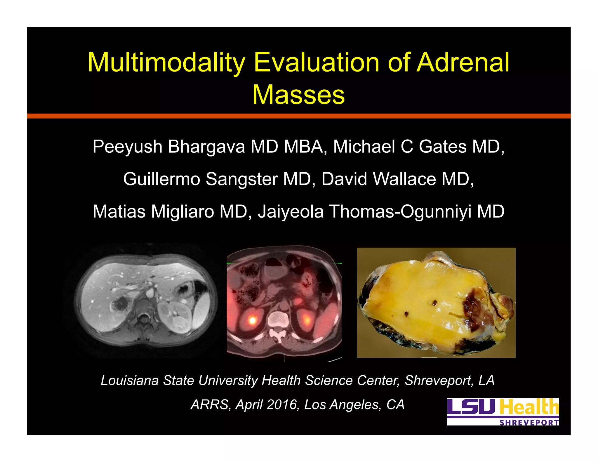
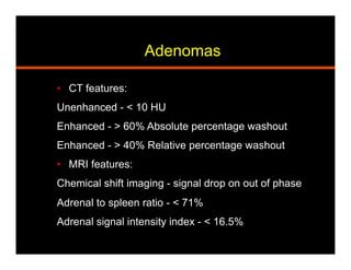
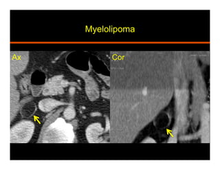
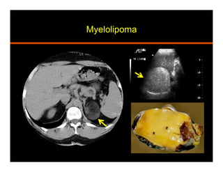
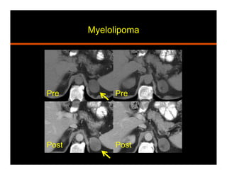
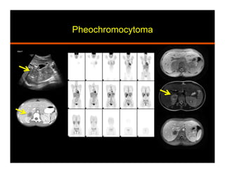
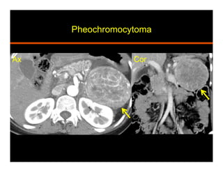
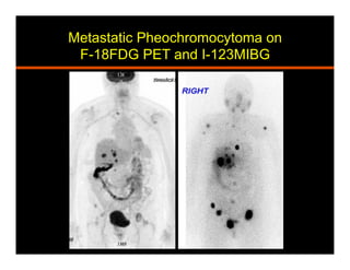
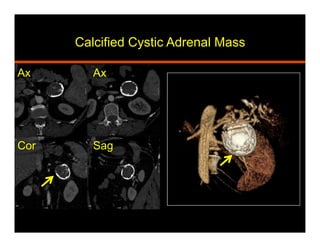
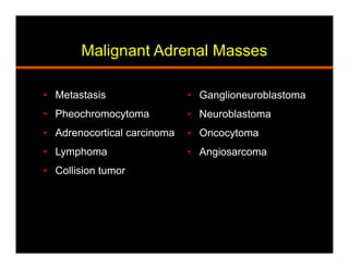
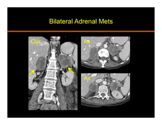
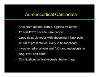
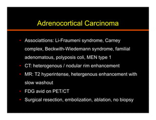
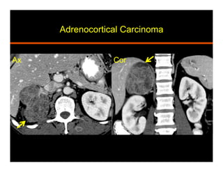
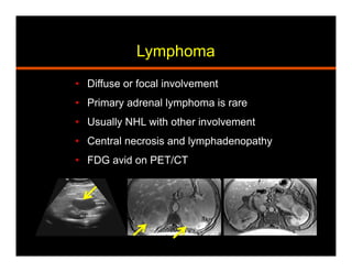
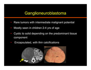
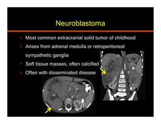
Multimodality Evaluation of Adrenal
Masses
Peeyush Bhargava MD MBA, Michael C Gates MD,
Guillermo Sangster MD, David Wallace MD,
Matias Migliaro MD, Jaiyeola Thomas-Ogunniyi MD
Louisiana State University Health Science Center, Shreveport, LA
ARRS, April 2016, Los Angeles, CA
Goals and Objectives
•  Review the spectrum of characteristic imaging
findings of adrenal masses using a
multimodality approach
•  Discuss the findings that can differentiate
between benign and malignant adrenal masses
•  Target Audience: Radiology residents and
practicing Radiologists
Benign Adrenal Masses
•  Adenoma
•  Incidentaloma
•  Hemorrhage
•  Myelolipoma
•  Hyperplasia
•  Pheochromocytoma
•  Collision Tumor
•  Ganglioneuroma
•  Lipoma
•  Cyst
•  Oncocytoma
•  Infection / Shock Adrenal
Adenomas
•  Most common adrenal tumor and incidentaloma
•  Benign, most non functional
•  70% lipid rich, 30% lipid poor
•  1-4 cm, well defined, solid, homogenous, smooth
•  Yellow to yellowish-brown in color
•  Vacuolated cells with intra-cytoplasmic lipid
Adenomas
•  CT features:
Unenhanced - < 10 HU
Enhanced - > 60% Absolute percentage washout
Enhanced - > 40% Relative percentage washout
•  MRI features:
Chemical shift imaging - signal drop on out of phase
Adrenal to spleen ratio - < 71%
Adrenal signal intensity index - < 16.5%
Adrenal Adenoma on CT
Lipid Rich
Adrenal Adenoma
MRI In/Out of Phase
ININ
OUT OUT
Absolute washout = (110 – 60 / 110 – 50) x 100 = 83%
Relative washout = (110 – 60 / 110 ) x 100 = 45%
Adenoma: APW, RPW
History: Incidental adrenal mass: 50 HU / 110 HU / 60 HU
Absolute washout = (110 – 60 / 110 – 50) x 100 = 83%
Relative washout = (110 – 60 / 110 ) x 100 = 45%
Incidentaloma
•  Adrenal tumors seen in 4 – 10 % of abdominal CTs
•  Most common lesion is an adenoma
•  Functional vs non-functional – 24 hr urine
metanepnrines, 17 hydroxycorticosteroids, 17
ketosteroids
•  CT / MRI characteristics – size, margins, HU / signal
drop on chemical shift / washout, homogeneity,
growth, history of cancer
•  PET/CT imaging / Biopsy
Algorithm for Adrenal Incidentaloma
Managing incidental findings on abdominal CT: white paper of the ACR incidental findings committee.
J Am Coll Radiol. 2010 Oct;7(10):754-73. doi: 10.1016/j.jacr.2010.06.013.
Hemorrhage
•  Traumatic (non accidental in peds) vs non traumatic
•  Non traumatic causes: coagulopathy, stress (sepsis,
surgery, burns, hypotension, pregnancy), venous
hypertension and hemorrhagic tumor
•  20% are bilateral, 1- 5 cm, well defined, round
Hemorrhage
•  CT: high attenuation (50-90 HU), does not
enhance, decreases in size and attenuation with
time, fat stranding, extension
•  MRI:
Acute (<7 days) – T1 iso to hypo, T2 markedly hypo
Subacute (1-7 wks) – T1 fat sat and T2 hyperintense
Chronic (after 7 wks) – T1 and T2 hypointense rim
Adrenal Hematoma
Sag
Ax
Sag
Ax Cor
Adrenal Hematoma
Cor Sag
Ax
Shock Adrenal (Hypoperfusion complex)
Ax Cor
Myelolipoma
•  Benign and non functioning tumor
•  Mature adipose and hematopoeitic tissue
•  Heterogeneous, pseudo-capsule, calcifications
•  US: hyperechoic lesion
•  CT: macroscopic fat (HU<30)
•  MRI: T1 hyperintense signal suppresses with fat sat
India ink (chemical shift) artifact at myeloma-adrenal
interface and within the mass on out of phase
Myelolipoma
Ax Cor
Myelolipoma
Myelolipoma
Pre Pre
PostPost
Hyperplasia
•  Benign and functional
•  Accounts for 2% cases of Cushings syndrome
and 30% cases of primary hyperaldosteronism
•  Sex steroid over production can lead to
feminization or virilization
•  Diffuse vs macronodular
Adrenal Hyperplasia (2 patients)
Pheochromocytoma
•  Arises from Chromaffin cells in adrenal medulla
•  Stores and releases catecholamines
•  Elevated serum / 24 hr urine metanephrines
•  Hypertension, palpitations, perspiration and headache
•  10% are: bilateral, extra adrenal (paragangliomas), occur
in children, and malignant (local invasion and mets)
•  Association with syndromes: MEN, NF 1, Von Hippel
Lindau, Tuberous sclerosis, Sturge-Weber
Pheochromocytoma
•  CT: homogeneous, well defined, soft tissue attenuation,
avid enhancement with delayed retention
•  MR: T2 bright (light bulb)
•  Take up I123 MIBG, In111 pentetreotide, and F18 FDG
•  Surgical resection
•  Pathology: yellow / tan, hemorrhage, cystic change,
central necrosis, myxoid degeneration. Nest of cells
(zellballen), Salt and pepper chromatin
Pheochromocytoma
Pheochromocytoma
Ax Cor
Metastatic Pheochromocytoma on
F-18FDG PET and I-123MIBG
Collision Tumor
•  Two coexisting histologically distinct tumors
•  Benign: adenoma and myelolipoma
•  Malignant: adenoma and metastasis
Cyst
•  Rare, benign, and non functional
•  Cystic, fluid attenuation, septations
•  Rim calcification, non enhancement
•  Prior hemorrhage or infarct
•  Aspirated if clinically indicated
•  Acellular turbid fluid with cholesterol crystals
•  Differentiate from cystic neoplasm (CT / MRI)
Calcified Cystic Adrenal Mass
Ax Ax
SagCor
Malignant Adrenal Masses
•  Ganglioneuroblastoma
•  Neuroblastoma
•  Oncocytoma
•  Angiosarcoma
•  Metastasis
•  Pheochromocytoma
•  Adrenocortical carcinoma
•  Lymphoma
•  Collision tumor
Metastasis
•  Common site after lung, liver, and bone
•  Primary: lung, breast, thyroid, colon, melanoma
•  Irregular, poorly defined mass, heterogeneous
•  Rim / nodular enhancement, central necrosis
•  Local invasion, hemorrhage
•  Rapid growth, other mets
•  T2 bright, no change on chemical shift imaging
•  FDG avid on PET/CT (SUV > 2.5 / liver)
Bilateral Adrenal Mets
Cor Ax
Ax
Adrenocortical Carcinoma
•  Arise from adrenal cortex, aggressive tumor
•  1st and 4th/5th decade, rare cancer
•  Large palpable mass with abdominal / flank pain
•  >6 cm at presentation, likely to be functional
•  Invasive (adrenal vein and IVC) and metastasis to
lung, liver, and bone
•  Calcification, central necrosis, hemorrhage
Adrenocortical Carcinoma
•  Associattions: Li-Fraumeni syndrome, Carney
complex, Beckwith-Wiedemann syndrome, familial
adenomatous, polyposis coli, MEN type 1
•  CT: heterogenous / nodular rim enhancement
•  MR: T2 hyperintense, hetergenous enhancement with
slow washout
•  FDG avid on PET/CT
•  Surgical resection, embolization, ablation, no biopsy
Adrenocortical Carcinoma
Ax Cor
Lymphoma
•  Diffuse or focal involvement
•  Primary adrenal lymphoma is rare
•  Usually NHL with other involvement
•  Central necrosis and lymphadenopathy
•  FDG avid on PET/CT
Ganglioneuroblastoma
•  Rare tumors with intermediate malignant potential
•  Mostly seen in children 2-4 yrs of age
•  Cystic to solid depending on the predominant tissue
component
•  Encapsulated, with thin calcifications
Neuroblastoma
•  Most common extracranial solid tumor of childhood
•  Arises from adrenal medulla or retroperitoneal
sympathetic ganglia
•  Soft tissue masses, often calcified
•  Often with disseminated disease
Contact Information
Peeyush Bhargava MD MBA
PGY 3 Radiology Resident
Phone: 832-374-2110
pbharg@lsuhsc.edu
Louisiana State University Health Science Center, Shreveport, LA

Adrenal Tumors

  • 1.
    Multimodality Evaluation ofAdrenal Masses Peeyush Bhargava MD MBA, Michael C Gates MD, Guillermo Sangster MD, David Wallace MD, Matias Migliaro MD, Jaiyeola Thomas-Ogunniyi MD Louisiana State University Health Science Center, Shreveport, LA ARRS, April 2016, Los Angeles, CA
  • 2.
    Goals and Objectives • Review the spectrum of characteristic imaging findings of adrenal masses using a multimodality approach •  Discuss the findings that can differentiate between benign and malignant adrenal masses •  Target Audience: Radiology residents and practicing Radiologists
  • 3.
    Benign Adrenal Masses • Adenoma •  Incidentaloma •  Hemorrhage •  Myelolipoma •  Hyperplasia •  Pheochromocytoma •  Collision Tumor •  Ganglioneuroma •  Lipoma •  Cyst •  Oncocytoma •  Infection / Shock Adrenal
  • 4.
    Adenomas •  Most commonadrenal tumor and incidentaloma •  Benign, most non functional •  70% lipid rich, 30% lipid poor •  1-4 cm, well defined, solid, homogenous, smooth •  Yellow to yellowish-brown in color •  Vacuolated cells with intra-cytoplasmic lipid
  • 5.
    Adenomas •  CT features: Unenhanced- < 10 HU Enhanced - > 60% Absolute percentage washout Enhanced - > 40% Relative percentage washout •  MRI features: Chemical shift imaging - signal drop on out of phase Adrenal to spleen ratio - < 71% Adrenal signal intensity index - < 16.5%
  • 6.
    Adrenal Adenoma onCT Lipid Rich
  • 7.
    Adrenal Adenoma MRI In/Outof Phase ININ OUT OUT
  • 8.
    Absolute washout =(110 – 60 / 110 – 50) x 100 = 83% Relative washout = (110 – 60 / 110 ) x 100 = 45% Adenoma: APW, RPW
  • 9.
    History: Incidental adrenalmass: 50 HU / 110 HU / 60 HU Absolute washout = (110 – 60 / 110 – 50) x 100 = 83% Relative washout = (110 – 60 / 110 ) x 100 = 45%
  • 10.
    Incidentaloma •  Adrenal tumorsseen in 4 – 10 % of abdominal CTs •  Most common lesion is an adenoma •  Functional vs non-functional – 24 hr urine metanepnrines, 17 hydroxycorticosteroids, 17 ketosteroids •  CT / MRI characteristics – size, margins, HU / signal drop on chemical shift / washout, homogeneity, growth, history of cancer •  PET/CT imaging / Biopsy
  • 11.
    Algorithm for AdrenalIncidentaloma Managing incidental findings on abdominal CT: white paper of the ACR incidental findings committee. J Am Coll Radiol. 2010 Oct;7(10):754-73. doi: 10.1016/j.jacr.2010.06.013.
  • 12.
    Hemorrhage •  Traumatic (nonaccidental in peds) vs non traumatic •  Non traumatic causes: coagulopathy, stress (sepsis, surgery, burns, hypotension, pregnancy), venous hypertension and hemorrhagic tumor •  20% are bilateral, 1- 5 cm, well defined, round
  • 13.
    Hemorrhage •  CT: highattenuation (50-90 HU), does not enhance, decreases in size and attenuation with time, fat stranding, extension •  MRI: Acute (<7 days) – T1 iso to hypo, T2 markedly hypo Subacute (1-7 wks) – T1 fat sat and T2 hyperintense Chronic (after 7 wks) – T1 and T2 hypointense rim
  • 14.
  • 15.
  • 16.
  • 17.
    Myelolipoma •  Benign andnon functioning tumor •  Mature adipose and hematopoeitic tissue •  Heterogeneous, pseudo-capsule, calcifications •  US: hyperechoic lesion •  CT: macroscopic fat (HU<30) •  MRI: T1 hyperintense signal suppresses with fat sat India ink (chemical shift) artifact at myeloma-adrenal interface and within the mass on out of phase
  • 18.
  • 19.
  • 20.
  • 21.
    Hyperplasia •  Benign andfunctional •  Accounts for 2% cases of Cushings syndrome and 30% cases of primary hyperaldosteronism •  Sex steroid over production can lead to feminization or virilization •  Diffuse vs macronodular
  • 22.
  • 23.
    Pheochromocytoma •  Arises fromChromaffin cells in adrenal medulla •  Stores and releases catecholamines •  Elevated serum / 24 hr urine metanephrines •  Hypertension, palpitations, perspiration and headache •  10% are: bilateral, extra adrenal (paragangliomas), occur in children, and malignant (local invasion and mets) •  Association with syndromes: MEN, NF 1, Von Hippel Lindau, Tuberous sclerosis, Sturge-Weber
  • 24.
    Pheochromocytoma •  CT: homogeneous,well defined, soft tissue attenuation, avid enhancement with delayed retention •  MR: T2 bright (light bulb) •  Take up I123 MIBG, In111 pentetreotide, and F18 FDG •  Surgical resection •  Pathology: yellow / tan, hemorrhage, cystic change, central necrosis, myxoid degeneration. Nest of cells (zellballen), Salt and pepper chromatin
  • 25.
  • 26.
  • 27.
  • 28.
    Collision Tumor •  Twocoexisting histologically distinct tumors •  Benign: adenoma and myelolipoma •  Malignant: adenoma and metastasis
  • 29.
    Cyst •  Rare, benign,and non functional •  Cystic, fluid attenuation, septations •  Rim calcification, non enhancement •  Prior hemorrhage or infarct •  Aspirated if clinically indicated •  Acellular turbid fluid with cholesterol crystals •  Differentiate from cystic neoplasm (CT / MRI)
  • 30.
    Calcified Cystic AdrenalMass Ax Ax SagCor
  • 31.
    Malignant Adrenal Masses • Ganglioneuroblastoma •  Neuroblastoma •  Oncocytoma •  Angiosarcoma •  Metastasis •  Pheochromocytoma •  Adrenocortical carcinoma •  Lymphoma •  Collision tumor
  • 32.
    Metastasis •  Common siteafter lung, liver, and bone •  Primary: lung, breast, thyroid, colon, melanoma •  Irregular, poorly defined mass, heterogeneous •  Rim / nodular enhancement, central necrosis •  Local invasion, hemorrhage •  Rapid growth, other mets •  T2 bright, no change on chemical shift imaging •  FDG avid on PET/CT (SUV > 2.5 / liver)
  • 33.
  • 34.
    Adrenocortical Carcinoma •  Arisefrom adrenal cortex, aggressive tumor •  1st and 4th/5th decade, rare cancer •  Large palpable mass with abdominal / flank pain •  >6 cm at presentation, likely to be functional •  Invasive (adrenal vein and IVC) and metastasis to lung, liver, and bone •  Calcification, central necrosis, hemorrhage
  • 35.
    Adrenocortical Carcinoma •  Associattions:Li-Fraumeni syndrome, Carney complex, Beckwith-Wiedemann syndrome, familial adenomatous, polyposis coli, MEN type 1 •  CT: heterogenous / nodular rim enhancement •  MR: T2 hyperintense, hetergenous enhancement with slow washout •  FDG avid on PET/CT •  Surgical resection, embolization, ablation, no biopsy
  • 36.
  • 37.
    Lymphoma •  Diffuse orfocal involvement •  Primary adrenal lymphoma is rare •  Usually NHL with other involvement •  Central necrosis and lymphadenopathy •  FDG avid on PET/CT
  • 38.
    Ganglioneuroblastoma •  Rare tumorswith intermediate malignant potential •  Mostly seen in children 2-4 yrs of age •  Cystic to solid depending on the predominant tissue component •  Encapsulated, with thin calcifications
  • 39.
    Neuroblastoma •  Most commonextracranial solid tumor of childhood •  Arises from adrenal medulla or retroperitoneal sympathetic ganglia •  Soft tissue masses, often calcified •  Often with disseminated disease
  • 40.
    Contact Information Peeyush BhargavaMD MBA PGY 3 Radiology Resident Phone: 832-374-2110 pbharg@lsuhsc.edu Louisiana State University Health Science Center, Shreveport, LA