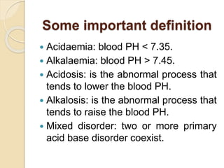
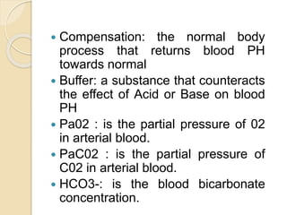
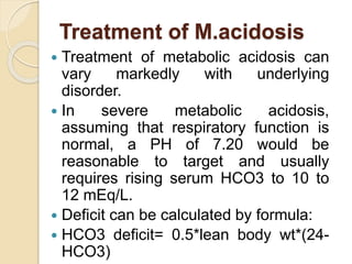
ACID BASE DISORDERS
DR KALYAN DEBNATH
PHASE -A
Dept of Anaesthesia , Analgesia
& Intensive care medicine
Some important definition
 Acidaemia: blood PH < 7.35.
 Alkalaemia: blood PH > 7.45.
 Acidosis: is the abnormal process that
tends to lower the blood PH.
 Alkalosis: is the abnormal process that
tends to raise the blood PH.
 Mixed disorder: two or more primary
acid base disorder coexist.
 Compensation: the normal body
process that returns blood PH
towards normal
 Buffer: a substance that counteracts
the effect of Acid or Base on blood
PH
 Pa02 : is the partial pressure of 02
in arterial blood.
 PaC02 : is the partial pressure of
C02 in arterial blood.
 HCO3-: is the blood bicarbonate
concentration.
PH
An acidaemia (low pH) result from
either a low HCO3(Metabolic) OR high
C02(Respiratory).
An alkalemia (high pH) result from
either a high HCO3(Metabolic) OR low
C02(Respiratory).
How body buffers work
 Immediately, buffers in
blood(NAHC03) change strong acid to
weak acid.
 After several minutes, this weak
acid decomposes to co2 carried by Hb
to be expired by lungs.
 After several hours, kidney
smoothly reabsorbs the HCO3
wasted.
Assess oxygenation
 Look at oxygenation (pa02 and Sa02)
 Look at Pao2/Fio2 ratio
 Normally, the ratio is around 1:400-
1:500.
 Less than 1:400—sugesstive of V-Q
mismatch or diffusion defect or
intracardiac shunt
 Less than 300 with bilateral lung
infiltrate in CXR: ARDS.
A-a gradient
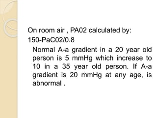
A-a gradient= PA02-Pa02
Here, PA02 is alveolar P02 & Pao2 is
arterial P02.
In general, A-a gradient can be calculated
by:
A-a gradient=[Fi02*(Patm-PH20)-
(PaC02/0.8)]-Pao2
On room air & at sea level, Fi02 is
0.21,Patm is 760 mmHg and PH20 is
47 mmHg.
On room air , PA02 calculated by:
150-PaC02/0.8
Normal A-a gradient in a 20 year old
person is 5 mmHg which increase to
10 in a 35 year old person. If A-a
gradient is 20 mmHg at any age, is
abnormal .
Metabolic Acidosis
Definition
 Metabolic acidosis can be defined as
a low arterial blood pH in conjunction
with low serum bicarbonate
concentration caused either by
increased acid generation or
decreased acid secretion or loss of
bicarbonate.
Types
A. M. Acidosis with increased anion gap
B. M. Acidosis with normal anion gap (
hyperchloraemic M. acidosis)
It is very important to calculate the Anion
gap if primary disorder is M. acidosis.
Anion Gap= (Na+ + K+) – (Cl- + HCO3)
Normal Anion Gap 12 – 16 mEq/L.
Metabolic acidosis with
hypokalaemia
 Diarrhoea
 RTA (type 1 & 2)
 Acetazolamide
 Partially treated DKA
Treatment of M.acidosis
 Treatment of metabolic acidosis can
vary markedly with underlying
disorder.
 In severe metabolic acidosis,
assuming that respiratory function is
normal, a PH of 7.20 would be
reasonable to target and usually
requires rising serum HCO3 to 10 to
12 mEq/L.
 Deficit can be calculated by formula:
 HCO3 deficit= 0.5*lean body wt*(24-
HCO3)
 Sodium bicarbonate can be given as oral
tablets, powder as a hypertonic
bicarbonate bolus or isotonic sodium
bicarbonate .For patients with volume
depletion , administration of three
ampoules of bicarbonate (each contain
50mEq of sodium bicarbonate)in 1L of
dextrose 5% in water(5%DA ) solution
will help both volume expansion and
alkalization.
 Replace 50% of deficit in 1st 24
hours and determine future
dosage based on response to
therapy and target bicarbonate.
 Renal replacement therapy is
employed for refractory severe
metabolic acidosis.
METABOLIC ALKALOSIS
• Metabolic alkalosis reflects an increase in
plasma HCO3.
• It is due to either gain of HCO3 or
extracellular volume contraction.
• It can be classified into Chloride responsive
or non responsive. for this spot urinary
chloride is to be checked.
• More than 20 mEq/l urinary chloride is
Chloride unresponsive and less than 20
mEq/l urinary chloride is Chloride
responsive.
Causes of chloride
responsive m. alkalosis
 Vomiting, NG suction
 Thiazide
 Loop diuretics
 Chronic hypercapnoea
 Villous adenoma of colon
Causes of chloride resistant
m. alkalosis
 Mineralocorticoid excess
(hyperaldosteronism, Cushing’s
syndrome, ACTH secreting tumor,
renin secreting tumor)
 Bartter syndrome, Gitelmann’s
syndrome
 Decreased effective circulating volume
in setting of heart failure / cirrhosis.
 Hypokalaemia.
M alkalosis associated with
alkali administration
 Sodium bicarbonate
 Lactate in ringers lactate
 Parenteral nutrition – acetate,
glutamate
 Citrate ( blood product, plasma
exchange)
 Milk Alkali syndrome
 dialysis
Treatment of M. Alkalosis
Chloride responsive alkalosis --
 The increase in bicarbonate resorption
can be counteracted by administration
of normal saline or by volume
expansion. Increased distal chloride
delivery will increase the bicarbonate
secretion in CT. effect can be
monitored by increase in urinary pH
(>7).
Chloride resistant alkalosis
 includes discontinuing diuretics, trail of
acetazolamide, dialysis.
 Acetazolamide is a carbonic anhydrase
inhibitor that increase excretion of
bicarbonate and potassium. Need to
monitor hypokalaemia.Typically seen in
edematous states (CHF, Cirrhosis,
nephrotic syndrome) following diuretic
therapy.
 dose 250mg to 375 mg once or twice
daily. . effect can be monitored by
increase in urinary pH (>7).
Treatment of R. acidosis
The goal is to increase the exhalation of CO2.
The treatments are :
I. Based on underlying cause.
II. By providing ventilation therapy.
III. IV administration of HCO3.
IV. Reversal of sedation or neuromuscular
relaxants.
V. Intubation and artificial ventilation, in severe
cases.
Treatment of R. alkalosis
 Correct the underlying disorder.
 Slow the rate of breathing.
 Administer of pain medication.
 Breathe into paper bag helps raise of
CO2 level, person rebreathes CO2
back in after breathing out.
 Hold the breath as long, as possible
and repeat the sequence 6 to 10
times.
Important consideration
about ABG sample
 If possible, take an ABG sample at
room air.
 Radial artery is preferred for colleting
the sample.
 Prefer to use 22 gauge needle.
 Avoid air bubbles.
 Cool the sample immediately.
Potential sampling error-
 Air contamination-spurious increase in
PO2.
 Duration of exposure is more
important than volume of air bubbles.
 Expel air immediately.
 Discard the sample if froth present.
 Venous sample- absence of flash of
blood on entry into vessel & absence
of autofilling of the syringe.
 Timing of measurement-blood cell
consume 02 , produce C02 & lower
pH. magnitude of changes depends
on time.
Acid base disorders

Acid base disorders

  • 1.
    ACID BASE DISORDERS DRKALYAN DEBNATH PHASE -A Dept of Anaesthesia , Analgesia & Intensive care medicine
  • 2.
    Some important definition Acidaemia: blood PH < 7.35.  Alkalaemia: blood PH > 7.45.  Acidosis: is the abnormal process that tends to lower the blood PH.  Alkalosis: is the abnormal process that tends to raise the blood PH.  Mixed disorder: two or more primary acid base disorder coexist.
  • 3.
     Compensation: thenormal body process that returns blood PH towards normal  Buffer: a substance that counteracts the effect of Acid or Base on blood PH  Pa02 : is the partial pressure of 02 in arterial blood.  PaC02 : is the partial pressure of C02 in arterial blood.  HCO3-: is the blood bicarbonate concentration.
  • 4.
    PH An acidaemia (lowpH) result from either a low HCO3(Metabolic) OR high C02(Respiratory). An alkalemia (high pH) result from either a high HCO3(Metabolic) OR low C02(Respiratory).
  • 5.
    How body bufferswork  Immediately, buffers in blood(NAHC03) change strong acid to weak acid.  After several minutes, this weak acid decomposes to co2 carried by Hb to be expired by lungs.  After several hours, kidney smoothly reabsorbs the HCO3 wasted.
  • 10.
    Assess oxygenation  Lookat oxygenation (pa02 and Sa02)  Look at Pao2/Fio2 ratio  Normally, the ratio is around 1:400- 1:500.  Less than 1:400—sugesstive of V-Q mismatch or diffusion defect or intracardiac shunt  Less than 300 with bilateral lung infiltrate in CXR: ARDS.
  • 11.
    A-a gradient A-a gradient=PA02-Pa02 Here, PA02 is alveolar P02 & Pao2 is arterial P02. In general, A-a gradient can be calculated by: A-a gradient=[Fi02*(Patm-PH20)- (PaC02/0.8)]-Pao2 On room air & at sea level, Fi02 is 0.21,Patm is 760 mmHg and PH20 is 47 mmHg.
  • 12.
    On room air, PA02 calculated by: 150-PaC02/0.8 Normal A-a gradient in a 20 year old person is 5 mmHg which increase to 10 in a 35 year old person. If A-a gradient is 20 mmHg at any age, is abnormal .
  • 13.
    Metabolic Acidosis Definition  Metabolicacidosis can be defined as a low arterial blood pH in conjunction with low serum bicarbonate concentration caused either by increased acid generation or decreased acid secretion or loss of bicarbonate.
  • 14.
    Types A. M. Acidosiswith increased anion gap B. M. Acidosis with normal anion gap ( hyperchloraemic M. acidosis) It is very important to calculate the Anion gap if primary disorder is M. acidosis. Anion Gap= (Na+ + K+) – (Cl- + HCO3) Normal Anion Gap 12 – 16 mEq/L.
  • 17.
    Metabolic acidosis with hypokalaemia Diarrhoea  RTA (type 1 & 2)  Acetazolamide  Partially treated DKA
  • 18.
    Treatment of M.acidosis Treatment of metabolic acidosis can vary markedly with underlying disorder.  In severe metabolic acidosis, assuming that respiratory function is normal, a PH of 7.20 would be reasonable to target and usually requires rising serum HCO3 to 10 to 12 mEq/L.  Deficit can be calculated by formula:  HCO3 deficit= 0.5*lean body wt*(24- HCO3)
  • 19.
     Sodium bicarbonatecan be given as oral tablets, powder as a hypertonic bicarbonate bolus or isotonic sodium bicarbonate .For patients with volume depletion , administration of three ampoules of bicarbonate (each contain 50mEq of sodium bicarbonate)in 1L of dextrose 5% in water(5%DA ) solution will help both volume expansion and alkalization.
  • 20.
     Replace 50%of deficit in 1st 24 hours and determine future dosage based on response to therapy and target bicarbonate.  Renal replacement therapy is employed for refractory severe metabolic acidosis.
  • 21.
    METABOLIC ALKALOSIS • Metabolicalkalosis reflects an increase in plasma HCO3. • It is due to either gain of HCO3 or extracellular volume contraction. • It can be classified into Chloride responsive or non responsive. for this spot urinary chloride is to be checked. • More than 20 mEq/l urinary chloride is Chloride unresponsive and less than 20 mEq/l urinary chloride is Chloride responsive.
  • 22.
    Causes of chloride responsivem. alkalosis  Vomiting, NG suction  Thiazide  Loop diuretics  Chronic hypercapnoea  Villous adenoma of colon
  • 23.
    Causes of chlorideresistant m. alkalosis  Mineralocorticoid excess (hyperaldosteronism, Cushing’s syndrome, ACTH secreting tumor, renin secreting tumor)  Bartter syndrome, Gitelmann’s syndrome  Decreased effective circulating volume in setting of heart failure / cirrhosis.  Hypokalaemia.
  • 24.
    M alkalosis associatedwith alkali administration  Sodium bicarbonate  Lactate in ringers lactate  Parenteral nutrition – acetate, glutamate  Citrate ( blood product, plasma exchange)  Milk Alkali syndrome  dialysis
  • 25.
    Treatment of M.Alkalosis Chloride responsive alkalosis --  The increase in bicarbonate resorption can be counteracted by administration of normal saline or by volume expansion. Increased distal chloride delivery will increase the bicarbonate secretion in CT. effect can be monitored by increase in urinary pH (>7).
  • 26.
    Chloride resistant alkalosis includes discontinuing diuretics, trail of acetazolamide, dialysis.  Acetazolamide is a carbonic anhydrase inhibitor that increase excretion of bicarbonate and potassium. Need to monitor hypokalaemia.Typically seen in edematous states (CHF, Cirrhosis, nephrotic syndrome) following diuretic therapy.  dose 250mg to 375 mg once or twice daily. . effect can be monitored by increase in urinary pH (>7).
  • 28.
    Treatment of R.acidosis The goal is to increase the exhalation of CO2. The treatments are : I. Based on underlying cause. II. By providing ventilation therapy. III. IV administration of HCO3. IV. Reversal of sedation or neuromuscular relaxants. V. Intubation and artificial ventilation, in severe cases.
  • 30.
    Treatment of R.alkalosis  Correct the underlying disorder.  Slow the rate of breathing.  Administer of pain medication.  Breathe into paper bag helps raise of CO2 level, person rebreathes CO2 back in after breathing out.  Hold the breath as long, as possible and repeat the sequence 6 to 10 times.
  • 31.
    Important consideration about ABGsample  If possible, take an ABG sample at room air.  Radial artery is preferred for colleting the sample.  Prefer to use 22 gauge needle.  Avoid air bubbles.  Cool the sample immediately.
  • 32.
    Potential sampling error- Air contamination-spurious increase in PO2.  Duration of exposure is more important than volume of air bubbles.  Expel air immediately.  Discard the sample if froth present.
  • 33.
     Venous sample-absence of flash of blood on entry into vessel & absence of autofilling of the syringe.  Timing of measurement-blood cell consume 02 , produce C02 & lower pH. magnitude of changes depends on time.