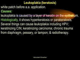
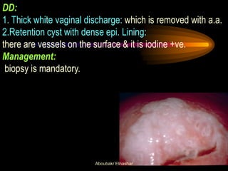
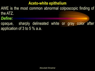
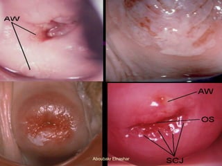
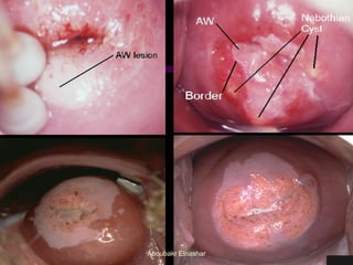
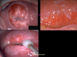
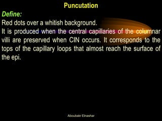
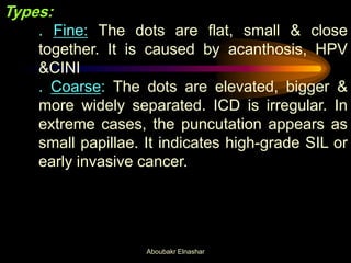
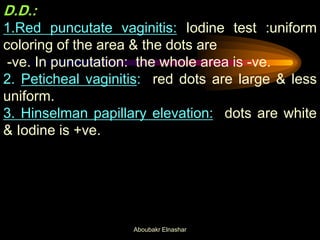
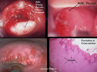
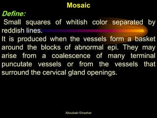
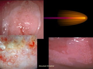
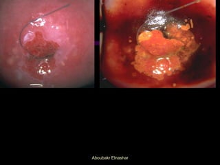
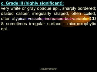
Aboubakr Elnashar
Leukoplakia (keratosis)
white patch before a.a. application.
Causes:
leukoplakia is caused by a layer of keratin on the epithelium.
Histologically, it shows hyperkeratosis or parakeratosis.
Several things can cause leukoplakia including HPV;
keratinizing CIN; keratinizing carcinoma; chronic trauma
from diaphragm, pessary, or tampon; & radiotherapy.
Aboubakr Elnashar
DD:
1. Thick white vaginal discharge: which is removed with a.a.
2.Retention cyst with dense epi. Lining:
there are vessels on the surface & it is iodine +ve.
Management:
biopsy is mandatory.
Aboubakr Elnashar
Aceto-white epithelium
AWE is the most common abnormal colposcopic finding of
the ATZ.
Define:
opaque, sharply delineated white or gray color after
application of 3 to 5 % a.a.
Aboubakr Elnashar
Aboubakr Elnashar
Mechanism:
1. coagulates the proteins of the nucleus &
cytoplasm & makes the protein opaque & white.
2. dehydrates the cells, the cytoplasmic volume is
reduced & the reflection is increased.
Duration:
variable, generally it appears after 20 secs &
disappears after 2 mins.
Aboubakr Elnashar
Effects of a.a.:
1.on the mucous: It coagulates mucous which can
then easily removed (mucolytic).
2.on the mature glycogen-producing epithelium:
no effect bec. The a.a. does not penetrate below the
outer one-third of the epithelium. The cells have very
small nuclei & a large amount of glycogen (not
protein)
3.on the col. epi.:
swell & become slightly opaque, particularly if the
beginning signs of metaplasia are present. So,it
makes its recognition easier.
Aboubakr Elnashar
4.on the immature metaplastic epi.:
Because they are very thin & have large nuclei. It is
not as white or opaque as CIN but instead appear
gray & filmy.
5.on dysplastic cells:
They contain large nuclei & large amounts of
chromatin( protein). They become white.
6.on gland openings of the TZ.:
are better outlined.
7.on puncutation & mosaic:
appear more prominent.
8.on vascular bed:
becomes less evident due to arteriolar spasm.
Aboubakr Elnashar
D.D:
Causes of acetowhite epi.
immature metaplasia,
healing or regenerating epi. ,
HPV infection,
CIN & invasive cancer.
D.D.depend on colour, margin, surface, vessels &
iodine staining.
1. Leukoplakia:
elevated white patch before a.a application.
Aboubakr Elnashar
Aboubakr Elnashar
2. Acetowhitness of normal metaplastic epi.:
occurs quickly & fades quickly. It is also more difficult to
reestablish after fading by reapplication of a.a. The
surface is shiny & translucent. The margin is indistinct, &
AWE blend gradually with the adjacent more mature sq.
epi.
3. Acetowhitness of CIN.:
It takes more time to appear. The effect is hastened &
aided by second a.a application. A definite & distinctive
margin is usually present. The intensity is more marked
than with physiologic variants.The surface is thicker &
more opaque &less shiny.
Aboubakr Elnashar
TTZ
Aboubakr Elnashar
Puncutation
Define:
Red dots over a whitish background.
It is produced when the central capillaries of the columnar
villi are preserved when CIN occurs. It corresponds to the
tops of the capillary loops that almost reach the surface of
the epi.
Aboubakr Elnashar
Types:
. Fine: The dots are flat, small & close
together. It is caused by acanthosis, HPV
&CINI
. Coarse: The dots are elevated, bigger &
more widely separated. ICD is irregular. In
extreme cases, the puncutation appears as
small papillae. It indicates high-grade SIL or
early invasive cancer.
Aboubakr Elnashar
D.D.:
1.Red puncutate vaginitis: Iodine test :uniform
coloring of the area & the dots are
-ve. In puncutation: the whole area is -ve.
2. Peticheal vaginitis: red dots are large & less
uniform.
3. Hinselman papillary elevation: dots are white
& Iodine is +ve.
Aboubakr Elnashar
Aboubakr Elnashar
Mosaic
Define:
Small squares of whitish color separated by
reddish lines.
It is produced when the vessels form a basket
around the blocks of abnormal epi. They may
arise from a coalescence of many terminal
puncutate vessels or from the vessels that
surround the cervical gland openings.
Aboubakr Elnashar
Types:
. Fine: The lines are fine & the fields are flat,
regular & small.
. Coarse: The lines are intense red &
pronounced & the fields are elevated,
irregular & bigger.
Puncutation & mosaic are often coexisting within
the ATZ & the causes are similar.
Aboubakr Elnashar
Aboubakr Elnashar
D.D.:
. Pseudomosaic: The squares are separated
by whitish lines more delicate & less defined
edges.
. Reticular TTZ.
. Geographic vaginitis :
Aboubakr Elnashar
Atypical vessels
They are an exaggeration of vascular abnormalities
described in puncutation & mosaic & occur as the lesion
becomes more severe in type.
They are irregular in size, shape, course & arrangement &
the ICD is greater than in normal epithelium. In benign
vascular patterns the branching is dichotomous or tree like
with large stems giving rise to smaller & smaller branches.
Aboubakr Elnashar
There is no particular pattern, the vessels being arranged in
a haphazard way. Atypical vessels show no decrease in
diameter of terminal branches & give rise to a coarse
network enclosing irregular avascular fields.
They are most characteristic of invasive cancer, but can
occur occasionally in high-grade SIL.
Aboubakr Elnashar
Iodine nonstaining
CIN & most HPV lesions fail to stain deeply when a solution
Schillers or Lugal,s iodine is applied because glycogen is
absent.
Certain subclinical HPV lesions containing glycogen
demonstrates less well-defined response, producing a
stippled or mosaic-like uptake of iodine that may be of value
in grading the significance of the disorder.
Iodine nonstaining is cause also by acanthotic epi..
Aboubakr Elnashar
Aboubakr Elnashar
Causes of the colposcopic abnormalities ( Campion et
al,1991)
Acetowhite epithelium
Increased cellular & nuclear density. Abnormal intracellular
keratins
Leukoplakia
Abnormal keratin production in the neoplastic process
Punctation & Mosaic
Alteration in epithelial capillaries.
Aboubakr Elnashar
Mosaic formation due to:
1. transformation of normal metaplasia,
2.capillary proliferative effect of HPV,
3.tumor angiogenic factor in severe neoplastic
transformation.
Atypical vessels
Specific alteration in epithelial capillaries, due to
angiogenesis, suggestive of invasive cancer.
Aboubakr Elnashar
Grading of abnormal colposcopic findings
1.Copplesson :
a. Grade I (insignificant):
Flat, acetowhite epi. , Borders not necessarily
sharp; semitransparent with or without fine
caliber, regularly shaped vessels, often with ill-
defined patterns, absence of atypical vessels,
small ICD.
b. Grade II (significant):
Flat, aceto-white epi. of greater opacity with sharp
borders; with or without dilated-caliber, regularly
shaped vessels; defined patterns; absence of
atypical vessels; usually increased ICD.
Aboubakr Elnashar
c. Grade III (highly significant):
very white or gray opaque epi., sharply bordered;
dilated caliber, irregularly shaped, often coiled,
often atypical vessels, increased but variable ICD
& sometimes irregular surface - microexophytic
epi.
Aboubakr Elnashar
2. Stafl:
Grading based on 4 factors:
1. Surface pattern
2. Color tone.
3. ICD.
4. Border of the lesion with normal tissue.
Aboubakr Elnashar
3. Reid s index
Colp.sign 0 1 2
Margin indistinct sharp peeling
Color snow white off white dull
Vessels thin absent
after AA
pun.or
mosaic
Iodine
staining
positive patial negative
Colp.
score
0-2
HPVorCIN1
3-5
CIN1or2
6-8
CIN2or3
Aboubakr Elnashar
4. Niekerk (1998)
Low grade High grade
•Acetowhite epithelium: shiny or snow dull, oyster white color
white,semitransparent
•Surface: flat irregular contour, microexophytic
•Demarcation: diffuse, irregular, sharp, straight line,
flocculated, feathered,
internal demarcation absent internal demarcation present
•Vessels: fine, regular shape, uniform coarse, dilated, increased ICD,
caliber, normal arborization, spaghetti bizarre, commas, corkscrews
changing calibers sharp bends
•Iodine: uniform mahogany brown mustard yellow, yellow or iodine -ve
Aboubakr Elnashar
Colposcopically suspect invasive cancer
Although rare. It is obvious after acetic acid
application with raised edge, irregular surface,
nodulation, ulceration, the mountain range
appearance & atypical blood vessels.
Aboubakr Elnashar
Aboubakr Elnashar
THANKS
Aboubakr Elnashar

ABNORMAL COLPOSCOPIC FINDINGS

  • 1.
  • 2.
    Leukoplakia (keratosis) white patchbefore a.a. application. Causes: leukoplakia is caused by a layer of keratin on the epithelium. Histologically, it shows hyperkeratosis or parakeratosis. Several things can cause leukoplakia including HPV; keratinizing CIN; keratinizing carcinoma; chronic trauma from diaphragm, pessary, or tampon; & radiotherapy. Aboubakr Elnashar
  • 3.
    DD: 1. Thick whitevaginal discharge: which is removed with a.a. 2.Retention cyst with dense epi. Lining: there are vessels on the surface & it is iodine +ve. Management: biopsy is mandatory. Aboubakr Elnashar
  • 4.
    Aceto-white epithelium AWE isthe most common abnormal colposcopic finding of the ATZ. Define: opaque, sharply delineated white or gray color after application of 3 to 5 % a.a. Aboubakr Elnashar
  • 5.
  • 6.
    Mechanism: 1. coagulates theproteins of the nucleus & cytoplasm & makes the protein opaque & white. 2. dehydrates the cells, the cytoplasmic volume is reduced & the reflection is increased. Duration: variable, generally it appears after 20 secs & disappears after 2 mins. Aboubakr Elnashar
  • 7.
    Effects of a.a.: 1.onthe mucous: It coagulates mucous which can then easily removed (mucolytic). 2.on the mature glycogen-producing epithelium: no effect bec. The a.a. does not penetrate below the outer one-third of the epithelium. The cells have very small nuclei & a large amount of glycogen (not protein) 3.on the col. epi.: swell & become slightly opaque, particularly if the beginning signs of metaplasia are present. So,it makes its recognition easier. Aboubakr Elnashar
  • 8.
    4.on the immaturemetaplastic epi.: Because they are very thin & have large nuclei. It is not as white or opaque as CIN but instead appear gray & filmy. 5.on dysplastic cells: They contain large nuclei & large amounts of chromatin( protein). They become white. 6.on gland openings of the TZ.: are better outlined. 7.on puncutation & mosaic: appear more prominent. 8.on vascular bed: becomes less evident due to arteriolar spasm. Aboubakr Elnashar
  • 9.
    D.D: Causes of acetowhiteepi. immature metaplasia, healing or regenerating epi. , HPV infection, CIN & invasive cancer. D.D.depend on colour, margin, surface, vessels & iodine staining. 1. Leukoplakia: elevated white patch before a.a application. Aboubakr Elnashar
  • 10.
  • 11.
    2. Acetowhitness ofnormal metaplastic epi.: occurs quickly & fades quickly. It is also more difficult to reestablish after fading by reapplication of a.a. The surface is shiny & translucent. The margin is indistinct, & AWE blend gradually with the adjacent more mature sq. epi. 3. Acetowhitness of CIN.: It takes more time to appear. The effect is hastened & aided by second a.a application. A definite & distinctive margin is usually present. The intensity is more marked than with physiologic variants.The surface is thicker & more opaque &less shiny. Aboubakr Elnashar
  • 12.
  • 13.
    Puncutation Define: Red dots overa whitish background. It is produced when the central capillaries of the columnar villi are preserved when CIN occurs. It corresponds to the tops of the capillary loops that almost reach the surface of the epi. Aboubakr Elnashar
  • 14.
    Types: . Fine: Thedots are flat, small & close together. It is caused by acanthosis, HPV &CINI . Coarse: The dots are elevated, bigger & more widely separated. ICD is irregular. In extreme cases, the puncutation appears as small papillae. It indicates high-grade SIL or early invasive cancer. Aboubakr Elnashar
  • 15.
    D.D.: 1.Red puncutate vaginitis:Iodine test :uniform coloring of the area & the dots are -ve. In puncutation: the whole area is -ve. 2. Peticheal vaginitis: red dots are large & less uniform. 3. Hinselman papillary elevation: dots are white & Iodine is +ve. Aboubakr Elnashar
  • 16.
  • 17.
    Mosaic Define: Small squares ofwhitish color separated by reddish lines. It is produced when the vessels form a basket around the blocks of abnormal epi. They may arise from a coalescence of many terminal puncutate vessels or from the vessels that surround the cervical gland openings. Aboubakr Elnashar
  • 18.
    Types: . Fine: Thelines are fine & the fields are flat, regular & small. . Coarse: The lines are intense red & pronounced & the fields are elevated, irregular & bigger. Puncutation & mosaic are often coexisting within the ATZ & the causes are similar. Aboubakr Elnashar
  • 19.
  • 20.
    D.D.: . Pseudomosaic: Thesquares are separated by whitish lines more delicate & less defined edges. . Reticular TTZ. . Geographic vaginitis : Aboubakr Elnashar
  • 21.
    Atypical vessels They arean exaggeration of vascular abnormalities described in puncutation & mosaic & occur as the lesion becomes more severe in type. They are irregular in size, shape, course & arrangement & the ICD is greater than in normal epithelium. In benign vascular patterns the branching is dichotomous or tree like with large stems giving rise to smaller & smaller branches. Aboubakr Elnashar
  • 22.
    There is noparticular pattern, the vessels being arranged in a haphazard way. Atypical vessels show no decrease in diameter of terminal branches & give rise to a coarse network enclosing irregular avascular fields. They are most characteristic of invasive cancer, but can occur occasionally in high-grade SIL. Aboubakr Elnashar
  • 23.
    Iodine nonstaining CIN &most HPV lesions fail to stain deeply when a solution Schillers or Lugal,s iodine is applied because glycogen is absent. Certain subclinical HPV lesions containing glycogen demonstrates less well-defined response, producing a stippled or mosaic-like uptake of iodine that may be of value in grading the significance of the disorder. Iodine nonstaining is cause also by acanthotic epi.. Aboubakr Elnashar
  • 24.
  • 25.
    Causes of thecolposcopic abnormalities ( Campion et al,1991) Acetowhite epithelium Increased cellular & nuclear density. Abnormal intracellular keratins Leukoplakia Abnormal keratin production in the neoplastic process Punctation & Mosaic Alteration in epithelial capillaries. Aboubakr Elnashar
  • 26.
    Mosaic formation dueto: 1. transformation of normal metaplasia, 2.capillary proliferative effect of HPV, 3.tumor angiogenic factor in severe neoplastic transformation. Atypical vessels Specific alteration in epithelial capillaries, due to angiogenesis, suggestive of invasive cancer. Aboubakr Elnashar
  • 27.
    Grading of abnormalcolposcopic findings 1.Copplesson : a. Grade I (insignificant): Flat, acetowhite epi. , Borders not necessarily sharp; semitransparent with or without fine caliber, regularly shaped vessels, often with ill- defined patterns, absence of atypical vessels, small ICD. b. Grade II (significant): Flat, aceto-white epi. of greater opacity with sharp borders; with or without dilated-caliber, regularly shaped vessels; defined patterns; absence of atypical vessels; usually increased ICD. Aboubakr Elnashar
  • 28.
    c. Grade III(highly significant): very white or gray opaque epi., sharply bordered; dilated caliber, irregularly shaped, often coiled, often atypical vessels, increased but variable ICD & sometimes irregular surface - microexophytic epi. Aboubakr Elnashar
  • 29.
    2. Stafl: Grading basedon 4 factors: 1. Surface pattern 2. Color tone. 3. ICD. 4. Border of the lesion with normal tissue. Aboubakr Elnashar
  • 30.
    3. Reid sindex Colp.sign 0 1 2 Margin indistinct sharp peeling Color snow white off white dull Vessels thin absent after AA pun.or mosaic Iodine staining positive patial negative Colp. score 0-2 HPVorCIN1 3-5 CIN1or2 6-8 CIN2or3 Aboubakr Elnashar
  • 31.
    4. Niekerk (1998) Lowgrade High grade •Acetowhite epithelium: shiny or snow dull, oyster white color white,semitransparent •Surface: flat irregular contour, microexophytic •Demarcation: diffuse, irregular, sharp, straight line, flocculated, feathered, internal demarcation absent internal demarcation present •Vessels: fine, regular shape, uniform coarse, dilated, increased ICD, caliber, normal arborization, spaghetti bizarre, commas, corkscrews changing calibers sharp bends •Iodine: uniform mahogany brown mustard yellow, yellow or iodine -ve Aboubakr Elnashar
  • 32.
    Colposcopically suspect invasivecancer Although rare. It is obvious after acetic acid application with raised edge, irregular surface, nodulation, ulceration, the mountain range appearance & atypical blood vessels. Aboubakr Elnashar
  • 33.
  • 34.