Downloaded 16 times
![IOSR Journal of Applied Chemistry (IOSR-JAC)
e-ISSN: 2278-5736.Volume 6, Issue 1 (Nov. – Dec. 2013), PP 01-09
www.iosrjournals.org
www.iosrjournals.org 1 | Page
Synthesis, Characterization of ZnS nanoparticles by
Coprecipitation method using various capping agents -
Photocatalytic activity and Kinetic study
DasariAyodhya, MaragoniVenkatesham, Amrutham Santoshi Kumari,
Kotu Girija Mangatayaru, Guttena Veerabhadram*
Department of Chemistry, University College of Science, Osmania University, Hyderabad-500007, India.
Abstract:ZnS nanoparticles are prepared by coprecipitation method using various capping agents like PVP
(polyvinylpyrrolidone), PVA (polyvinylalcohol) and PEG-4000 (polyethyleneglycol). These are characterized by
UV-Visible spectra, X-ray diffraction (XRD) studies, Fourier Transform Infra-red spectra (FTIR) and
Transmission electron microscopy (TEM). UV-Visible absorption spectra are used to find the optical band gap
and the values obtained have been found to be in the range of 3.80-4.00eV. The particle size of nanoparticles
calculated from XRD pattern has been in the range of 2-4 nm. It is also observed that the particle size of
nanoparticle is affected by the nature of capping agent. Photo catalytic degradation of xylenol orange (XO) by
the nanoparticles shows that these act as photo catalysts under sunlight irradiation. The XO dye was degraded
more than 87.24, 83.42 and 73.05% in the presence of PEG-4000, PVA and PVP capped ZnS nanoparticles in
120, 150 and 180 min. respectively. The kinetics of catalyzed by synthesized ZnS nanoparticles with XO dye
follows pseudo-first order kinetics with reasonable apparent rate constants.
Keywords: Coprecipitation method, Kinetic study, Photocatalytic activity, Xylenol orange, ZnS nanoparticle.
I. Introduction
Nanoparticles or quantum dots are defined as small particles with 1-100 nm in diameter at least in one
dimension. As the diametersof the particles approach their Bohr diameter, the optical properties begin to change
and quantum confinement effect begins to play a much more important role. Itbrings a great difference in
physical and electronic properties of the nanometer scale particles compare to bulk materials. Among the family
of semiconductors, II-VI groupsemiconductorcompounds have immense technological importance in various
applied fields of science and technology. For instance, ZnS [1-2], CdS[3], ZnO [4], CdTe [5] etc., are important
because of their excellent electronic and optical properties for optoelectronic applications. Among those ZnS is
an important member in II-VIgroup semiconductors having a larger value of band gap energy [6]. It has two
structures: a cubic form and a hexagonal form [7-8]. Transition from bulk to nanoparticles lead to the display of
quantum mechanical properties and an increased dominance of surface atoms which increases the chemical
reactivity of a material. Notable examples include the tunable band gap [9] and catalytic behavior [10]of
nanoparticles. For nano crystals prepared by solution based chemical methods, a capping agent who adsorbs to
the nano crystals surface, generally is added both to control the size of nano crystals and to prevent
agglomeration of synthesized nano crystals. Polymers are chosen as good host materials because they usually
exhibit long-term stability and possess flexible reprocess ability. In addition, the small size and high optical
activity of ZnS nanoparticles make them interesting for optoelectronic applications operating in the ultraviolet
region [11-14].
In past decade, semiconductor micro/nanoparticles have been synthesized through various ways
including hydrothermal process [15], micro-emulsion method [16], sol-gel method [17], chemical co-
precipitation method [18], sonochemical method [19], microwave irradiation [20] and solvothermal method [21]
etc. However, these methods normally consist of two or more steps and rigorous conditions, such as high
pressure or high temperature, usually are required [15-16, 22]. Fabrication of inorganic nanoparticles in solid
polymer matrices has attracted considerable interest because, the combination of inorganic particles and
polymers provide a simple route for the preparation of stable and processable materialshaving the promising
properties of both components [23].
ZnS nanoparticles could be used as good photo catalysts due to rapid generation of the electron-hole
pairs by photo-excitation and highly negative reduction potentials of excited electrons; as conduction band
position of ZnS in aqueous solution is higher than that of other semiconductors such as TiO2 and ZnO[24].
Since, a larger ratio of surface to volume of a catalyst would facilitate a better catalytic activity [25-26]; the size
controlled synthesis of ZnS nanostructures to produce a larger ratio of surface to volume is of great importance.
The enhanced surface to volume ratio causes increase of surface states, which change the activity of electrons](https://image.slidesharecdn.com/a0610109-150425012754-conversion-gate02/75/Synthesis-Characterization-of-ZnS-nanoparticles-by-Coprecipitation-method-using-various-capping-agents-Photocatalytic-activity-and-Kinetic-study-1-2048.jpg)
![Synthesis, Characterization of ZnS nanoparticles by Coprecipitation method using various capping
www.iosrjournals.org 2 | Page
and holes, affecting the chemical reaction dynamics. The size quantization increases the band gap of photo
catalysts to enhance the redox potential of conduction band electrons and valence band holes [27].
In the present work, ZnS nanoparticles are prepared by coprecipitation technique and PVP, PVA and
PEG-4000 are used as capping and stabilizing agents, which modify surface of nanoparticles and prevents the
growth of the particle to larger size. The effect of capping agent on optical absorption spectra has been
investigated. XRD, FTIR and TEM studies are made for these samples. To explore possible application of the
ZnS nanoparticles, the catalytic degradation of XO is carried out under the exposure of sunlight.
II. Experimental
2.1. Preparation of ZnS nanoparticles
ZnS Nanoparticles were synthesized using PVP, PVAand PEG-4000 as capping agents by simple
coprecipitation technique. The principle involved in this technique is the precipitation of metal ions with sulfide
ions in the solution. 0.1 M aqueous solution of Zincacetate dihydrate and 0.1 M aqueous solution of
Sodiumsulfide were mixed in the presence of various capping agent solutions like PVP, PVAand PEG-4000.
First, solutions of 0.1 M Zinc acetatedihydrate, 0.1M Sodium sulfide and 1% by weight of capping agents were
prepared in double distilled water. 15 ml of zinc acetate and 15 ml of 1% of PVP were mixed together and
stirred for 30 minuteson a magnetic stirrer to get a homogeneous solution. This was followed by drop wise
addition of appropriate amount of 0.1 M Sodium sulfide under vigorous stirring for 1 hour. A white colour
precipitate was obtained which was separated by centrifugation and washed several times with double distilled
water. The precipitate was dried in oven at 80o
C for 4 hours to get powder sample. Using the same method, ZnS
Nanoparticles were prepared using other capping agents also.
2.2. Characterization of ZnS nanoparticles
The absorption spectra of ZnS nanoparticles were recorded with an UV-Visible spectrophotometer
(UV-3600 series, Shimadzu) in the range of 200-800 nm. X-ray diffraction (XRD) measurement of ZnS
nanoparticles was carried out on X’pert Pro X-ray diffractometer (Panalytical B.V., Netherlands) operating at 40
kV and a current of 30 mA at a scan rate of 0.388 min-1
to determine the nano crystalline phase and structure.
The FTIR spectra of the samples were recorded with Shimadzu spectrophotometer in the range of 4000-400 cm-1
using KBr pellet technique. The size and morphology of the nanoparticles were determined by TEM (HITACHI
H-7500). For sample preparation, dilute drops of suspensions were allowed to dry slowly on carbon-coated
copper grids for TEM measurements.
2.3. Degradation of Xylenol Orange (C31H28N2Na4O13S)
Xylenol orange (3,3′-bis[N,N-bis(carboxymethyl)aminomethyl]-o-cresolsulfonephthalein) (XO), a
water-soluble dye of the triphenylmethane group, was tested for the degradation by ZnS nanoparticles. The
degradation of XOwas carried out in the presence of sunlight at 350
C. A 30 mg sample of ZnS Nanoparticles
was dispersed in a 30 ml of double distilled water under ultrasound irradiation. Then the solution was mixed
with 30 ml of 5×10⁻⁵
M XO (0.0038 gr./lit.) solution. The solution was stirred in dark at room temperature for
one hour to make the absorption/ desorption between XO and catalysts and to reach equilibrium. Then, the
solution was stirred by the magnetic stirrer under the sunlight.2 ml of the solution was withdrawn from the
reaction mixture during the study of catalysis for every 30 minutes time interval to record UV-Visible
absorption spectrafor the photodegradation of the XO dye.
The bleaching of XO catalyzed with ZnS Nanoparticles capped with PVP, PVA and PEG-4000 was
studied under sunlight. The absorbance of XO samples at λmax= 570 nm was measured by a UV-Visible
spectrophotometer. The decrease of absorbance values of samples at λmax of dye after irradiation at definite time
intervals should be the rate of decolourization and therefore, photodegradation efficiency of the dye. The
degradation efficiency was calculated as:
% D = 100 × [(Ao-At)/Ao]
Where, Ao and At are the initial and at time t absorbance of the sample respectively and t is the
irradiation time of sample.
III. Results And Discussion
3.1. UV-Visible measurements
UV-Visible spectra are very much helpful in identifying the nanomaterials. The optical absorption
spectra of ZnS Nanoparticles with different capping agents like PVP, PVA and PEG-4000 are shown in Fig.1.
The study of optical absorption is important to understand the behavior of semiconductor nanoparticles. A
fundamental property of semiconductors is the band gap – the energy separation between the filled valence band
and the empty conduction band. Optical excitation of electrons across the band gap is strongly allowed,
producing an abrupt increase in absorption at the wavelength corresponding to the band gap energy (Eg). This](https://image.slidesharecdn.com/a0610109-150425012754-conversion-gate02/75/Synthesis-Characterization-of-ZnS-nanoparticles-by-Coprecipitation-method-using-various-capping-agents-Photocatalytic-activity-and-Kinetic-study-2-2048.jpg)
![Synthesis, Characterization of ZnS nanoparticles by Coprecipitation method using various capping
www.iosrjournals.org 3 | Page
feature in the optical spectrum is known as the optical absorption edge. It is evident that, the samples exhibit a
strong absorption wavelength at 312 nm for PVP capped ZnS, 321 nm for PVA capped ZnS and 327 nm for
PEG-4000 capped ZnS Nanoparticles suggesting blue shift with respect to the bulk arising from quantum
confinement effect of the nanoparticles. The band gap energy of the samples corresponding to the absorption
edge is found to be 3.98 eV, 3.86 eV and 3.79 eV respectively. The band gap of bulk ZnS is 3.68 eV at 300 K.
The quantum confinement effect allows one to tune the emission and excitation wavelengths of
nanoparticles by tuning particle size D(E).The particle size is given by the following expression [28].
𝐷 𝐸 =
0.32 − 2.9 Eg − 3.49
3.50 − Eg
Where, Eg is the band gap in eV and D(E) is the diameter of the nanoparticle in nm.The obtained band
gap and particle size values for different samples are shown in Table 1. From the table, it is clear that the values
of optical band gap decreases with the increase in the particle size.
Fig. 1 UV-Visible absorption spectra ofZnS nanoparticles.
Table 1. Particle size calculations from band gap energy.
3.2. X-ray diffraction measurements
Fig.2 shows the typical powder XRD patterns of prepared ZnS nanoparticles using different capping
agents like PVP, PVA and PEG-4000 bycoprecipitation technique. In each of these patterns three reflections
from (111), (220) and (311) planes are observed indicating the cubic zinc blende structure (cubic, β-ZnS). It is
found that the change in the capping molecule do nothave any effect on the crystal structure of nanoparticles.
The typical broadening of the three diffraction peaks is also observed, implying that the size of ZnS
nanoparticles is very small. The broad diffraction peaks are attributed to the characteristic small particle effect
[29]. The peak broadening at lower angle is more meaningful for the calculation of particle size. Therefore size
of nanoparticles has been calculated using Debye-Scherrer formula using (111) reflection from the XRD pattern.
Debye-Scherrer formula for particle size determination is given by [30]:
𝐷 =
0.94 𝜆
𝛽 𝐶𝑂𝑆Ѳ
Where, D is the particle size, λ is the wavelength of X-rays (1.54056Ao
), β is the full width at half
maximum after correcting the instrument peak broadening (β expressed in radians) and Ѳ is the Bragg’s angle.
S.No Sample Wavelength (nm) Band gap (eV) Particle size (nm)
1 ZnS-PVP 312 3.98 3.5
2 ZnS-PVA 321 3.86 4.0
3 ZnS-PEG 327 3.79 4.3](https://image.slidesharecdn.com/a0610109-150425012754-conversion-gate02/75/Synthesis-Characterization-of-ZnS-nanoparticles-by-Coprecipitation-method-using-various-capping-agents-Photocatalytic-activity-and-Kinetic-study-3-2048.jpg)
![Synthesis, Characterization of ZnS nanoparticles by Coprecipitation method using various capping
www.iosrjournals.org 4 | Page
The values of particle size obtained from XRD for different capping agents are listed in Table 2. From
the table, it is clear that the particle size of PVP capped ZnS nanoparticles is more when compared to PVA and
less with PEG.XRD broadening could be due to other contributions like strain (ε) and stress. The lattice
parameter (a) of the unit cells is calculated according to the relation [31]:
1
𝑑2
=
1
𝑎2
(ℎ2
+ 𝑘2
+ 𝑙2
)
Where, d is the interplaner spacing of the atomic planes as determined from the position of the peak
(111), lattice parameter is estimated in the range of 5.35 – 5.43A0
. These values are smaller compared to the
bulk value of 5.48Ao
. As already mentioned the XRD peak broadening could also be due to the strain in addition
to the crystalline size of the particles. Hence an attempt has been made to estimate the average strain of the ZnS
nanoparticles using Stokes-Wilson equation:
𝑆𝑡𝑟𝑎𝑖𝑛 (∈) =
𝛽
4 𝑡𝑎𝑛Ѳ
The Dislocation density (δ) was also calculated from the relation [32-33]:
𝐷𝑖𝑠𝑙𝑜𝑐𝑎𝑡𝑖𝑜𝑛 𝐷𝑒𝑛𝑠𝑖𝑡𝑦 (δ) =
15 ∈
𝑎𝐷
Where, ε is average strain, ′𝑎′ is the lattice parameter and D is average crystal size.The average strain
and the dislocation densities values are given in Table 2. The lattice parameter, strains and dislocation densities
are found to decrease in the orderPEG-4000, PVA and PVP.
Fig. 2 XRD pattern of ZnS nanoparticles with various capping agents.
S.No Sample Avg.Lattice parameters
(a) in Ao
Avg.Strain (ℰ)
in Ao
Avg. Dislocation
density(δ)
Avg. Particle
size(nm)
1 ZnS-PVP 5.43 0.42 0.46 2.60
2 ZnS-PVA 5.38 0.56 0.91 2.45
3 ZnS-PEG 5.42 0.37 0.45 3.25
Table 2. Values of particle size, lattice parameter (a), strain (ℰ) and dislocation density (δ) from XRD spectra
3.3. Fourier Transform Infra-red measurements](https://image.slidesharecdn.com/a0610109-150425012754-conversion-gate02/75/Synthesis-Characterization-of-ZnS-nanoparticles-by-Coprecipitation-method-using-various-capping-agents-Photocatalytic-activity-and-Kinetic-study-4-2048.jpg)
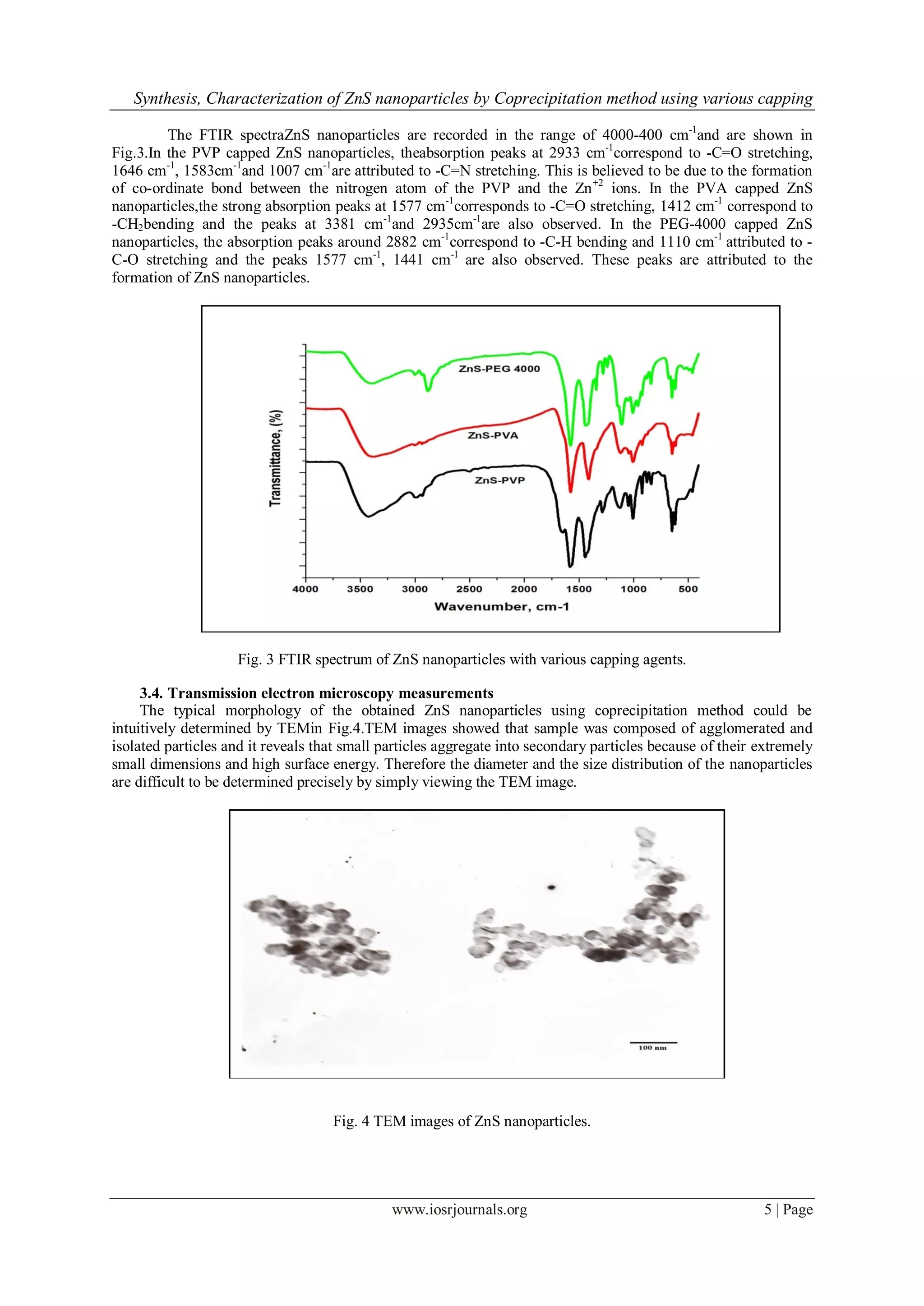
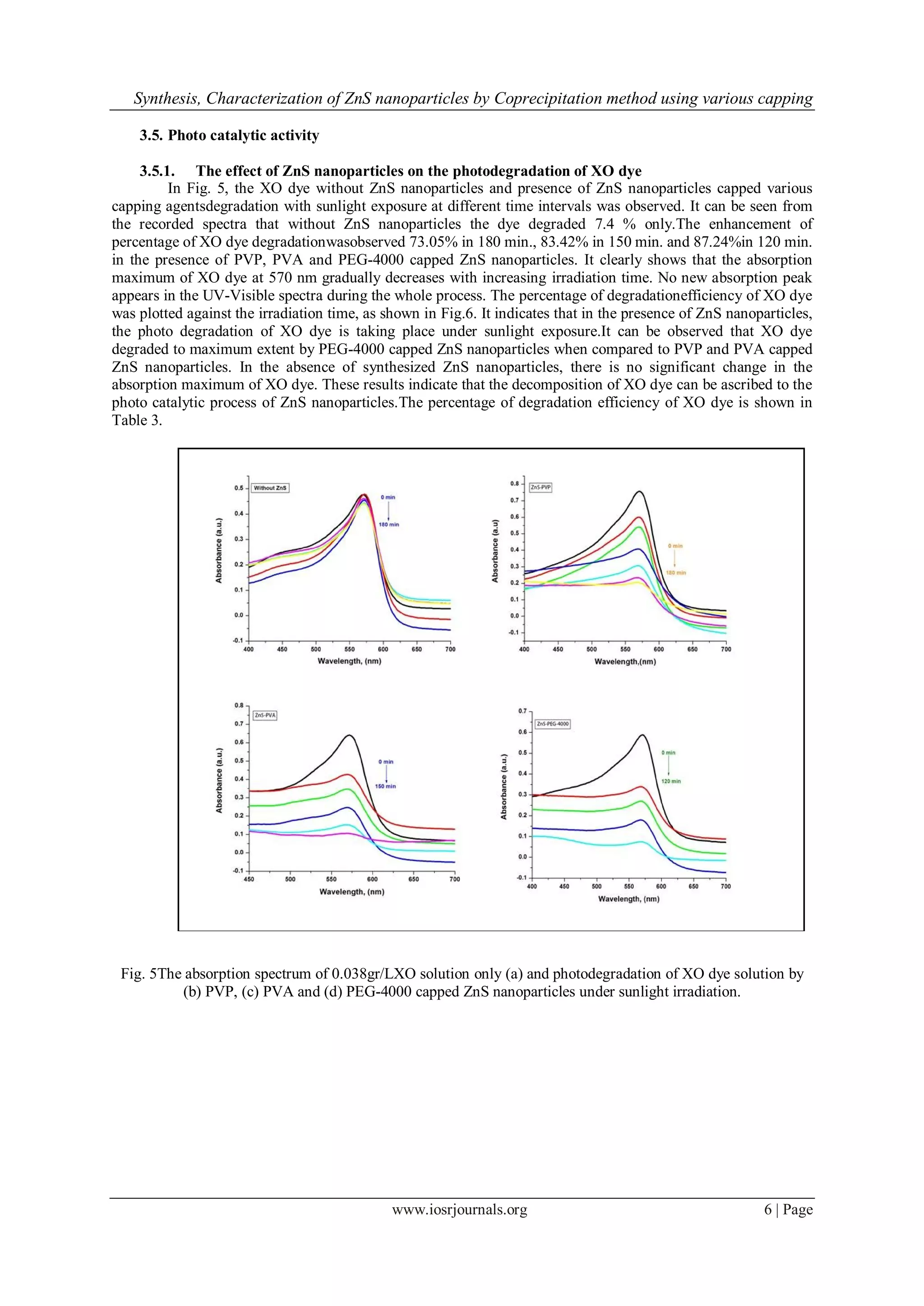
![Synthesis, Characterization of ZnS nanoparticles by Coprecipitation method using various capping
www.iosrjournals.org 7 | Page
Fig. 6 The effect of XO dye degradation by various capping agents capped ZnS nanoparticles.
S.No Time (min) % of Degradation of Dye
ZnS-PVP ZnS-PVA ZnS-PEG-4000
1 0 0 0 0
2 30 20.74 33.28 42.34
3 60 28.66 45.78 54.25
4 90 46.23 61.40 69.55
5 120 59.44 76.25 87.24
6 150 69.22 83.42 -
7 180 73.05 - -
Table 3.Degradation efficiency of dye in the presence of ZnS nanoparticles with various capping agents.
3.5.2. The effect of capping agents and kinetic rate constants
The kinetics of XO photo degradation catalyzed by synthesized ZnS nanoparticles was studied under
sunlight. The photo catalytic degradation of various organic compounds such as dyes in the presence of a
heterogeneous photo catalyst can be formally described by the Langmuir-Hinshelwood Kinetics model [34].
Rate (r) =dC/dt =k KC/1+KC
For low concentrations of dyes (KC<<1), neglecting KC in the denominator and integrating with respect to time
t, the above equation can be simplified to the pseudo-first order kinetic model equation.
ln Co/Ct =k Kt =Kapp t then the equation is modified as ln Ao/At =Kapp t
Where, dC/dt is the rate of dye degradation (mg/L. min.), k is the reaction rate constant (min-1
), K is the
adsorption co-efficient of the dye on to the photo catalyst particle (L/mg.) and Kapp(min-1
) is the apparent rate
constant calculated from the curves. Fig.7shows the ln Ao/At values plotted against time of degradation ofXO
dyewith different capping agents catalyzed by ZnS nanoparticles. The apparent rate constants of degradation
with different capping agents with XO dye, Kappwere determined from the slopes of the plotsand are in
accordance to the proposed pseudo-first order kinetic model. The apparent rate constants are given in Table 4,
which show photo degradation of XO dye decreases from PEG-4000 to PVA and PVP.](https://image.slidesharecdn.com/a0610109-150425012754-conversion-gate02/75/Synthesis-Characterization-of-ZnS-nanoparticles-by-Coprecipitation-method-using-various-capping-agents-Photocatalytic-activity-and-Kinetic-study-7-2048.jpg)
![Synthesis, Characterization of ZnS nanoparticles by Coprecipitation method using various capping
www.iosrjournals.org 8 | Page
Fig. 7 Kinetic study of photo degradation of XO in the presence of ZnS nanoparticles.
Sample Rate constants
ZnS-PVP 7.4×10⁻³
min-1
ZnS-PVA 11.7×10⁻³
min-1
ZnS-PEG-4000 15.5×10⁻³
min-1
Table 4. Values of apparent rate constants Kapp (min-1
) for the degradation of 5×10⁻⁵
M XO in the presence of
ZnS nanoparticles synthesized using various capping agents.
IV. Conclusion
The simple synthesis of the ZnS nanoparticles by coprecipitation method using various capping agents
like PVP, PVA and PEG-4000 is reported. The UV-Visiblespectra revealed that there is a blue shift of
absorption band from the bulk. FTIR spectra supported theformation of ZnS nanoparticles.In XRD spectra the
particle size (2-4 nm) was calculated from the Debye-Scherrer formula. TEM analysis indicates that the ZnS
nanoparticles are small in size.The photo catalytic degradation ofXO illustrates that this newly synthesized
PEG-4000 capped ZnS nanoparticles could be used as a promising catalytic degradation material than PVP
capped ZnS and PVA capped ZnS nanoparticles. The kinetic rate constant of degradation is higher in the
presence of PEG-4000 capped ZnS nanoparticles.
Acknowledgements
The authors would like to acknowledge Department of Biotechnology lab, Osmania University for
providing necessary facilities. One of the authors D.Ayodhya wishes to thank University Grants Commission for
the award of Junior Research Fellowship.
References
[1]. A.D. Dinsmore, D.S. Hsu, H.F. Gray, S.B. Qadri, Y. Tian, B.R. Ratna, Mn-doped ZnS nanoparticles as efficient low-voltage
cathodoluminescent phosphors. Appl. Phys. Lett., 75, 1999, 802-804.
[2]. R. Maity, K.K. Chattopadhyay, Synthesis and optical characterization of ZnS and ZnS:Mnnanocrystalline thin films by chemical
route. Nanotechnology, 15, 2004, 812-816.
[3]. J. Tittel, W. Gohde, F. Koberling, Th. Basche, A. Kornowski, H. Weller, A. Eychmuller, Fluorescence Spectroscopy on Single
CdSNanocrystals. J. Phys. Chem. B, 101, 1997, 3013-3016.
[4]. S. Mahamuni, K. Borgohain, B.S. Bendre, V.J. Leppert, S.H. Risbud, Spectroscopic and structural characterization of
electrochemically grown ZnO quantum dots. J. Appl. Phys., 85, 1999, 2861-2865.
[5]. A.L. Rogach, NanocrystallineCdTe and CdTe(S) particles: wet chemical preparation, size-dependent optical properties and
perspectives of optoelectronic applications. Mater. Sci. Eng. B, 69-70, 2000, 435-440.
[6]. L. Wang, X. Xu, X. Yuan, Preparation and photoluminescent properties of doped nanoparticles of ZnS by solid-state reaction.
Journal of Luminescence, 130, 2010, 137-140.](https://image.slidesharecdn.com/a0610109-150425012754-conversion-gate02/75/Synthesis-Characterization-of-ZnS-nanoparticles-by-Coprecipitation-method-using-various-capping-agents-Photocatalytic-activity-and-Kinetic-study-8-2048.jpg)
![Synthesis, Characterization of ZnS nanoparticles by Coprecipitation method using various capping
www.iosrjournals.org 9 | Page
[7]. S. Saravanakumar, M. Abdul Khadar, S.K. Dhara, T.R. Ravindran, K.G.M. Nair, Photoluminescence and Raman studies of ZnS
nanoparticles implanted with Cu+ ions. Nuclear Instruments and Methods in Physics Research B, 251, 2006, 435-440.
[8]. Z. Deng, J. Qi, Y. Zhang, Q. Liao, Y. Huang, Growth mechanism and optical properties of ZnS nanotetrapods. Nanotechnology, 18,
2007, 475603 (4pp).
[9]. H. Fendler, F.C. Meldrum, The Colloid Chemical Approach to Nanostructured Materials. Adv. Mater., 7, 1995, 607-632.
[10]. N. Lopez, T.V.W. Janssens, B.S. Clausen, Y. Xu, M. Mavrikakis, T. Bligaard, J.K. Norskov, On the origin of the catalytic activity
of gold nanoparticles for low-temperature CO oxidation. J. Catal., 223, 2004, 232-235.
[11]. A.P. Alivisatos, Semiconductor Clusters, Nanocrystals, and Quantum Dots.Science, 271, 1996, 933-937.
[12]. H.S. Yang, P.H. Holloway, B.B. Ratna, Photoluminescent and electroluminescent properties of Mn-doped ZnS nanocrystals. J.
Appl. Phys., 93, 2003, 586-592.
[13]. J.P. Borah, K.C. Sarma, Optical and Optoelectronic Properties of ZnS Nanostructured Thin Film. Acta. Phys. Polon. A, 114, 2008,
713-719.
[14]. C. Wang, Q. Li, B. Hu, Optoelectronic characterization of ZnS/PS systems. Chin. Opt. Lett., 7, 2009, 432-434.
[15]. R. Maity, U.N. Maiti, M.K. Mitra, K.K. Chattopadhyay, Synthesis and optical characterization of polymer-capped nanocrystalline
ZnS thin films by chemical process. Physica E, 33, 2006, 104-109.
[16]. X. Cheng, Q. Zhao, Y. Yang, S.C. Tjong, R.K.Y. Li, A facile method for the synthesis of ZnS/polystyrene composite particles and
ZnS hollow micro-spheres. J. Mater. Sci., 45, 2010, 777-782.
[17]. J.L. Yuan, K. Kajiyoshi, K. Yanagisawa, H. Sasaoka, K. Nishimura, Fabrication of silica nanocoatings on ZnS-type phosphors via a
sol–gel route using cetyltrimethylammonium chloride dispersant. Mater.Lett., 60, 2006, 1284-1286.
[18]. I. Shafiq, A. Sharif, L.C. Sing, ZnSxSe1−x nanowire arrays with tunable optical properties grown on ZnS nanoribbon substrates.
Physica E, 41, 2009, 739-745.
[19]. X.H. Liao, J.J. Zhu, H.Y. Chen, Microwave synthesis of nanocrystalline metal sulfides in formaldehyde solution. Mater. Sci. Eng.
B, 85, 2001, 85-89.
[20] J.J. Zhu, M. Zhou, J. Xu, X. Liao, Preparation of CdS and ZnS nanoparticles using microwave irradiation. Mater.Lett., 47, 2001, 25-
29.
[21]. Q. Zhao, L. Hou, R. Huang, Synthesis of ZnS nanorods by a surfactant-assisted soft chemistry method. Inorg. Chem. Commun., 6,
2003, 971-973.
[22]. C. Wang, Y.H. Ao, P.F. Wang, S.H. Zhang, J. Qian, J. Hou, A simple method for large-scale preparation of ZnS nanoribbon film
and its photocatalytic activity for dye degradation. Appl. Surf. Sci., 256, 2010, 4125-4128.
[23]. J. Pyun, K. Matyjaszewski, Synthesis of Nanocomposite Organic/Inorganic Hybrid Materials Using Controlled/“Living” Radical
Polymerization. Chem. Mater., 13, 2001, 3436-3448.
[24]. J.Y. Liao, K.C. Ho, A photovoltaic cell incorporating a dye-sensitized ZnS/ZnO composite thin film and a hole-injecting PEDOT
layer. Sol. Energy Mater. Sol. Cells., 86, 2005, 229-241.
[25]. Y. Li, X.Y. He, M.H. Cao, Micro-emulsion-assisted synthesis of ZnS nanospheres and their photocatalytic activity.Mater. Res.
Bull., 43, 2008, 3100-3110.
[26]. H.F. Shi, X.K. Li, D.F. Wang, Y.P. Yuan, Z.G. Zou, J.H. Ye, NaNbO3 Nanostructures: Facile Synthesis, Characterization, and Their
Photocatalytic Properties. Catal.Lett., 132, 2009, 205-212.
[27]. A.J. Hoffman, G. Mills, H. Yee, M.R. Hoffmann, Q-sized cadmium sulfide: synthesis, characterization and efficiency of photo
initiation of polymerization of several vinylic monomers. J. Phys. Chem., 96, 1992, 5546-5552.
[28]. J.F. Suyver, S.F. Wuister, J.J. Kelly, A. Meijerink, Synthesis and Photoluminescence of Nanocrystalline ZnS:Mn2+
. Nano Lett., 1,
2001, 429-433.
[29]. S.B. Qadri, E.F. Skelton, D. Hsu, A.D. Dinsmore, J. Yang, H.F. Gray, B.R. Ratna, Size-induced transition-temperature reduction in
nanoparticles of ZnS. Phys. Rev. B, 60, 1999, 9191-9193.
[30]. A. Guinier, X-ray diffraction(SanFrancisco, CA, 1963).
[31]. Tran ThiQuynhHoa, Le Van Vu, Ta DinhCanh, Nguyen Ngoc Long, Preparation of ZnS nanoparticles by hydrothermal method.
Journal of Physics: Conf. Ser., 187, 2009, 012081.
[32]. M. Miyake, K. Murase, T. Hirato, Y. Awakura, Hall effect measurements on CdTe layers electrodeposited from acidic aqueous
electrolyte. J. Electroanal Chem., 562, 2004, 247-253.
[33]. K. Nakamoto, Infrared spectra of inorganic and coordination compounds (Wiley, New York, 1963).
[34]. H. Al-Ekabi, N. Serpone, Kinetic Studies in Heterogeneous Photocatalysis. 1. Photocatalytic Degradation of Chlorinated Phenols in
Aerated Aqueous Solutions over TiO2 Supported on a Glass Matrix.J. Phys. Chem., 92, 1988, 5726-5731.](https://image.slidesharecdn.com/a0610109-150425012754-conversion-gate02/75/Synthesis-Characterization-of-ZnS-nanoparticles-by-Coprecipitation-method-using-various-capping-agents-Photocatalytic-activity-and-Kinetic-study-9-2048.jpg)

The document discusses the synthesis and characterization of ZnS nanoparticles using the coprecipitation method with various capping agents (PVP, PVA, and PEG-4000). It details the optical properties, particle sizes, and photocatalytic activities of the nanoparticles, showing that their size and degradation efficiency vary with the type of capping agent used. The degradation of the dye xylenol orange under sunlight indicates that the ZnS nanoparticles exhibit effective photocatalytic properties, with significant dye degradation observed over specified time intervals.


![Thin_Film_Technology_introduction[1]](https://cdn.slidesharecdn.com/ss_thumbnails/1b4496c8-2102-411b-8465-a3dd3f398327-150205034538-conversion-gate02-thumbnail.jpg?width=640&height=640&fit=bounds)