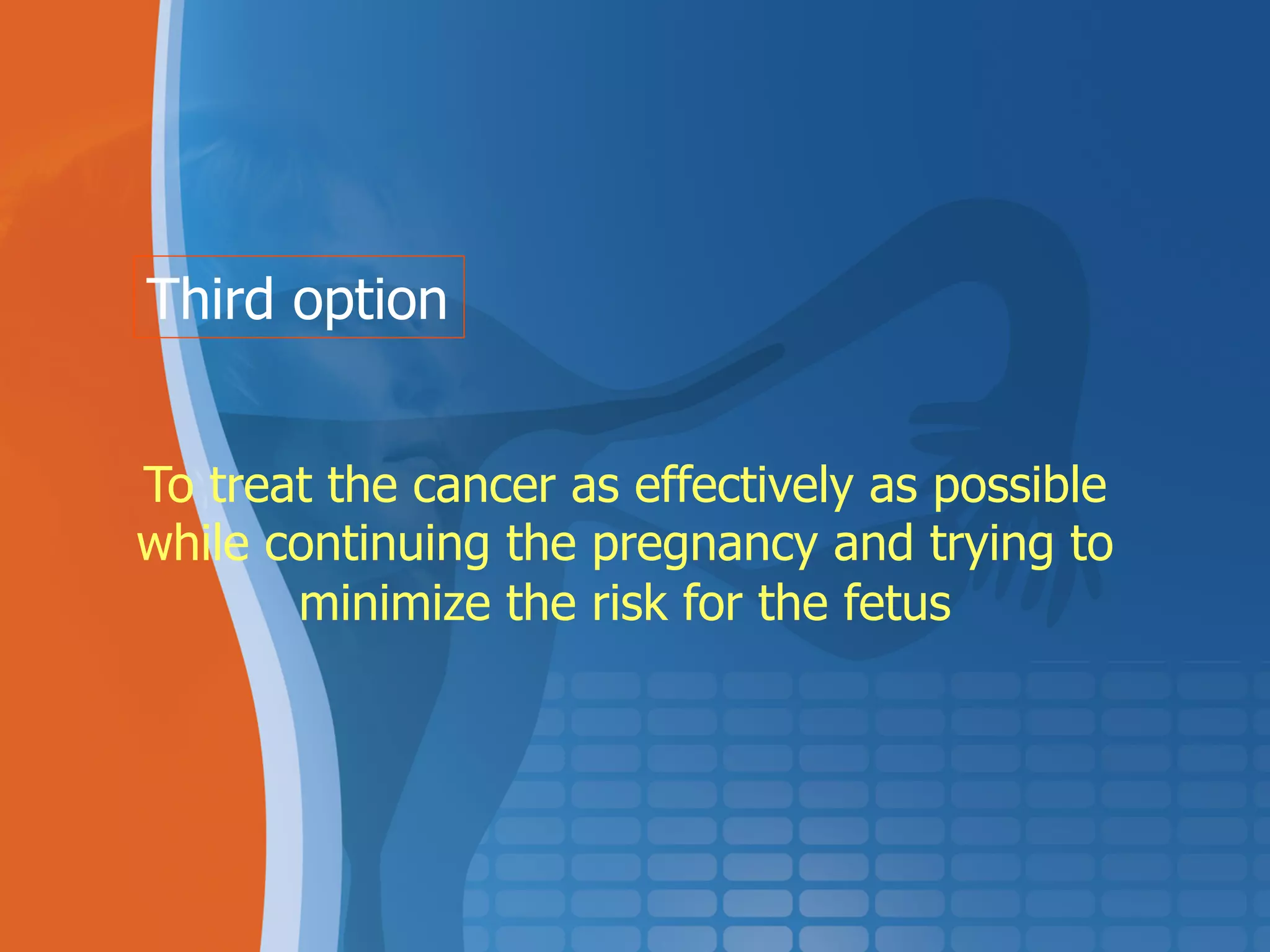
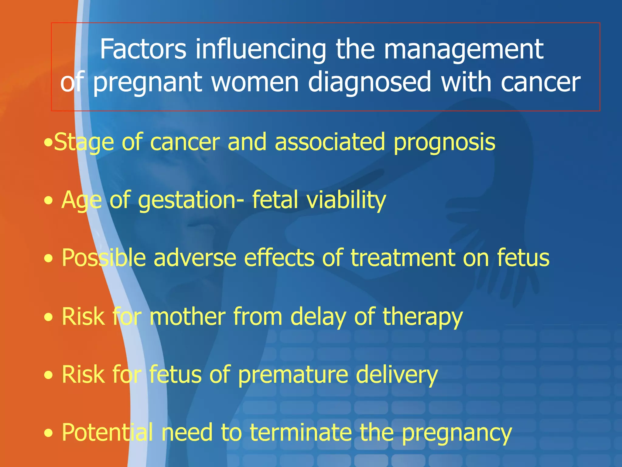
Cancer in Pregnancy
March 2012
Prafull Ghatage
Gynecologic Oncologist
Tom Baker Cancer Centre
Calgary, CANADA.
Learning Objectives
•  Incidence
•  Investigations and management issues
•  Risks of surgery
•  Risks of radiotherapy
•  Risks of chemotherapy
•  Termination - ? Necessary / Advantageous
•  If delivery - ? how / ? When
•  Future pregnancies
The occurrence of cancer in pregnancy
is rare,
about 1 case per 1000 deliveries
The Dilemma
? Save the mother
? Save the baby
Malignant disease in pregnancy
complicates the management of
both cancer
and the pregnancy.
Is the potential life of an unborn child
more important than prolonging a life of a young woman?
And whose decision is this ?
?
Fetus Mother
Pregnancy
Risk
Management of cancer in pregnancy
There are not many options and
none of them are ideal
For women diagnosed with cancer
waiting for 40 weeks
could be a death sentence
particularly with high-grade, aggressive
or metastatic cancers.
To delay treatment until the child
can be safely delivered
•  For mother this poses the risk that may be hard to quantify
•  It also means that she will have to care for a very premature
baby while coping with the side-effects of cancer treatment
This option is more viable
the lower the risk posed by the cancer and
the more advanced the pregnancy
First option
To terminate the pregnancy to allow normal
treatment to go ahead
•  This may be the safest option for the mother s health
•  Unacceptable to some mothers
More likely to be considered early in pregnancy
Second option
To treat the cancer as effectively as possible
while continuing the pregnancy and trying to
minimize the risk for the fetus
Third option
Problems in treatment of cancer in pregnancy
•  Late diagnosis
•  Damaging effects of radiotherapy
•  Consequences of chemotherapy
Delay in diagnostics
•  Presenting symptoms often attributed to pregnancy
•  Anatomical and physiological changes of
pregnancy may compromise the physical examination
•  Tumor markers are increased in pregnancy
(beta HCG, AFP, CA 125... )
•  Imaging techniques or invasive procedures
Cancer in pregnancy is often detected later
because the symptoms are masked by other,
usually physiological, body changes
Cancer in young women
Site Age 15-44 (%)
Cervix 35
Ovary 15
Lymphoma 23
Thyroid 50
Melanoma 27
Breast 15
Leukemia 10
Cancer incidence in Pregnancy
Site Est. incidence / 1000 pregnancies
Cervix
Non-invasive
Invasive
1.3-1.7
1.0
Breast 0.33 – 0.7
Melanoma 0.14
Ovary 0.10 ( 0.01%)
Thyroid Unknown
Lymphoma 0.01
Leukemia 0.01
Colon 0.20
Factors influencing the management
of pregnant women diagnosed with cancer
• Stage of cancer and associated prognosis
•  Age of gestation- fetal viability
•  Possible adverse effects of treatment on fetus
•  Risk for mother from delay of therapy
•  Risk for fetus of premature delivery
•  Potential need to terminate the pregnancy
Diagnostic procedures that are
SAFE in pregnancy:
•  Ultrasound
• Magnetic resonance imaging (MRI)
Difficulties in diagnostics & staging
Some techniques are unreliable
•  Mammogram
•  Blood tests- tumor markers
Some techniques are dangerous
•  CT scan
•  Radioisotope investigations
•  Cervical conisation
Effective Radiation dose with
background radiation exposure
Procedure Radiation
dose in
millisieverts
Comparable
to
background
radiation for:
Additional lifetime
risk of fetal cancer
CXR 0.1 5 years Low
Abdominal
XR
2.2 9 months Very low
CT abd/
Pelvis
15 5 years Low
CT abd/pelvis
with contrast
30 10 years Moderate
Mammogram 0.4 7 weeks Very low
1 cGy = 1J/kg; 1 Sv = 1J/kg.
Three stages of Embryogenesis
•  I – First 2 weeks – Blastocyst resistant to teratogens.
Blastocyst has NOT differentiated.
•  II – Organogenesis – 3rd to 8th week. Maximal
teratogenesis. Ends by 13th week
•  III – Increase in fetal and organ size. CNS development
complete by 16 weeks. Brain and gonadal tissue
will continue to develop. Teratogens will cause
IUGR but no organ malformation
Radiotherapy
•  Contraindicated in pregnancy
•  Possible in early pregnancy with lead
shielding
•  Maybe also consider in late pregnancy for
the chest with shielding
Risks of radiotherapy
Therapeutic doses of 5000-6000 cGy
expose the fetus to 10 cGy in early pregnancy
and 200 cGy or more in later pregnancy
Doses over 2.5-5 cGy pose high risk
for malformation early in pregnancy
With 10 cGy the risk is 50%
Conception to days 9/10 Lethal
Weeks 2-6 Malformation
Growth retardation
Weeks 12-16 Mental and growth
retardation, microcephaly
Weeks 20-25 to birth Sterility, malignancies,
genetic disorders
Effects of radiotherapy
Risks of chemotherapy
Almost all drugs cross the placental
barrier to some extent
As chemotherapeutic drugs work
by inhibiting cell division,
they pose a risk to the developing fetus.
Risks of chemotherapy
•  Spontaneous abortion
•  Malformations
•  Teratogenesis
•  Mutations
•  Carcinogenesis
•  Organ toxicity
•  Retarded development
First trimester
•  Most likely in the 1st trimester.
•  Fetal malformation rate 12.7-17% with single-drug
regimens and up to 25% with combination regimens
(cf - general population rate 1-3%)
•  Low birth weight ~ 40%
Second and third trimester
•  Relatively low risk
•  It is preferable to wait until the development of CNS
is complete, around 16 weeks
Risks of chemotherapy
Delivery
If a baby is delivered within 2 weeks of the last
chemotherapy dose, there is a risk of a neutropenic baby
being born to a neutropenic mother
Breastfeeding
Breast feeding is not advisable for women who have
recently been on chemotherapy
Risks of chemotherapy
Lancet Oncology ,Amant et al, Feb 2012
•  68 pregnancies
•  236 cycles of chemotherapy
•  Safe in the 2nd and 3rd trimester
•  No association with CNS, Cardiac or Auditory morbidity.
Risks of chemotherapy
Pre-invasive and invasive cervical
cancer in pregnancy
Incidence of cervical pre-invasive
and invasive cancer in pregnant women
is similar to the incidence in general population
Pregnant women (4230) 0.17%
Non-pregnant women (107230) 0.18%
Bokhman JV, 1998.
The disease has been detected
during the pregnancy or postpartum
period
in 1.7 to 3.1%.
In reproductive age ≈10%
Creasman WT et al., 1970
Screening for invasive cervical cancer
should be performed during
the first antenatal examination
Harper DM, Roach MS. J Fam Pract, 1996; 42: 79-83
Management of abnormal cervical smear
during pregnancy
Pregnancy is not a contraindication for a pap smear
Abnormal cytology (5%)
Colposcopy
Biopsy
Indications for colposcopy
•  Clinically SUSPICIOUS cervix
•  Recurrent and otherwise unexplained
BLEEDING
•  ABNORMAL pap
The aim of colposcopic examination
during the pregnancy
is to exclude invasion
Normal cervix in pregnancy
Normal cervix at 24 weeks
HPV in pregnancy
CIN III in pregnancy (ASCPP)
Microglandular Hyperplasia
Microinvasive cancer (ASCCP)
Early invasive cancer (ASCCP)
Decidual reaction
Conization in pregnancy
•  MICROINVASION confirmed by biopsy
•  Pap suggestive of INVASION
•  ??? Unsatisfactory colposcopic
examination in a histologically proven
high grade lesion
Management after the histological finding in pregnancy
CIN Microinvasive cancer Invasive cancer
Conization
Postpone further Radical
diagnostic and hysterectomy
therapeutic procedures or
for post-partum period radiotherapy
Targeted biopsy
Treatment of cervical cancer in pregnancy
is affected
•  by the stage of the disease
•  by the age of gestation
•  mother’s belief regarding pregnancy termination
•  future childbearing desires
The treatment of invasive cervical cancer
in pregnancy
should proceed without regard for the fetus,
unless the lesion is diagnosed at a stage
close to fetal viability
Stage Ib/ IIa
Cervical cancer in pregnancy
I trimester: Surgery with embryo in utero
III trimester: Radical Caesarean hysterectomy
II trimester ? Medical and ethical problem
Invasive cervical cancer in second trimester
Before 20-24 weeks
Evacuating pregnancy by hysterotomy
and immediately after radical hysterectomy
After 24-28 weeks
Waiting for fetal maturity
Delay of treatment for 2-10 weeks
•  Small tumor
•  Stage < IIB
•  Gestational age > 20 weeks
van Villet W i sar. Eur J Obst Gynec Reprod Biol, 1998; 79: 153-7
Adnexal masses during pregnancy
1:1000 deliveries
Most masses are benign. Malignant tumors are
generally low grade and stage with survival of
75%
Ovarian cancer 1 per 10.000 – 100.000 births
Ovarian tumors and the pregnancy
Most frequent types of ovarian tumors
in pregnancy
Benign cystic teratoma ................. 36%
Serous cystadenoma ................ 25%
Mucinous cystadenoma ................. 12%
Corpus luteum cyst ................. 5.5%
Malignant tumors ................ 4%
Malignant ovarian tumors and pregnancy
In non-pregnant woman 20% ovarian tumors
are malignant.
In pregnancy this percentage is
decreased to 5% ( 3% - 9.7%)
- - Epithelial carcinomas 33-65%
- - Germ-cell tumors 17-40%
- - Sex cord-stromal tumors 9-13%
Malignant ovarian tumors and pregnancy
•  Only 16% of ovarian tumors detected in the first
trimester
•  20% diagnosed during CS or after delivery
•  Almost 25% have an acute presentation (torsion)
If there are no complications, the best timing for surgery
of persistent ovarian mass in pregnancy is between
16 to18 weeks of gestation
If adnexal mass is < 8 cm, unilateral,
mobile and asymptomatic:
- observation and repeat U/S at 14 to 16
weeks.
If adnexal mass is > 8 cm, solid or of complex
appearance, bilateral or persists into 2nd
trimester:
- laparotomy
Management of ovarian mass in pregnancy
Breast cancer
•  3% of breast cancers is associated with pregnancy
•  In the reproductive period patients, breast cancer
associated with pregnancy in 14% cases
•  The incidence of breast cancer in pregnancy is 0.03
(1: 3000-1:10 000 pregnancies)
•  Pregnant women have a 2.5 fold higher risk to
present with advanced cancer
Breast cancer in pregnancy
•  Delay in starting the treatment is not recommended
•  Mastectomy with axillary lymph node dissection
does not jeopardise pregnancy
•  Conservative surgery ?
•  Chemotherapy can be administered in pregnancy
•  There is no consensus regarding radiotherapy
Survival is equal as in non-pregnant patients
if the stage of the disease is considered
Breast cancer in pregnancy
•  Later pregnancies do not influence overall survival
•  Next pregnancy should not be planned at least
for 2 years after treatment
The patient, her partner and her doctor
are required
to take a difficult decision without always a
clear answer
(rights of the fetus ≠ rights of the mother)
When should therapeutic abortion
be recommended?
Therapeutic abortion- general considerations
- Absence of guidelines.
- Final decision is not always easy
- Issue becomes more important when cancer
diagnosis is made during the first trimester
Most important parameters are:
- the stage
- the indication for treatment
- the curability of the disease.
Recommendations for therapeutic abortion
during the first trimester
1. Primary aggressive breast cancer
2. Advanced breast cancer
3. Stage III-IV aggressive NHL or
Hodgkin s disease
4. Acute leukemia
Conclusion
•  Malignancy is rare in pregnancy
•  Consideration of mother and fetus
•  Close coordination also required between
patient, obstetrician, neonatologist and
oncologist
Cancer in pregnancy march 2012 ghatage co

Cancer in pregnancy march 2012 ghatage co

  • 1.
    Cancer in Pregnancy March2012 Prafull Ghatage Gynecologic Oncologist Tom Baker Cancer Centre Calgary, CANADA.
  • 2.
    Learning Objectives •  Incidence • Investigations and management issues •  Risks of surgery •  Risks of radiotherapy •  Risks of chemotherapy •  Termination - ? Necessary / Advantageous •  If delivery - ? how / ? When •  Future pregnancies
  • 3.
    The occurrence ofcancer in pregnancy is rare, about 1 case per 1000 deliveries
  • 4.
    The Dilemma ? Savethe mother ? Save the baby Malignant disease in pregnancy complicates the management of both cancer and the pregnancy.
  • 5.
    Is the potentiallife of an unborn child more important than prolonging a life of a young woman? And whose decision is this ? ?
  • 6.
  • 7.
    Management of cancerin pregnancy There are not many options and none of them are ideal
  • 8.
    For women diagnosedwith cancer waiting for 40 weeks could be a death sentence particularly with high-grade, aggressive or metastatic cancers.
  • 9.
    To delay treatmentuntil the child can be safely delivered •  For mother this poses the risk that may be hard to quantify •  It also means that she will have to care for a very premature baby while coping with the side-effects of cancer treatment This option is more viable the lower the risk posed by the cancer and the more advanced the pregnancy First option
  • 10.
    To terminate thepregnancy to allow normal treatment to go ahead •  This may be the safest option for the mother s health •  Unacceptable to some mothers More likely to be considered early in pregnancy Second option
  • 11.
    To treat thecancer as effectively as possible while continuing the pregnancy and trying to minimize the risk for the fetus Third option
  • 12.
    Problems in treatmentof cancer in pregnancy •  Late diagnosis •  Damaging effects of radiotherapy •  Consequences of chemotherapy
  • 13.
    Delay in diagnostics • Presenting symptoms often attributed to pregnancy •  Anatomical and physiological changes of pregnancy may compromise the physical examination •  Tumor markers are increased in pregnancy (beta HCG, AFP, CA 125... ) •  Imaging techniques or invasive procedures
  • 14.
    Cancer in pregnancyis often detected later because the symptoms are masked by other, usually physiological, body changes
  • 15.
    Cancer in youngwomen Site Age 15-44 (%) Cervix 35 Ovary 15 Lymphoma 23 Thyroid 50 Melanoma 27 Breast 15 Leukemia 10
  • 16.
    Cancer incidence inPregnancy Site Est. incidence / 1000 pregnancies Cervix Non-invasive Invasive 1.3-1.7 1.0 Breast 0.33 – 0.7 Melanoma 0.14 Ovary 0.10 ( 0.01%) Thyroid Unknown Lymphoma 0.01 Leukemia 0.01 Colon 0.20
  • 17.
    Factors influencing themanagement of pregnant women diagnosed with cancer • Stage of cancer and associated prognosis •  Age of gestation- fetal viability •  Possible adverse effects of treatment on fetus •  Risk for mother from delay of therapy •  Risk for fetus of premature delivery •  Potential need to terminate the pregnancy
  • 18.
    Diagnostic procedures thatare SAFE in pregnancy: •  Ultrasound • Magnetic resonance imaging (MRI)
  • 19.
    Difficulties in diagnostics& staging Some techniques are unreliable •  Mammogram •  Blood tests- tumor markers Some techniques are dangerous •  CT scan •  Radioisotope investigations •  Cervical conisation
  • 20.
    Effective Radiation dosewith background radiation exposure Procedure Radiation dose in millisieverts Comparable to background radiation for: Additional lifetime risk of fetal cancer CXR 0.1 5 years Low Abdominal XR 2.2 9 months Very low CT abd/ Pelvis 15 5 years Low CT abd/pelvis with contrast 30 10 years Moderate Mammogram 0.4 7 weeks Very low 1 cGy = 1J/kg; 1 Sv = 1J/kg.
  • 21.
    Three stages ofEmbryogenesis •  I – First 2 weeks – Blastocyst resistant to teratogens. Blastocyst has NOT differentiated. •  II – Organogenesis – 3rd to 8th week. Maximal teratogenesis. Ends by 13th week •  III – Increase in fetal and organ size. CNS development complete by 16 weeks. Brain and gonadal tissue will continue to develop. Teratogens will cause IUGR but no organ malformation
  • 22.
    Radiotherapy •  Contraindicated inpregnancy •  Possible in early pregnancy with lead shielding •  Maybe also consider in late pregnancy for the chest with shielding
  • 23.
    Risks of radiotherapy Therapeuticdoses of 5000-6000 cGy expose the fetus to 10 cGy in early pregnancy and 200 cGy or more in later pregnancy Doses over 2.5-5 cGy pose high risk for malformation early in pregnancy With 10 cGy the risk is 50%
  • 24.
    Conception to days9/10 Lethal Weeks 2-6 Malformation Growth retardation Weeks 12-16 Mental and growth retardation, microcephaly Weeks 20-25 to birth Sterility, malignancies, genetic disorders Effects of radiotherapy
  • 25.
    Risks of chemotherapy Almostall drugs cross the placental barrier to some extent As chemotherapeutic drugs work by inhibiting cell division, they pose a risk to the developing fetus.
  • 26.
    Risks of chemotherapy • Spontaneous abortion •  Malformations •  Teratogenesis •  Mutations •  Carcinogenesis •  Organ toxicity •  Retarded development
  • 27.
    First trimester •  Mostlikely in the 1st trimester. •  Fetal malformation rate 12.7-17% with single-drug regimens and up to 25% with combination regimens (cf - general population rate 1-3%) •  Low birth weight ~ 40% Second and third trimester •  Relatively low risk •  It is preferable to wait until the development of CNS is complete, around 16 weeks Risks of chemotherapy
  • 28.
    Delivery If a babyis delivered within 2 weeks of the last chemotherapy dose, there is a risk of a neutropenic baby being born to a neutropenic mother Breastfeeding Breast feeding is not advisable for women who have recently been on chemotherapy Risks of chemotherapy
  • 29.
    Lancet Oncology ,Amantet al, Feb 2012 •  68 pregnancies •  236 cycles of chemotherapy •  Safe in the 2nd and 3rd trimester •  No association with CNS, Cardiac or Auditory morbidity. Risks of chemotherapy
  • 30.
    Pre-invasive and invasivecervical cancer in pregnancy
  • 31.
    Incidence of cervicalpre-invasive and invasive cancer in pregnant women is similar to the incidence in general population Pregnant women (4230) 0.17% Non-pregnant women (107230) 0.18% Bokhman JV, 1998.
  • 32.
    The disease hasbeen detected during the pregnancy or postpartum period in 1.7 to 3.1%. In reproductive age ≈10% Creasman WT et al., 1970
  • 33.
    Screening for invasivecervical cancer should be performed during the first antenatal examination Harper DM, Roach MS. J Fam Pract, 1996; 42: 79-83
  • 34.
    Management of abnormalcervical smear during pregnancy Pregnancy is not a contraindication for a pap smear Abnormal cytology (5%) Colposcopy Biopsy
  • 35.
    Indications for colposcopy • Clinically SUSPICIOUS cervix •  Recurrent and otherwise unexplained BLEEDING •  ABNORMAL pap
  • 36.
    The aim ofcolposcopic examination during the pregnancy is to exclude invasion
  • 37.
  • 38.
  • 39.
  • 41.
    CIN III inpregnancy (ASCPP)
  • 42.
  • 43.
  • 44.
  • 45.
  • 46.
    Conization in pregnancy • MICROINVASION confirmed by biopsy •  Pap suggestive of INVASION •  ??? Unsatisfactory colposcopic examination in a histologically proven high grade lesion
  • 47.
    Management after thehistological finding in pregnancy CIN Microinvasive cancer Invasive cancer Conization Postpone further Radical diagnostic and hysterectomy therapeutic procedures or for post-partum period radiotherapy Targeted biopsy
  • 48.
    Treatment of cervicalcancer in pregnancy is affected •  by the stage of the disease •  by the age of gestation •  mother’s belief regarding pregnancy termination •  future childbearing desires
  • 49.
    The treatment ofinvasive cervical cancer in pregnancy should proceed without regard for the fetus, unless the lesion is diagnosed at a stage close to fetal viability
  • 50.
  • 51.
    Cervical cancer inpregnancy I trimester: Surgery with embryo in utero III trimester: Radical Caesarean hysterectomy II trimester ? Medical and ethical problem
  • 52.
    Invasive cervical cancerin second trimester Before 20-24 weeks Evacuating pregnancy by hysterotomy and immediately after radical hysterectomy After 24-28 weeks Waiting for fetal maturity
  • 53.
    Delay of treatmentfor 2-10 weeks •  Small tumor •  Stage < IIB •  Gestational age > 20 weeks van Villet W i sar. Eur J Obst Gynec Reprod Biol, 1998; 79: 153-7
  • 54.
    Adnexal masses duringpregnancy 1:1000 deliveries Most masses are benign. Malignant tumors are generally low grade and stage with survival of 75% Ovarian cancer 1 per 10.000 – 100.000 births Ovarian tumors and the pregnancy
  • 55.
    Most frequent typesof ovarian tumors in pregnancy Benign cystic teratoma ................. 36% Serous cystadenoma ................ 25% Mucinous cystadenoma ................. 12% Corpus luteum cyst ................. 5.5% Malignant tumors ................ 4%
  • 56.
    Malignant ovarian tumorsand pregnancy In non-pregnant woman 20% ovarian tumors are malignant. In pregnancy this percentage is decreased to 5% ( 3% - 9.7%) - - Epithelial carcinomas 33-65% - - Germ-cell tumors 17-40% - - Sex cord-stromal tumors 9-13%
  • 57.
    Malignant ovarian tumorsand pregnancy •  Only 16% of ovarian tumors detected in the first trimester •  20% diagnosed during CS or after delivery •  Almost 25% have an acute presentation (torsion) If there are no complications, the best timing for surgery of persistent ovarian mass in pregnancy is between 16 to18 weeks of gestation
  • 58.
    If adnexal massis < 8 cm, unilateral, mobile and asymptomatic: - observation and repeat U/S at 14 to 16 weeks. If adnexal mass is > 8 cm, solid or of complex appearance, bilateral or persists into 2nd trimester: - laparotomy Management of ovarian mass in pregnancy
  • 59.
    Breast cancer •  3%of breast cancers is associated with pregnancy •  In the reproductive period patients, breast cancer associated with pregnancy in 14% cases •  The incidence of breast cancer in pregnancy is 0.03 (1: 3000-1:10 000 pregnancies) •  Pregnant women have a 2.5 fold higher risk to present with advanced cancer
  • 60.
    Breast cancer inpregnancy •  Delay in starting the treatment is not recommended •  Mastectomy with axillary lymph node dissection does not jeopardise pregnancy •  Conservative surgery ? •  Chemotherapy can be administered in pregnancy •  There is no consensus regarding radiotherapy Survival is equal as in non-pregnant patients if the stage of the disease is considered
  • 61.
    Breast cancer inpregnancy •  Later pregnancies do not influence overall survival •  Next pregnancy should not be planned at least for 2 years after treatment
  • 62.
    The patient, herpartner and her doctor are required to take a difficult decision without always a clear answer (rights of the fetus ≠ rights of the mother) When should therapeutic abortion be recommended?
  • 63.
    Therapeutic abortion- generalconsiderations - Absence of guidelines. - Final decision is not always easy - Issue becomes more important when cancer diagnosis is made during the first trimester Most important parameters are: - the stage - the indication for treatment - the curability of the disease.
  • 64.
    Recommendations for therapeuticabortion during the first trimester 1. Primary aggressive breast cancer 2. Advanced breast cancer 3. Stage III-IV aggressive NHL or Hodgkin s disease 4. Acute leukemia
  • 65.
    Conclusion •  Malignancy israre in pregnancy •  Consideration of mother and fetus •  Close coordination also required between patient, obstetrician, neonatologist and oncologist