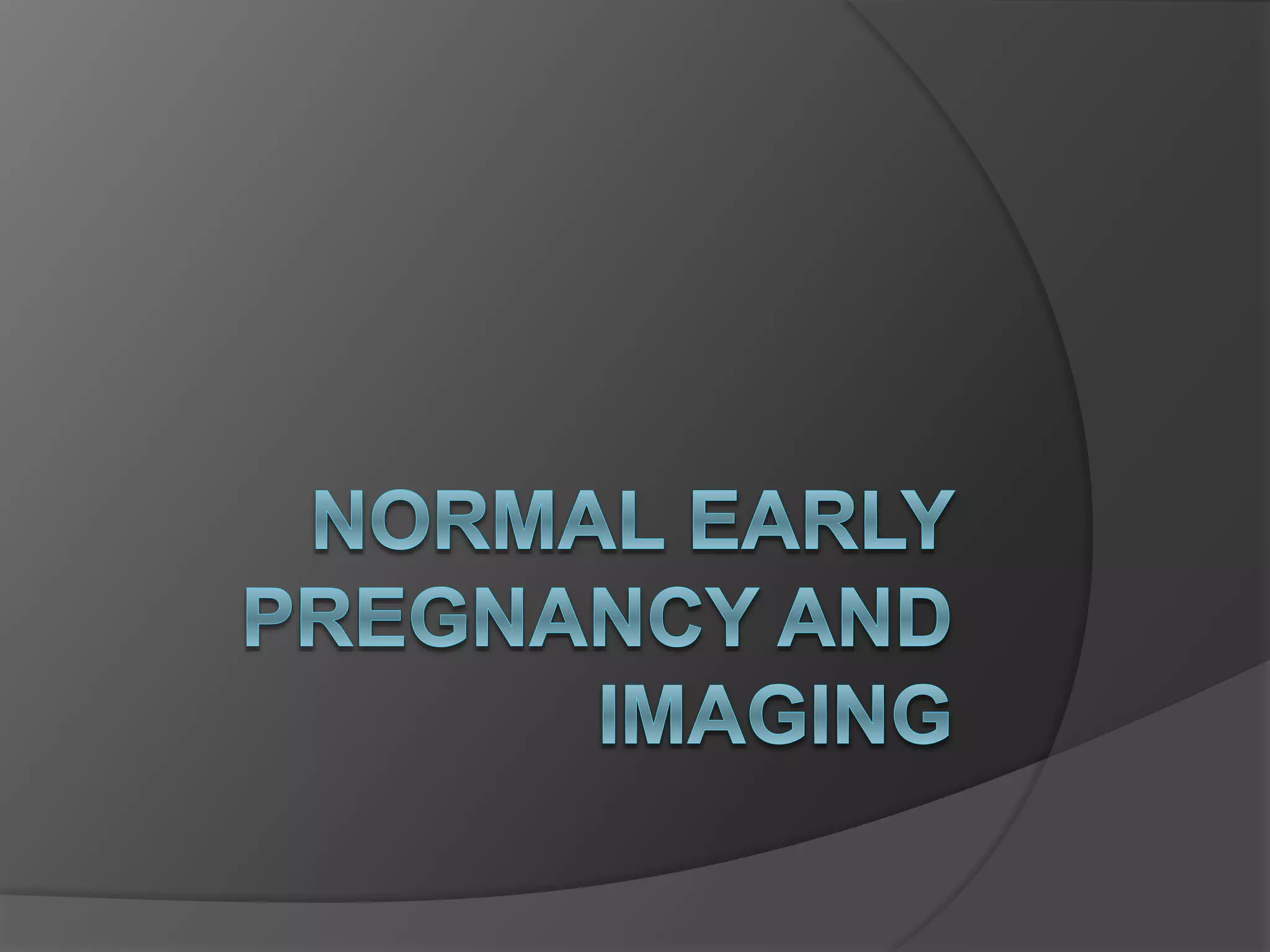
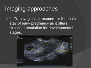
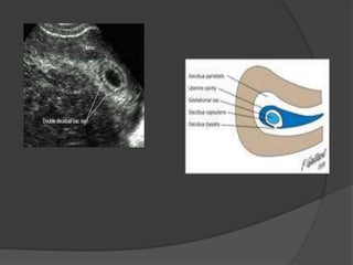
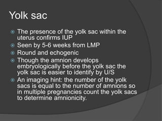
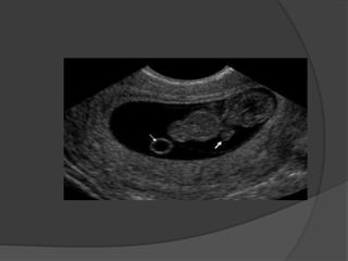
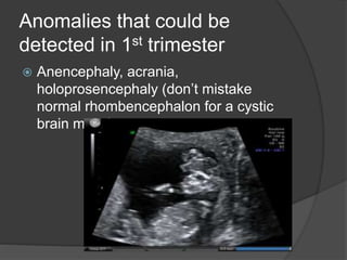
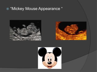
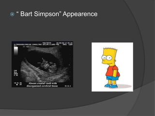
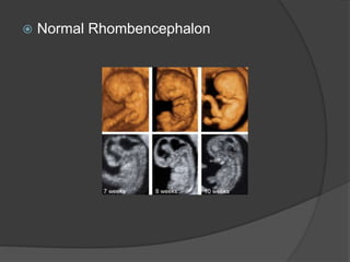
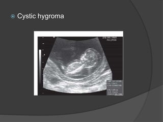
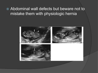
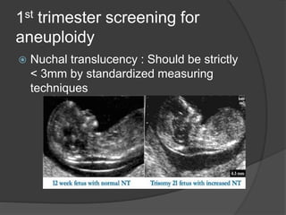
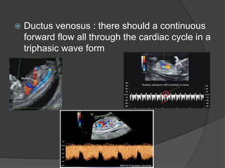
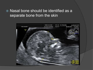
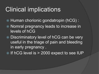
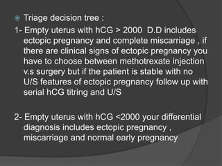
Transvaginal ultrasound is the main imaging approach in the first trimester. The double decidual sac sign appears by 4-5 weeks and confirms an intrauterine pregnancy. By 5-6 weeks, a yolk sac and embryonic heartbeat can be seen when the crown-rump length reaches 5mm. Anomalies like anencephaly can be detected. Nuchal translucency measurement and assessment of ductus venosus flow are used for first trimester aneuploidy screening. An empty uterus with hCG over 2000 suggests ectopic pregnancy or miscarriage.
2. 1st trimester : the period from fertilization of
the ovum to the start of the 13th post
menstrual week.
3. Imaging approaches
1- Transvaginal ultrasound : is the main
stay of early pregnancy as it offers
excellent resolution for developmental
stages.
4. 2- Transabdominal ultrasound : excellent for
rapid confirmation of a live IUP
5. 3- Doppler : the use of Doppler should be avoided in 1st
trimester due to concerns regarding heating effect and
embryonic damage and some recent studies correlating
the abuse of Doppler in 1st trimester and autism. The
usual Doppler sign is the “ring of fire” which is a non
specific finding as it can be found in ectopic pregnancy
and in the corpus luteum as well as normal chorionic
tissue
6. 4- M-Mode : is used to document
cardiac activity in the embryo
7. 5- MRI : current recommendations are to
avoid MRI studies as the its effect on embryo
at such early stage is not sufficiently studied
8. 6- CT : as an ionizing radiation it’s
contraindicated in 1st trimester except in
grave indications as in trauma
10. Embryologic stages
1- Embryo formation :
-Ovum + sperm = zygote
-Cleavage of zygote = morula
-Central fluid filling of the morula= blastocystic cavity
that separates trophoblast from inner cell mass and
is divided into
- trophoblast : embryonic part of the placenta
- Inner cell mass ( embryoblast) : Primordium of
embryo
- Embryoblast forms a bilaminar embryonic disc
- The embryonic disc transforms into a 3D C-shaped
embryo be the beginning of 6 week post menstrual
11.
12. Timing of some embryonic
events
Neural tube closes by day 26 (5th-6th
weeks)
13. Heart is partitioned by the end of 8th week
but cardiac activity can be detected prior to
that when the embryo reaches a length of 5-
6 mm
15. Embryo gives the recognizable human form
by the end of 10 weeks
16. Physiologic Bowel herniation : because the bowel
grows rapidly than the rest of the embryo it
herniates at the base of the cord then it
undergoes rotation within the cord then returns to
abdominal cavity . Please Note that the liver never
herniates normally.
17. Multiple Pregnancy
Multiple pregnancies and types of twinning depends on
the number of zygotes and timing of division and they are
:
1- Dizygotic twins : two fertilized ova and they are all
dichorionic , diamniotic
2- Monozygotic twins : the amnionicity and chorionicity
depends by the time of zygote division
- Before 3rd day post conception : diamniotic, dichorionic
- 4th-8th day post conception : monochorionic , diamniotic
- Cleavage of the inner cell mass of blastocyst after 8th day
post conception : monochorionic, monoamniotic.
- Incomplete cleavage of embryonic disc after 13th day post
conception : conjoined twins
22. Normal imaging milestones
1- Double decidual sac Sign (DDSS) : is the earliest
sign of IUP seen by 4-5 wks from LMP by TV U/S
formed of two echogenic rings and it’s where the
fertilized ovum implants itself into the decidualized
endometrium and formed of :
- Decidua Parietalis : outer ring lining uterine cavity
- Decidua capsularis : Inner ring covering the free
margin of the G.S
- Decidua Basalis : which the endometial base of the
sac
Decidua basalis + chorion frondosum = placenta
23.
24. Yolk sac
The presence of the yolk sac within the
uterus confirms IUP
Seen by 5-6 weeks from LMP
Round and echogenic
Though the amnion develops
embryologically before the yolk sac the
yolk sac is easier to identify by U/S
An imaging hint: the number of the yolk
sacs is equal to the number of amnions so
in multiple pregnancies count the yolk sacs
to determine amnionicity.
25.
26. The Embryo
First seen as a focal thickening in the yolk
sac
“ Diamond ring Sign”: Embryo appears as
the echogenic (diamond) on top if the yolk
sac (ring)
“ Double Bleb “ sign : yolk sac and amniotic
sac and the embryo is in the amniotic sac
Distinct embryo with cardiac activity is seen
by 5-6 weeks after LMP and a CRL of 5
mm is the discriminatory value of the
presence of cardiac activity known as the “
5 Alive “ rule.
31. Some Imaging Protocols
When your measuring the Mean Sac
Diameter (MSD) : measure only the
sonolucent area and don’t include the
echogenic chorionic rim, also measure the
sac in three different diameters and take
their average .
32. Crown – Rump length: is the most accurate
means of dating pregnancy that should be
used once an embryo is visible , remember
not to include the yolk sac in your
measurement and only measure the longest
axis of the embryo
33. Normal 1st trimesteric
Measurement
MSD increases by about 1mm per day
Sac Diameter should be about 1 cm longer
than CRL
Cord length at this stage is almost as the
embryonic length
Embryonic heart rate :
-< 6week = 110-115 bpm
- By 8 weeks = 144-159 bpm
- >9 weeks = 137-144 bpm
- A rate <90 bpm is considered embryonic
bradycardia
34. Anomalies that could be
detected in 1st trimester
Anencephaly, acrania,
holoprosencephaly (don’t mistake
normal rhombencephalon for a cystic
brain mass)
39. Abdominal wall defects but beware not to
mistake them with physiologic hernia
40. 1st trimester screening for
aneuploidy
Nuchal translucency : Should be strictly
< 3mm by standardized measuring
techniques
41. Ductus venosus : there should a continuous
forward flow all through the cardiac cycle in a
triphasic wave form
42. Nasal bone should be identified as a
separate bone from the skin
43. Questions you should be able
to answer by the end of your
scan
Is there an IUP?
Is there a definite ectopic pregnancy or
suspicious sign of it?
How many gestations are there?
If there are multiple sacs what is the
chorionicty and amnionicity?
Is there a yolk sac?
Is there an embryo?
44. Clinical implications
Human chorionic gondatropin (hCG) :
Nomral pregnancy leads to increase in
levels of hCG
Discriminatory level of hCG can be very
useful in the triage of pain and bleeding
in early pregnancy .
If hCG level is > 2000 expect to see IUP
45. Triage decision tree :
1- Empty uterus with hCG > 2000 D.D includes
ectopic pregnancy and complete miscarriage , if
there are clinical signs of ectopic pregnancy you
have to choose between methotrexate injection
v.s surgery but if the patient is stable with no
U/S features of ectopic pregnancy follow up with
serial hCG titring and U/S
2- Empty uterus with hCG <2000 your differential
diagnosis includes ectopic pregnancy ,
miscarriage and normal early pregnancy