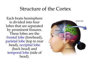
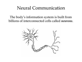
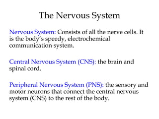
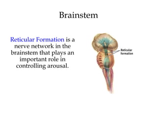
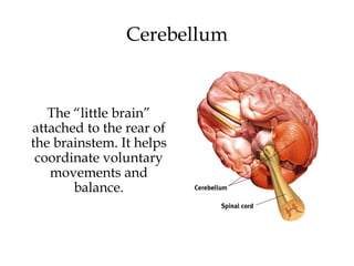
Psychology
(9th
edition)
David Myers
PowerPoint Slides
Aneeq Ahmad
Henderson State University
Worth Publishers, © 2010
The Biology of Mind
Chapter 2
The Biology of Mind
Neural Communication
 Neurons
 How Neurons Communicate
 How Neurotransmitters Influence Us
The Nervous System
 The Peripheral Nervous System
 The Central Nervous System
The Endocrine System
The Brain
• The Tools of Discovery: Having our Head
Examined
• Older Brain Structures
• The Cerebral Cortex
• Our Divided Brain
• Right-Left Differences in the Intact Brain
Neural Communication
The body’s information system is built from
billions of interconnected cells called neurons.
Neural Communication
Note the similarities in the above brain regions, which are all
engaged in information processing.
Neurobiologists and other investigators
understand that humans and animals operate
similarly when processing information.
Neuron
A nerve cell, or a neuron, consists of many
different parts.
Parts of a Neuron
Cell Body: Life support center of the neuron.
Dendrites: Branching extensions at the cell body.
Receive messages from other neurons.
Axon: Long single extension of a neuron, covered with
myelin [MY-uh-lin] sheath to insulate and speed up
messages through neurons.
Terminal Branches of axon: Branched endings of an
axon that transmit messages to other neurons.
Action Potential
A neural impulse. A brief
electrical charge that
travels down an axon and
is generated by the
movement of positively
charged atoms in and out
of channels in the axon’s
membrane.
Threshold
Threshold: Each neuron receives
excitatory and inhibitory signals from
many neurons. When the excitatory
signals minus the inhibitory signals
exceed a minimum intensity (threshold)
the neuron fires an action potential.
Action Potential Properties
All-or-None Response: A strong stimulus can
trigger more neurons to fire, and to fire more
often, but it does not affect the action potentials
strength or speed.
Intensity of an action potential remains the
same throughout the length of the axon.
Synapse
Synapse [SIN-aps] a junction between the axon
tip of the sending neuron and the dendrite or
cell body of the receiving neuron. This tiny gap
is called the synaptic gap or cleft.
Neurotransmitters
Neurotransmitters
(chemicals) released
from the sending
neuron travel across the
synapse and bind to
receptor sites on the
receiving neuron,
thereby influencing it to
generate an action
potential.
Reuptake
Neurotransmitters in
the synapse are
reabsorbed into the
sending neurons
through the process of
reuptake. This process
applies the brakes on
neurotransmitter
action.
How Neurotransmitters Influence Us
Serotonin pathways are
involved with mood
regulation.
From Mapping the Mind, Rita Carter, © 1989
University of California Press
Dopamine Pathways
Dopamine pathways
are involved with
diseases such as
schizophrenia and
Parkinson’s disease.
From Mapping the Mind, Rita Carter, © 1989
University of California Press
Neurotransmitters
Lock & Key Mechanism
Neurotransmitters bind to the receptors of the
receiving neuron in a key-lock mechanism.
Agonists
Antagonists
Nervous System
Central
Nervous
System
(CNS)
Peripheral
Nervous
System
(PNS)
The Nervous System
Nervous System: Consists of all the nerve cells. It
is the body’s speedy, electrochemical
communication system.
Central Nervous System (CNS): the brain and
spinal cord.
Peripheral Nervous System (PNS): the sensory and
motor neurons that connect the central nervous
system (CNS) to the rest of the body.
The Nervous System
Kinds of Neurons
Sensory Neurons carry incoming information from the
sense receptors to the CNS. Motor Neurons carry
outgoing information from the CNS to muscles and
glands. Interneurons connect the two neurons.
Sensory Neuron
(Bipolar)
Interneuron Neuron
(Unipolar)
Motor Neuron
(Multipolar)
Peripheral Nervous System
Somatic Nervous System: The division of the
peripheral nervous system that controls the body’s
skeletal muscles.
Autonomic Nervous System: Part of the PNS that
controls the glands and other muscles.
The Nerves
Nerves consist of neural “cables” containing many
axons. They are part of the peripheral nervous
system and connect muscles, glands, and sense
organs to the central nervous system.
Autonomic Nervous System (ANS)
Sympathetic Nervous System: Division of the
ANS that arouses the body, mobilizing its
energy in stressful situations.
Parasympathetic Nervous System: Division of
the ANS that calms the body, conserving its
energy.
Autonomic Nervous System (ANS)
Sympathetic NS
“Arouses”
(fight-or-flight)
Parasympathetic NS
“Calms”
(rest and digest)
Central Nervous System
The Brain and Neural Networks
Complex Neural Network
Interconnected neurons form networks in the
brain. Theses networks are complex and modify
with growth and experience.
Central Nervous System
The Spinal Cord and Reflexes
Simple Reflex
The Endocrine System
The Endocrine
System is the body’s
“slow” chemical
communication
system.
Communication is
carried out by
hormones
synthesized by a set
of glands.
Hormones
Hormones are chemicals synthesized by the
endocrine glands that are secreted in the
bloodstream. Hormones affect the brain and many
other tissues of the body.
For example, epinephrine (adrenaline) increases
heart rate, blood pressure, blood sugar, and
feelings of excitement during emergency
situations.
Pituitary Gland
Is called the “master gland.” The anterior pituitary
lobe releases hormones that regulate other glands.
The posterior lobe regulates water and salt
balance.
Thyroid & Parathyroid Glands
Regulate metabolic and calcium rate.
Adrenal Glands
Adrenal glands consist of the adrenal medulla and
the cortex. The medulla secretes hormones
(epinephrine and norepinephrine) during stressful
and emotional situations, while the adrenal cortex
regulates salt and carbohydrate metabolism.
Gonads
Sex glands are located in different places in men
and women. They regulate bodily development
and maintain reproductive organs in adults.
The Brain:
Older Brain Structures
The Brainstem is the oldest part of the brain, beginning
where the spinal cord swells and enters the skull. It is
responsible for automatic survival functions.
Brainstem
The Medulla [muh-
DUL-uh] is the base of
the brainstem that
controls heartbeat and
breathing.
Brainstem
The Thalamus [THAL-
uh-muss] is the brain’s
sensory switchboard,
located on top of the
brainstem. It directs
messages to the sensory
areas in the cortex and
transmits replies to the
cerebellum and
medulla.
Brainstem
Reticular Formation is a
nerve network in the
brainstem that plays an
important role in
controlling arousal.
The “little brain”
attached to the rear of
the brainstem. It helps
coordinate voluntary
movements and
balance.
Cerebellum
The Brain
Techniques to Study the Brain
A brain lesion
experimentally
destroys brain tissue to
study animal behaviors
after such destruction.
Hubel (1990)
Clinical Observation
Clinical observations have shed light on a
number of brain disorders. Alterations in brain
morphology due to neurological and
psychiatric diseases are now being catalogued.
TomLanders/BostonGlobe
Electroencephalogram (EEG)
An amplified recording of the electrical waves
sweeping across the brain’s surface, measured
by electrodes placed on the scalp.
AJPhoto/PhotoResearchers,Inc.
PET Scan
PET (positron emission
tomography) Scan is a
visual display of brain
activity that detects a
radioactive form of
glucose while the brain
performs a given task.
CourtesyofNationalBrookhavenNationalLaboratories
MRI Scan
MRI (magnetic resonance
imaging) uses magnetic
fields and radio waves to
produce computer-
generated images that
distinguish among
different types of brain
tissue. Top images show
ventricular enlargement in
a schizophrenic patient.
Bottom image shows brain
regions when a
participants lies.
Both photos from Daniel Weinberger, M.D., CBDB, NIMH
James Salzano/ Salzano Photo Lucy Reading/ Lucy Illustrations
The Limbic System is a
doughnut-shaped
system of neural
structures at the border
of the brainstem and
cerebrum, associated
with emotions such as
fear, aggression and
drives for food and sex.
It includes the
hippocampus, amygdala,
and hypothalamus.
The Limbic System
Amygdala
The Amygdala [ah-MIG-
dah-la] consists of two lima
bean-sized neural clusters
linked to the emotions of
fear and anger.
Hypothalamus
The Hypothalamus lies
below (hypo) the
thalamus. It directs
several maintenance
activities like eating,
drinking, body
temperature, and
control of emotions. It
helps govern the
endocrine system via
the pituitary gland.
Rats cross an electrified
grid for self-stimulation
when electrodes are
placed in the reward
(hypothalamus) center
(top picture). When the
limbic system is
manipulated, a rat will
navigate fields or climb
up a tree (bottom
picture).
Reward Center
SanjivTalwar,SUNYDownstate
The Cerebral Cortex
The intricate fabric of interconnected neural cells that covers
the cerebral hemispheres. It is the body’s ultimate control
and information processing center.
Structure of the Cortex
Each brain hemisphere
is divided into four
lobes that are separated
by prominent fissures.
These lobes are the
frontal lobe (forehead),
parietal lobe (top to rear
head), occipital lobe
(back head) and
temporal lobe (side of
head).
Functions of the Cortex
The Motor Cortex is the area at the rear of the
frontal lobes that control voluntary movements.
The Sensory Cortex (parietal cortex) receives
information from skin surface and sense organs.
Visual Function
The functional MRI scan
shows the visual cortex
is active as the subject
looks at faces.
CourtesyofV.P.Clark,K.Keill,J.Ma.
Maisog,S.Courtney,L.G.
Ungerleider,andJ.V.Haxby,
NationalInstituteofMentalHealth
Auditory Function
The functional MRI scan
shows the auditory
cortex is active in
patients who hallucinate.
More intelligent animals have increased
“uncommitted” or association areas of the cortex.
Association Areas
Language
Aphasia is an impairment of language, usually
caused by left hemisphere damage either to
Broca’s area (impaired speaking) or to Wernicke’s
area (impaired understanding).
Specialization & Integration
Brain activity when hearing, seeing, and
speaking words
The brain is sculpted by our genes but also by our
experiences.
Plasticity refers to the brain’s ability to modify
itself after some types of injury or illness.
The Brain’s Plasticity
Our Divided Brain
Our brain is divided into two hemispheres.
The left hemisphere processes reading, writing,
speaking, mathematics, and comprehension
skills. In the 1960s, it was termed as the
dominant brain.
Splitting the Brain
A procedure in which the two hemispheres of the
brain are isolated by cutting the connecting fibers
(mainly those of the corpus callosum) between them.
Corpus Callosum
MartinM.Rother
CourtesyofTerenceWilliams,UniversityofIowa
Split Brain Patients
With the corpus callosum severed, objects (apple)
presented in the right visual field can be named.
Objects (pencil) in the left visual field cannot.
Divided Consciousness
Try This!
Try drawing one shape with your left hand and one
with your right hand, simultaneously.
BBC
Right-Left Differences in the Intact
Brain
People with intact brains also show left-right
hemispheric differences in mental abilities.
A number of brain scan studies show normal
individuals engage their right brain when
completing a perceptual task and their left brain
when carrying out a linguistic task.

Chapter 2, Myers Psychology 9e

  • 1.
    Psychology (9th edition) David Myers PowerPoint Slides AneeqAhmad Henderson State University Worth Publishers, © 2010
  • 2.
    The Biology ofMind Chapter 2
  • 3.
    The Biology ofMind Neural Communication  Neurons  How Neurons Communicate  How Neurotransmitters Influence Us The Nervous System  The Peripheral Nervous System  The Central Nervous System
  • 4.
    The Endocrine System TheBrain • The Tools of Discovery: Having our Head Examined • Older Brain Structures • The Cerebral Cortex • Our Divided Brain • Right-Left Differences in the Intact Brain
  • 5.
    Neural Communication The body’sinformation system is built from billions of interconnected cells called neurons.
  • 6.
    Neural Communication Note thesimilarities in the above brain regions, which are all engaged in information processing. Neurobiologists and other investigators understand that humans and animals operate similarly when processing information.
  • 7.
    Neuron A nerve cell,or a neuron, consists of many different parts.
  • 8.
    Parts of aNeuron Cell Body: Life support center of the neuron. Dendrites: Branching extensions at the cell body. Receive messages from other neurons. Axon: Long single extension of a neuron, covered with myelin [MY-uh-lin] sheath to insulate and speed up messages through neurons. Terminal Branches of axon: Branched endings of an axon that transmit messages to other neurons.
  • 9.
    Action Potential A neuralimpulse. A brief electrical charge that travels down an axon and is generated by the movement of positively charged atoms in and out of channels in the axon’s membrane.
  • 10.
    Threshold Threshold: Each neuronreceives excitatory and inhibitory signals from many neurons. When the excitatory signals minus the inhibitory signals exceed a minimum intensity (threshold) the neuron fires an action potential.
  • 11.
    Action Potential Properties All-or-NoneResponse: A strong stimulus can trigger more neurons to fire, and to fire more often, but it does not affect the action potentials strength or speed. Intensity of an action potential remains the same throughout the length of the axon.
  • 12.
    Synapse Synapse [SIN-aps] ajunction between the axon tip of the sending neuron and the dendrite or cell body of the receiving neuron. This tiny gap is called the synaptic gap or cleft.
  • 13.
    Neurotransmitters Neurotransmitters (chemicals) released from thesending neuron travel across the synapse and bind to receptor sites on the receiving neuron, thereby influencing it to generate an action potential.
  • 14.
    Reuptake Neurotransmitters in the synapseare reabsorbed into the sending neurons through the process of reuptake. This process applies the brakes on neurotransmitter action.
  • 15.
    How Neurotransmitters InfluenceUs Serotonin pathways are involved with mood regulation. From Mapping the Mind, Rita Carter, © 1989 University of California Press
  • 16.
    Dopamine Pathways Dopamine pathways areinvolved with diseases such as schizophrenia and Parkinson’s disease. From Mapping the Mind, Rita Carter, © 1989 University of California Press
  • 17.
  • 18.
    Lock & KeyMechanism Neurotransmitters bind to the receptors of the receiving neuron in a key-lock mechanism.
  • 19.
  • 20.
  • 21.
  • 22.
    The Nervous System NervousSystem: Consists of all the nerve cells. It is the body’s speedy, electrochemical communication system. Central Nervous System (CNS): the brain and spinal cord. Peripheral Nervous System (PNS): the sensory and motor neurons that connect the central nervous system (CNS) to the rest of the body.
  • 23.
  • 24.
    Kinds of Neurons SensoryNeurons carry incoming information from the sense receptors to the CNS. Motor Neurons carry outgoing information from the CNS to muscles and glands. Interneurons connect the two neurons. Sensory Neuron (Bipolar) Interneuron Neuron (Unipolar) Motor Neuron (Multipolar)
  • 25.
    Peripheral Nervous System SomaticNervous System: The division of the peripheral nervous system that controls the body’s skeletal muscles. Autonomic Nervous System: Part of the PNS that controls the glands and other muscles.
  • 26.
    The Nerves Nerves consistof neural “cables” containing many axons. They are part of the peripheral nervous system and connect muscles, glands, and sense organs to the central nervous system.
  • 27.
    Autonomic Nervous System(ANS) Sympathetic Nervous System: Division of the ANS that arouses the body, mobilizing its energy in stressful situations. Parasympathetic Nervous System: Division of the ANS that calms the body, conserving its energy.
  • 28.
    Autonomic Nervous System(ANS) Sympathetic NS “Arouses” (fight-or-flight) Parasympathetic NS “Calms” (rest and digest)
  • 29.
    Central Nervous System TheBrain and Neural Networks Complex Neural Network Interconnected neurons form networks in the brain. Theses networks are complex and modify with growth and experience.
  • 30.
    Central Nervous System TheSpinal Cord and Reflexes Simple Reflex
  • 31.
    The Endocrine System TheEndocrine System is the body’s “slow” chemical communication system. Communication is carried out by hormones synthesized by a set of glands.
  • 32.
    Hormones Hormones are chemicalssynthesized by the endocrine glands that are secreted in the bloodstream. Hormones affect the brain and many other tissues of the body. For example, epinephrine (adrenaline) increases heart rate, blood pressure, blood sugar, and feelings of excitement during emergency situations.
  • 33.
    Pituitary Gland Is calledthe “master gland.” The anterior pituitary lobe releases hormones that regulate other glands. The posterior lobe regulates water and salt balance.
  • 34.
    Thyroid & ParathyroidGlands Regulate metabolic and calcium rate.
  • 35.
    Adrenal Glands Adrenal glandsconsist of the adrenal medulla and the cortex. The medulla secretes hormones (epinephrine and norepinephrine) during stressful and emotional situations, while the adrenal cortex regulates salt and carbohydrate metabolism.
  • 36.
    Gonads Sex glands arelocated in different places in men and women. They regulate bodily development and maintain reproductive organs in adults.
  • 37.
    The Brain: Older BrainStructures The Brainstem is the oldest part of the brain, beginning where the spinal cord swells and enters the skull. It is responsible for automatic survival functions.
  • 38.
    Brainstem The Medulla [muh- DUL-uh]is the base of the brainstem that controls heartbeat and breathing.
  • 39.
    Brainstem The Thalamus [THAL- uh-muss]is the brain’s sensory switchboard, located on top of the brainstem. It directs messages to the sensory areas in the cortex and transmits replies to the cerebellum and medulla.
  • 40.
    Brainstem Reticular Formation isa nerve network in the brainstem that plays an important role in controlling arousal.
  • 41.
    The “little brain” attachedto the rear of the brainstem. It helps coordinate voluntary movements and balance. Cerebellum
  • 42.
    The Brain Techniques toStudy the Brain A brain lesion experimentally destroys brain tissue to study animal behaviors after such destruction. Hubel (1990)
  • 43.
    Clinical Observation Clinical observationshave shed light on a number of brain disorders. Alterations in brain morphology due to neurological and psychiatric diseases are now being catalogued. TomLanders/BostonGlobe
  • 44.
    Electroencephalogram (EEG) An amplifiedrecording of the electrical waves sweeping across the brain’s surface, measured by electrodes placed on the scalp. AJPhoto/PhotoResearchers,Inc.
  • 45.
    PET Scan PET (positronemission tomography) Scan is a visual display of brain activity that detects a radioactive form of glucose while the brain performs a given task. CourtesyofNationalBrookhavenNationalLaboratories
  • 46.
    MRI Scan MRI (magneticresonance imaging) uses magnetic fields and radio waves to produce computer- generated images that distinguish among different types of brain tissue. Top images show ventricular enlargement in a schizophrenic patient. Bottom image shows brain regions when a participants lies. Both photos from Daniel Weinberger, M.D., CBDB, NIMH James Salzano/ Salzano Photo Lucy Reading/ Lucy Illustrations
  • 47.
    The Limbic Systemis a doughnut-shaped system of neural structures at the border of the brainstem and cerebrum, associated with emotions such as fear, aggression and drives for food and sex. It includes the hippocampus, amygdala, and hypothalamus. The Limbic System
  • 48.
    Amygdala The Amygdala [ah-MIG- dah-la]consists of two lima bean-sized neural clusters linked to the emotions of fear and anger.
  • 49.
    Hypothalamus The Hypothalamus lies below(hypo) the thalamus. It directs several maintenance activities like eating, drinking, body temperature, and control of emotions. It helps govern the endocrine system via the pituitary gland.
  • 50.
    Rats cross anelectrified grid for self-stimulation when electrodes are placed in the reward (hypothalamus) center (top picture). When the limbic system is manipulated, a rat will navigate fields or climb up a tree (bottom picture). Reward Center SanjivTalwar,SUNYDownstate
  • 51.
    The Cerebral Cortex Theintricate fabric of interconnected neural cells that covers the cerebral hemispheres. It is the body’s ultimate control and information processing center.
  • 52.
    Structure of theCortex Each brain hemisphere is divided into four lobes that are separated by prominent fissures. These lobes are the frontal lobe (forehead), parietal lobe (top to rear head), occipital lobe (back head) and temporal lobe (side of head).
  • 53.
    Functions of theCortex The Motor Cortex is the area at the rear of the frontal lobes that control voluntary movements. The Sensory Cortex (parietal cortex) receives information from skin surface and sense organs.
  • 54.
    Visual Function The functionalMRI scan shows the visual cortex is active as the subject looks at faces. CourtesyofV.P.Clark,K.Keill,J.Ma. Maisog,S.Courtney,L.G. Ungerleider,andJ.V.Haxby, NationalInstituteofMentalHealth
  • 55.
    Auditory Function The functionalMRI scan shows the auditory cortex is active in patients who hallucinate.
  • 56.
    More intelligent animalshave increased “uncommitted” or association areas of the cortex. Association Areas
  • 57.
    Language Aphasia is animpairment of language, usually caused by left hemisphere damage either to Broca’s area (impaired speaking) or to Wernicke’s area (impaired understanding).
  • 58.
    Specialization & Integration Brainactivity when hearing, seeing, and speaking words
  • 59.
    The brain issculpted by our genes but also by our experiences. Plasticity refers to the brain’s ability to modify itself after some types of injury or illness. The Brain’s Plasticity
  • 60.
    Our Divided Brain Ourbrain is divided into two hemispheres. The left hemisphere processes reading, writing, speaking, mathematics, and comprehension skills. In the 1960s, it was termed as the dominant brain.
  • 61.
    Splitting the Brain Aprocedure in which the two hemispheres of the brain are isolated by cutting the connecting fibers (mainly those of the corpus callosum) between them. Corpus Callosum MartinM.Rother CourtesyofTerenceWilliams,UniversityofIowa
  • 62.
    Split Brain Patients Withthe corpus callosum severed, objects (apple) presented in the right visual field can be named. Objects (pencil) in the left visual field cannot.
  • 63.
  • 64.
    Try This! Try drawingone shape with your left hand and one with your right hand, simultaneously. BBC
  • 65.
    Right-Left Differences inthe Intact Brain People with intact brains also show left-right hemispheric differences in mental abilities. A number of brain scan studies show normal individuals engage their right brain when completing a perceptual task and their left brain when carrying out a linguistic task.

Editor's Notes

  • #8 Preview Question 1: What are neurons, and how do they transmit information?
  • #16 Preview Question 2: How do neurotransmitters affect our mood and behavior?
  • #22 Preview Question 3: What are the major divisions of the nervous system, and what are their basic functions?
  • #32 Preview Question 4: How does the endocrine system-the body’s slower information system-transmit its messages?
  • #38 Preview Question 5: What are the functions of the brainstem and its associated structures?
  • #48 Preview Question 6: What are the functions of limbic system structures?
  • #53 Preview Question 7: What are the four lobes of the cerebral cortex, and where are they located?
  • #54 Preview Question 8: What are the functions of the cerebral cortex?
  • #62 Preview Question 9: What is a split brain, and what does it reveal about brain functioning?