1. Fractures. Dislocations. Thermal injuries.
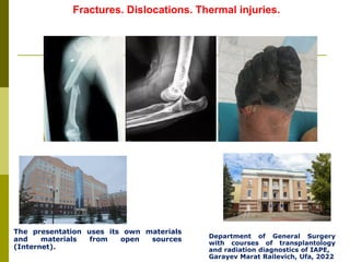
The presentation uses its own materials
and materials from open sources
(Internet).
Department of General Surgery
with courses of transplantology
and radiation diagnostics of IAPE,
Garayev Marat Railevich, Ufa, 2022
2. By origin:
1. Congenital - in the prenatal development
2. Acquired fractures - in the childbirth and continue in the coming years
By reasons:
1. Trauma (at falling, impact, compression, rotation, abruption)
2. Pathological (osteomyelitis, tumors, metabolic disorders)
Fractures - a violation of anatomical integrity of bone
Classification:
3. Condition of skin and mucous membranes:
1. Closed - without skin damage and mucous membranes
2. Opened - with damage of the skin and mucous membranes
By completeness of fracture:
1. Complete
2. Incomplete:
a) cracks
b) subperiosteal
(in children by the type of
“greenstick fracture")
Classification (continued)
4. Localization:
1. Diaphyseal
2. Metaphyseal
3. Epiphyseal
4. Intraarticular
By fracture line:
1. Transverse
2. Longitudinal
3. Slanted
4. Spiral
5. Splintered
6. Separated
7. Impacted
8. Compressed
By character of displacement:
1. Without a displacement
2. With the displacement:
a) in length: with shortening and lengthening of the limb
b) by angle:
abduction - the angle is turned outward
adduction - angle of the fracture is turned inward
c) rotational - shift of bone fragments on the axis
Classification (continued)
5. By complexity:
1. Simple
2. Combined (fractures of several bones)
3. Mixed (fracture with other trauma: burn, frozen disease etc.)
By complications:
1. Bleeding
2. Traumatic shock
3. Damages of head and spinal cord
4. Damages of internal organs
By healing:
1. Primary hematoma
2. Primary bone callous (4-6 weeks)
3. Secondary bone callous (5-6 weeks)
Classification (continued)
6. Indirect attributes:
1. Pain
2. Swelling and hematoma
3. Deformation
4. Infringement of function
5. Change the length of finiteness (shortening, lengthening)
Authentic attributes:
1. Abnormal mobility
2. Crepitation (bone crunch)
3. Visualization of bone fragments (open fracture)
Clinical picture
8. It’s based in clinical picture and radiography in 2 projections.
If necessary, it is possible to perform radiography in additional
projections, CT.
Diagnostics
9.
10.
11.
12. At treatment of fractures should be holding 4 principles:
1. Reposition
2. Immobilization (fixation)
3. Functional treatment
4. Stimulation formation of bone callous
Treatment
13. 1.Reposition - comparison of fragments in correct position carried
out after estimation of radiological character the displacement and
good anesthesia (by regional anesthesia or narcosis)
Distinguish: one-stage reposition and long reposition
One-stage reposition: at fractures of small bones or at small displacement under
the corner.
At fractures of big bones (femoral, bones of shin, humeral) with displacement of
bones on length, the one-stage reposition is impracticable because will be
resistance of muscles. In such cases is carry out long reposition by skeletal
extension.
14. 2.Fixation – it’s maintenance of immovability the fragments
for healing of fracture
Distinguish 3 kinds of fixation the fragments:
plaster bandages,
extension
and operative method
Plaster bandages: should fix of 2 joints, at fracture of
femoral and humeral bones - 3 joints
Kinds:
1. Circular bandage
2. Splint bandage
3. Corset bandage (on the trunk)
Plaster bandages should not squeeze the tissues and should not break
the blood circulation (fingers are leaving the open for control of blood
supply). If presence the wound on the finiteness in the plaster bandage
left the window.
20. Methods of extension
1. Adhesive bandage
2. Skeletal traction
The finiteness for stretching is placed in special splint (Bohler frame) and
suspended a cargo (8-12 kg at fracture of the hip, 2-4 kg at fracture of the
tibia). Skeletal traction is used in cases when the one-stage reduction of bone
fragments is impossible.
At traction is saved the mobility in joints which prevents the muscle atrophy
and violation of trophism.
However, a skeletal traction has a drawback - need for compliance of bed
rest for a long time.
So often, the skeletal traction is carried out to complete repositioning of the
bone fragments and then transferred to the plaster method.
21. REDUCING A SUPRACONYLAR FRACTURE
ON A BOHLER-BRAUN FRAME is only
necessary if there is very severe angulation. It is
one of the few correct uses of this frame.
Examples of immobilization with methods of extension
24. Surgical treatment of fractures
All types of operations on fractures are called the osteosynthesis
and divided into 3 groups:
1. Intramedullary osteosynthesis, when the metal rod is introduced into the
medullary canal;
2. Extramedullary osteosynthesis, when the fragments are joined outside the
medullary canal with the plates, screws, wires, etc.;
3. Extrafocal osteosynthesis using the Ilizarov apparatus, Gudushauri apparatus.
On the other, the compression-distraction method, when the stimulation of
callus formation is achieved by dosed compression or distraction the region of
fracture.
33. 3. Functional treatment
The functional treatment is used for all types of fractures and methods of
treatment. This is preservation of functional activity of the limbs during the
maturation of bone callus.
These include a comparison of fragments in the physiologically adequate limb
position, ability to preserve a limb function without compromising the healing
process, thus preventing an improper symphysis of fractures, contractures and false
joint.
34. Complications of fractures
• Direct
• Remote
Direct – it is a traumatic shock, damage of soft tissues by
fragments, bleeding.
Remote - wrong coalescence of fractures, osteomyelitis,
pseudoarthrosis (false joint), ankyloses.
35. Delayed consolidation of fractures – coalescence available, but
slowed by time.
Reasons:
general: deficiency of vitamins, calcium, advanced age, concomitant
diseases;
local: fault of immobilization, partial interposition of soft tissue between
the fragments.
False joint - coalescence between the fragments is completely absent.
Reasons: osteomyelitis, complete interposition of soft tissues between
the fragments.
36. Treatment
At delayed consolidation necessary to extend the period of gypsum,
appointed a general treatment (calcium, vitamins, etc.).
At false joint - surgical treatment:
remove of soft tissue between the bone fragments;
2) resection of affected bone fragments with fixation their by Ilizarov’s
apparatus.
37. Dislocation – violation of the congruence of the articular surfaces of the bones, both
with violation of the integrity of the joint capsule, and without violation, under the
influence of mechanical forces (trauma) or destructive processes in the joint (arthrosis,
arthritis).
Classification
By character of contact of articular surfaces:
- Complete - articular surfaces are not in contact with each other;
- Partial (subluxation) - articular surfaces are keep the partial contact.
By origin:
- Congenital
- Traumatic
Habitual dislocation – it’s when damaged the ligaments and joint
capsule. This dislocation is more than 1 time in the same joint.
38. 1. Pain;
2. Involuntary limb position in which the pain is the smallest;
3. If you trying to change a position of limb it's takes the same position -
a symptom of springy fixation;
4. Limited range of motion or impossibility of motion in the joint;
5. Deformation of joint;
6. Changing the length of limb.
Clinical picture
39. 1. Clinic
2. Arthrogram
Treatment
• Reposition under local or general anesthesia.
(Methods of reposition by Kocher, Janelidze, Hippocrates)
• Fixation (immobilization) of limbs at to 2-3 weeks
• Surgical treatment is carried out at longstanding, chronic dislocations, at the
habitual dislocation
Diagnosis
43. Kocher's method. The arm is pulled (traction)
in the direction of red arrow. Now the limb is externally rotated (red arrow).
Next, the arm is brought close to the body (adduction; red
arrow). The shoulder relocates at this moment and a pop
like sensation is felt. If this is not felt or the shoulder does
not relocate at this moment then internal rotation should
not be done or else a fracture of the humerus may occur.
Lastly the limb is internally rotated (red arrow) to
stabilize the dislocation.
44. At the Hippocratic method the limb is gently rotated along
with simultaneous traction.
45. By the depth of the lesion:
Grade I - superficial burn, manifested by hyperemia, slight swelling
of the skin
II degree - burn of the upper layer of the skin, manifested by
hyperemia, serous bubbles, puffiness of the skin, pain sensitivity is
preserved
III a degree - a burn of the entire thickness of the skin, the wound
is covered with a scab, there are bubbles with cloudy liquid at the
edges, pain sensitivity is reduced
III b degree - burn of the entire thickness of the skin with a
transition to subcutaneous tissue, the wound is covered with a thick
layer of dark brown scab
IV degree - burn of the underlying tissues: tendons, muscles,
bones, the bottom of the wound is insensitive to pain.
Burns are considered superficial I - IIIа degrees, deep - IIIб -
IV degrees.
By localization:
burns of the respiratory tract
burns of mucous membranes
burns of the skin
combined burns
Burns are tissue injuries caused by exposure to thermal,
chemical, electrical or radiation energy.
Classification of burns:
47. 1 - the "palm" rule: the surface
of a person's palm is
approximately equal to 1% of the
body area.
2 - the rule of the "nine": areas
of the human body are multiples of
nine.
So, the head and neck make up
9% of the body area, the upper
limbs - 9% each, the lower leg and
foot - 9%, the thigh - 9%, the
chest in front - 9%, the back -
9%, the abdomen - 9%, the
lumbar and gluteal region - 9%
and another 1% is the perineum.
3 - the Vilyavin method - using
special measuring grids (where 1
cell is 1% of the body surface).
Methods determination of the affected area:
48. Burns up to 15% can occur without general manifestations.
With burns of more than 15% of the body, burn disease develops,
occurring in 4 stages:
stage I - burn shock, begins from the moment of the burn, can
last up to 24 - 48 hours. It is accompanied by pain syndrome,
hypovolemia, a decrease in BCC due to fluid loss.
Stage II is the stage of burn toxemia: from 24-48 hours to 1-2
weeks, due to massive absorption of tissue breakdown products into
the bloodstream, which, against the background of hypovolemia, is
accompanied by toxic liver and kidney damage, high fever, anemia,
leukocytosis, acidosis increase. With large areas of burns, oliguria,
anuria, and uremia develop. A decrease in urine of less than 50
ml/hour is a poor prognostic sign.
Burn disease
49. Stage III - septic stage - develops from 2 to 3 weeks. Almost all
burn wounds become infected, while Pseudomonas aeruginosa infection
is very dangerous, difficult to treat. Sepsis often develops,
accompanied by chills, hectic fever, anemia, exhaustion of patients,
immunity decreases.
Stage IV is the stage of recovery (the wound is cleaned, covered
with granulations, marginal epithelialization begins). At this stage,
plastic closure of wounds is performed.
Burn disease
50. it does not always depend on the area of the burn. With the mass admission of
patients with burns , medical sorting and the following prognostic rules can be
applied:
rule one hundred: the sum of the digits of the age and area of the burn. Up to 80
units - the forecast is favorable, 80-100 units - doubtful, more than 100 units -
unfavorable. This rule does not take into account the depth of the burn.
the Franc index: 1% of a superficial burn is taken as 1 unit, 1% of a deep burn is
3 units, with a burn of the respiratory tract, 30 units are added to the amount. With a
total Franc index of up to 70 units, the forecast is favorable, 70-90 units - doubtful,
more than 90 units - unfavorable.
Forecast
51. Signs of a burn of the respiratory tract are: singed hair in the nose, soot on the
tongue and teeth, hoarseness of voice, cough, shortness of breath, wheezing in the
lungs. With fibrobronchoscopy - soot, tracheobronchitis phenomena,
hypersecretion, swelling of the mucous membrane of the trachea and bronchi.
Diagnosis of burn shock: Unlike other forms of shock in burn shock, blood
pressure can remain normal for a long time due to powerful pain impulses. In the
first minutes and hours, excitement, motor restlessness, chills and muscle trembling
are noted, followed by apathy. The skin is pale. The CVD is sharply reduced. With
severe shock, BP falls.
Distinguish:
mild shock - burn area up to 20% and Franc index up to 70 units;
severe shock - with a burn area of 20 to 40%, the Franc index is 70-130 units;
shock of an extremely severe degree - the burn area is more than 40%, the Franc
index is more than 130 units.
Diagnostics
52. First aid - removal of the victim from the flame zone, extinguish the
fire on the clothes. If possible, cool the burn surface (in everyday life -
with a stream of cold water until the pain disappears). Sterile bandages
are applied. Narcotic analgesics are administered to prevent shock.
In case of burns of II-IV degree, emergency prevention of tetanus is
carried out (in the emergency room of the hospital).
During the shock period - infusion therapy, transfusion of blood
components (according to indications), plasma, blood substitutes,
rheopolyglucine, narcotic analgesics, neuroleptanalgesia are used.
During the period of burn toxemia - detoxification therapy,
transfusion of blood components (according to indications), plasma,
polyglucine, rheopolyglucine, detoxification doses, albumin, crystalloids,
sodium bicarbonate, glucose solution, anticoagulants. With the
phenomena of anuria, plasmapheresis, hemodialysis (artificial kidney) is
performed.
In the septic stage, broad-spectrum antibiotics are used, immune
forces are corrected, hormone therapy is carried out.
Treatment (general)
53. Treatment (local)
Burns of the first degree - do not require local treatment. It is possible to
use aerosols, creams to reduce pain and accelerate skin recovery.
Burns of II-IIIa degree with localization on the face - open management
with the use of aerosols, sea buckthorn oil, atarvmatic mesh ointment
dressings. Burns of the brush - bandages "gloves" with water-soluble
ointments "Levosin", "Levomekol", "Dioxicol", "Mafenid-acetate", which must
be changed after 2-3 days. In case of burns of the trunk, extremities -
bandages with these ointments, in the absence of suppuration, change them
after 3 to 5 days, since with frequent changes of bandages, granulations are
damaged. In case of suppuration of wounds, necrectomies are performed,
treatment with antiseptics. Bandages are removed after their abundant
wetting with antiseptic solutions to prevent damage to regenerating tissues.
54. Treatment (local) (continued)
Deep burns of the IIIb-IV degree: at first, wet bandages with antiseptics
are used, scab drying. With prolonged preservation of the scab (2-3 weeks),
substructural suppuration develops, which can lead to septic complications.
Early (4-5 days), delayed (8-10 days) and phased (2-3 weeks) necrectomy
are necessary for their prevention. Necrectomy is performed in the operating
room, under anesthesia. The resulting wound is closed with ointment
dressings, allo- and xenotransplant, or skin grafting is performed.
In case of extensive burns of the trunk, the management of patients is
optimal in the conditions of cambustiological departments (in Ufa on the
basis of GKB No. 18) on special beds of the "Clinitron" type.
55. They arise as a result of exposure to the skin or mucous membranes
of acids and alkalis, under the action of chemical warfare agents.
Under the action of acids, coagulation necrosis develops, alkalis -
colliquation necrosis.
Example: Example:
Acid burn Alkali burn
Treatment
First aid - copious washing of the affected area with running water,
bandages with neutralizing solutions: in case of acid burn - sodium
bicarbonate (soda), in case of alkali burn - acetic, boric or citric acid.
Speedy transportation to the hospital.
Chemical burns
56. Electrical injury
Develops when exposed to an electric current. At the same time,
small thermal burns, the so-called "current signs", are formed at the
site of the input and output of the current. Electric current can pass
through the body in any direction, and its passage through the centers
of the cardiovascular, respiratory and nervous systems is most
dangerous, which can lead to cardiac arrest and death.
In mild cases, the victims get off with fright, short-term loss of
consciousness may occur. With moderate lesions, shock develops,
respiratory arrest, cardiac fibrillation may occur.With severe lesions,
instant death is possible.
57. Electrical injury – Treatment
First aid: the victim is released from the action of the current, with
respiratory arrest and cardiac fibrillation, artificial ventilation of the
lungs, external heart massage are necessary.
Next: 100% hospitalization.
If necessary, defibrillation of the heart is performed, hardware
ventilation, cardiac drugs are administered, oxygen therapy is
performed.
58. - tissue damage caused by the action of low temperature.
Frostbite
Local cold damage - frostbite of tissues.
There are 4 degrees of frostbite:
Grade I - hyperemia, swelling of the skin, hypersensitivity of the
skin. The changes are reversible.
Grade II - damage to the surface layers of the skin, bubbles with
blood-serous contents are formed, the bottom of the bubbles is
sensitive to mechanical irritation.
Grade III - damage to the skin and the underlying fatty tissue, large
blisters with hemorrhagic exudate are formed, their bottom is
insensitive to mechanical irritation.
IV degree - damage to the skin, tendons, muscles, bones. Tissue
necrosis leads to mummification or wet gangrene. The necrosis line,
or demarcation zone, is formed within 2 weeks or more.
60. Frostbite occurs in 2 stages:
I - the hidden stage, begins with a feeling of cold, burning in the
area of frostbite, then comes a complete loss of sensitivity of the
frostbitten area. During this period, it is impossible to determine the
depth and area of tissue necrosis.
II - reactive stage, develops after warming the affected area, and
only by the end of the week it is possible to accurately determine the
boundaries of frostbite.
Clinical picture
61. Treatment
Warming of the sick and frostbitten area, it is forbidden to rub the
frostbitten areas with snow. The patient is given hot tea,
antispasmodics, novocaine blockades are prescribed to eliminate cold
vascular spasm. In the II-IV degrees of frostbite, emergency
prevention of tetanus is carried out.
Local treatment consists in opening the blisters under aseptic
conditions, bandaging with antiseptics, with frostbite of III-IV degrees
- necrectomy / amputation is performed after the formation of the
demarcation zone.
62. General overcooling – develops with prolonged exposure to cold on the body
as a whole, proceeds in 4 phases.
Phase I - there is a feeling of cold, tremor, pallor of the skin, "goose
bumps". The body temperature is kept at +36 +37ºC.
Phase II - body temperature decreases by 1-2 degrees. At the same time,
there is pallor of the face, cyanosis, stiffness of movements in the joints, the
skin feels cold as pain.
Phase III - the temperature drops to +34+27ºC. At the same time, pain
sensitivity decreases to its complete disappearance, bradycardia is noted,
apathy and drowsiness develop, muscle tremor turns into muscle rigor.
Phase IV - body temperature drops below +27ºC. At the same time, the
functions of the organs are gradually suspended, breathing, pulse, blood
pressure are barely detected, reflexes are absent, pain is not felt.In the future,
due to the suppression of the activity of the central nervous system, death
occurs.
Treatment: urgent compensation of heat loss is necessary, for which the
victim is placed in a warm room, wrapped up, intravenously injected with a
glucose solution heated to +40 ° C, you can warm the victim in a hot (t to
+40 ° C) bath. A solution of soda is injected, infusion therapy is performed,
and diuresis is stimulated.
General overcooling (hypotermia)
63. Thanks for your attention!
I am ready to answer your
questions.