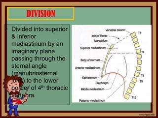
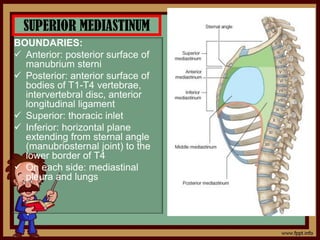
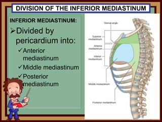
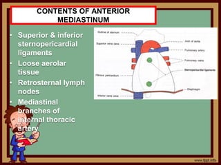
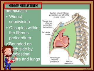
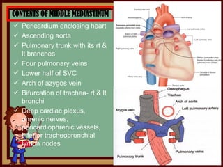
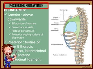
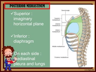
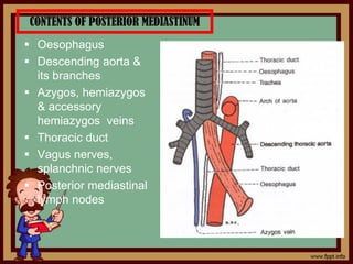
MEDIASTINUM
BY:
PHANINDRA PRASAD POUDEL
ASSISTANT PROFESSOR
DEPARTMENT OF ANATOMY
DEFINATION
• Space between rt & lt
lungs & pleural sacs
• Extension:
Thoracic inlet above
to diaphragm below
Sternum anteriorly to
thoracic vertebrae
behind
BOUNDARIES
• Anteriorly: posterior
surface of sternum
• Posteriorly: anterior
surface of bodies of 12
thoracic vertebrae,
intervertebral disc,
anterior longitudinal
ligament
• Superior: thoracic inlet
• Inferior: upper surface of
diaphragm
• On each side: mediastinal
pleura of lungs
DIVISION
• Divided into superior
& inferior
mediastinum by an
imaginary plane
passing through the
sternal angle
(manubriosternal
joint) to the lower
border of 4th thoracic
vertebra.
SUPERIOR MEDIASTINUM
BOUNDARIES:
 Anterior: posterior surface of
manubrium sterni
 Posterior: anterior surface of
bodies of T1-T4 vertebrae,
intervertebral disc, anterior
longitudinal ligament
 Superior: thoracic inlet
 Inferior: horizontal plane
extending from sternal angle
(manubriosternal joint) to the
lower border of T4
 On each side: mediastinal
pleura and lungs
CONTENTS
• Arranged into 3 groups
a. Retrosternal:
 Origin of
sternothyroid &
sternohyoid
muscles
 Thymus gland
 Rt & lt
brachiocephalic
veins, Superior
vena cava, Left
superior
intercostal vein
CONTENTS…
b. Intermediate
structures:
Arch of aorta with
its 3 large
branches;
brachiocephalic
trunk, left common
carotid & left
subclavian arteries
Phrenic nerves,
vagus nerves &
cardiac nerves
CONTENTS…
c. Prevertebral
structures:
 Trachea with
paratracheal &
tracheosophageal
lymph nodes
 Esophagus
 Left recurrent
laryngeal nerve
 Thoracic duct
 Origin of longus colli
from T3
CLINICAL ANATOMY:
• Prevertebral fascia of neck:
 Extends into superior mediastinum & attached to T4
 Infection in neck may extend into superior mediastinum
• Pretracheal fascia of neck:
 Extends upto superior mediastinum & attached to the
tunica adventitia of arch of aorta
 Infection in between the pretracheal & prevertebral
fascia extends into superior mediastinum
 Results into infection in the superior mediastinum,
mediastinitis
II CLASS
INFERIOR MEDIASTINUM
BOUNDARIES:
 Anterior: posterior surface of
the body of sternum
 Posterior: anterior surface of
the bodies of T4-T12
vertebrae, intervertebral disc,
anterior longitudinal ligament
 Superior: horizontal plane
extending from sternal angle
(manubriosternal joint) to the
lower border of T4
 Inferior: diaphragm
 On each side: mediastinal
pleura and lungs
DIVISION OF THE INFERIOR MEDIASTINUM
INFERIOR MEDIASTINUM:
Divided by
pericardium into:
Anterior
mediastinum
Middle mediastinum
Posterior
mediastinum
ANTERIOR MEDIASTINUM
BOUNDARIES:
 Anterior : posterior
surface of body of
sternum
 Posterior : fibrous
pericardium
 Superior : transthoracic
plane
 Inferior : diaphragm
 On each side :
mediastinal pleura and
lungs
CONTENTS OF ANTERIOR
MEDIASTINUM
• Superior & inferior
sternopericardial
ligaments
• Loose aerolar
tissue
• Retrosternal lymph
nodes
• Mediastinal
branches of
internal thoracic
artery
MIDDLE MEDIASTINUM
BOUNDARIES:
Widest
subdivision
Occupies within
the fibrous
pericardium
Bounded on
each side by
mediastinal
pleura and lungs
CONTENTS OF MIDDLE MEDIASTINUM
 Pericardium enclosing heart
 Ascending aorta
 Pulmonary trunk with its rt &
lt branches
 Four pulmonary veins
 Lower half of SVC
 Arch of azygos vein
 Bifurcation of trachea- rt & lt
bronchi
 Deep cardiac plexus,
phrenic nerves,
pericardiophrenic vessels,
inferior tracheobronchial
lymph nodes
POSTERIOR MEDIASTINUM
BOUNDARIES:
 Anterior : above
downwards
 Bifurcation of trachea
 Pulmonary vessels
 Fibrous pericardium
 Posterior sloping surface of
diaphragm
 Posterior : bodies of
lower 8 thoracic
vertebrae, intervertebral
discs, anterior
longitudinal ligament
POSTERIOR MEDIASTINUM
Superior :
imaginary
horizontal plane
Inferior :
diaphragm
On each side :
mediastinal
pleura and lungs
CONTENTS OF POSTERIOR MEDIASTINUM
 Oesophagus
 Descending aorta &
its branches
 Azygos, hemiazygos
& accessory
hemiazygos veins
 Thoracic duct
 Vagus nerves,
splanchnic nerves
 Posterior mediastinal
lymph nodes
CLINICAL ANATOMY
a. Connection of posterior mediastinum with the
spaces around the neck via superior mediastinum
o Connection with retropharyngeal space, space
between trachea & esophagus, spaces around carotid
sheath of neck
o Infections of neck may gravitate into superior &
posterior mediastinum
b. Compression of mediastinal structures by
any form of tumours results into mediastinal
syndrome with various symptoms:
i. Obstruction of superior venacava gives rise to
engorgement of veins of upper half of the body
ii. Pressure over the trachea causes dyspnea &
cough
iii. Pressure over the esophagus causes dysphagia
iv. Pressure over the left recurrent laryngeal nerve
causes hoarseness of voice
v. Pressure over the intercostal nerve results into
intercostal neuralgia
vi. Pressure over the vertebral column causes the erosion
of vertebral bodies
vii. Pressure over the phrenic nerve causes the paralysis
of diaphragm
HOME ASSIGNMENT
1. Draw a well labeled diagram showing
the subdivisions of Mediastinum.
2. What are the boundaries and contents
of Inferior mediastinum?
THE END

4.Mediastinum.pdf

  • 1.
  • 2.
    DEFINATION • Space betweenrt & lt lungs & pleural sacs • Extension: Thoracic inlet above to diaphragm below Sternum anteriorly to thoracic vertebrae behind
  • 3.
    BOUNDARIES • Anteriorly: posterior surfaceof sternum • Posteriorly: anterior surface of bodies of 12 thoracic vertebrae, intervertebral disc, anterior longitudinal ligament • Superior: thoracic inlet • Inferior: upper surface of diaphragm • On each side: mediastinal pleura of lungs
  • 4.
    DIVISION • Divided intosuperior & inferior mediastinum by an imaginary plane passing through the sternal angle (manubriosternal joint) to the lower border of 4th thoracic vertebra.
  • 5.
    SUPERIOR MEDIASTINUM BOUNDARIES:  Anterior:posterior surface of manubrium sterni  Posterior: anterior surface of bodies of T1-T4 vertebrae, intervertebral disc, anterior longitudinal ligament  Superior: thoracic inlet  Inferior: horizontal plane extending from sternal angle (manubriosternal joint) to the lower border of T4  On each side: mediastinal pleura and lungs
  • 6.
    CONTENTS • Arranged into3 groups a. Retrosternal:  Origin of sternothyroid & sternohyoid muscles  Thymus gland  Rt & lt brachiocephalic veins, Superior vena cava, Left superior intercostal vein
  • 7.
    CONTENTS… b. Intermediate structures: Arch ofaorta with its 3 large branches; brachiocephalic trunk, left common carotid & left subclavian arteries Phrenic nerves, vagus nerves & cardiac nerves
  • 8.
    CONTENTS… c. Prevertebral structures:  Tracheawith paratracheal & tracheosophageal lymph nodes  Esophagus  Left recurrent laryngeal nerve  Thoracic duct  Origin of longus colli from T3
  • 9.
    CLINICAL ANATOMY: • Prevertebralfascia of neck:  Extends into superior mediastinum & attached to T4  Infection in neck may extend into superior mediastinum • Pretracheal fascia of neck:  Extends upto superior mediastinum & attached to the tunica adventitia of arch of aorta  Infection in between the pretracheal & prevertebral fascia extends into superior mediastinum  Results into infection in the superior mediastinum, mediastinitis
  • 10.
  • 11.
    INFERIOR MEDIASTINUM BOUNDARIES:  Anterior:posterior surface of the body of sternum  Posterior: anterior surface of the bodies of T4-T12 vertebrae, intervertebral disc, anterior longitudinal ligament  Superior: horizontal plane extending from sternal angle (manubriosternal joint) to the lower border of T4  Inferior: diaphragm  On each side: mediastinal pleura and lungs
  • 12.
    DIVISION OF THEINFERIOR MEDIASTINUM INFERIOR MEDIASTINUM: Divided by pericardium into: Anterior mediastinum Middle mediastinum Posterior mediastinum
  • 13.
    ANTERIOR MEDIASTINUM BOUNDARIES:  Anterior: posterior surface of body of sternum  Posterior : fibrous pericardium  Superior : transthoracic plane  Inferior : diaphragm  On each side : mediastinal pleura and lungs
  • 14.
    CONTENTS OF ANTERIOR MEDIASTINUM •Superior & inferior sternopericardial ligaments • Loose aerolar tissue • Retrosternal lymph nodes • Mediastinal branches of internal thoracic artery
  • 15.
    MIDDLE MEDIASTINUM BOUNDARIES: Widest subdivision Occupies within thefibrous pericardium Bounded on each side by mediastinal pleura and lungs
  • 16.
    CONTENTS OF MIDDLEMEDIASTINUM  Pericardium enclosing heart  Ascending aorta  Pulmonary trunk with its rt & lt branches  Four pulmonary veins  Lower half of SVC  Arch of azygos vein  Bifurcation of trachea- rt & lt bronchi  Deep cardiac plexus, phrenic nerves, pericardiophrenic vessels, inferior tracheobronchial lymph nodes
  • 17.
    POSTERIOR MEDIASTINUM BOUNDARIES:  Anterior: above downwards  Bifurcation of trachea  Pulmonary vessels  Fibrous pericardium  Posterior sloping surface of diaphragm  Posterior : bodies of lower 8 thoracic vertebrae, intervertebral discs, anterior longitudinal ligament
  • 18.
    POSTERIOR MEDIASTINUM Superior : imaginary horizontalplane Inferior : diaphragm On each side : mediastinal pleura and lungs
  • 19.
    CONTENTS OF POSTERIORMEDIASTINUM  Oesophagus  Descending aorta & its branches  Azygos, hemiazygos & accessory hemiazygos veins  Thoracic duct  Vagus nerves, splanchnic nerves  Posterior mediastinal lymph nodes
  • 20.
    CLINICAL ANATOMY a. Connectionof posterior mediastinum with the spaces around the neck via superior mediastinum o Connection with retropharyngeal space, space between trachea & esophagus, spaces around carotid sheath of neck o Infections of neck may gravitate into superior & posterior mediastinum
  • 21.
    b. Compression ofmediastinal structures by any form of tumours results into mediastinal syndrome with various symptoms: i. Obstruction of superior venacava gives rise to engorgement of veins of upper half of the body ii. Pressure over the trachea causes dyspnea & cough iii. Pressure over the esophagus causes dysphagia
  • 22.
    iv. Pressure overthe left recurrent laryngeal nerve causes hoarseness of voice v. Pressure over the intercostal nerve results into intercostal neuralgia vi. Pressure over the vertebral column causes the erosion of vertebral bodies vii. Pressure over the phrenic nerve causes the paralysis of diaphragm
  • 23.
    HOME ASSIGNMENT 1. Drawa well labeled diagram showing the subdivisions of Mediastinum. 2. What are the boundaries and contents of Inferior mediastinum? THE END