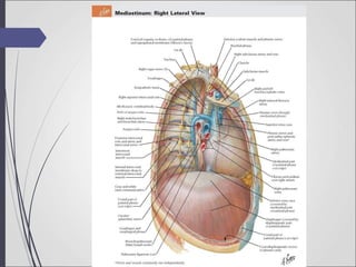
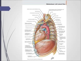
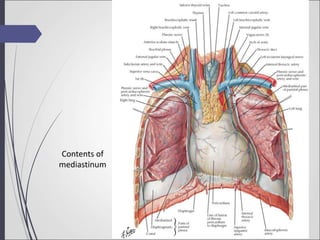
Anatomy
Presentation
by :- Ajay T Raj
1st year MBBS
Roll No. 5
Mediastinum
Introduction
 Mediastinum is the middle space left in the thoracic cavity in
between the lungs.
 The mediastinum is the median septum of the thorax
between the two lungs.
 Its most important content is the heart enclosed in the
pericardium.
Boundaries
 Interiorly :- Sternum
 Posteriorly :-Vertebral column
 Superiorly :- Thoracic inlet
 Inferiorly :- Diaphragm
 On each side :- Mediastinal pleura
Divisions
 The mediastinum is divided into two by an imaginary plane
passing through the sternal angle anteriorly and the lower
border of the body of the fourth thoracic vertebra
posteriorly.
 The mediastinum is divided into
Superior mediastinum and
Inferior mediastinum.
Superior Mediastinum
Boundaries
 Anteriorly:- Manubrium sterni
 Posteriorly:- Upper four thoracic vertebrae.
 Superiorly:- Plane of the thoracic inlet.
 Inferiorly:- An imaginary plane passing through the sternal
angle in front, and the lower border of the body
of the fourth thoracic vertebra behind.
 On each side:- Mediastinal pleura.
Superior Mediastinum(continued)
Contents
 Trachea and oesophagus.
 Muscles :- Sternohyoid, sternothyriod, lower ends of longus
colli.
 Arteries:- Arch of aorta, brachiocephalic artery, left common
carotid artery, left subclavian artery
 Veins:- Right and left brachiocephalic veins, upper half of the
superior vena cava, left superior intercostal vein
 Nerves :- Vagus, phrenic, cardiac nerves of both sides, left
recurrent laryngeal nerve
 Thoracic duct
 Lymph nodes:- Paratracheal, brachiocephalic and
tracheobronchial.
Inferior Mediastinum
 The inferior mediastinum is divided into anterior, middle
and posterior mediastinum by the pericardium
 The area in front of pericardium : Anterior Mediastinum
 The area behind the pericardium : posterior mediastinum
 The pericardium and its contents in the middle
mediastinum.
Anterior Mediastinum
Boundaries
 Anteriorly :- Body of sternum
 Posteriorly :- pericardium
 Superiorly Imaginary plane passing separating the superior
mediastinum from the inferior mediastinum.
 Inferiorly:- Superior surface of the diaphragm
 On each side :- Mediastinal pleura
Inferior Mediastinum(continued)
Anterior Mediastinum
Contents
 Sternopericardial ligaments
 Lymph nodes and lymphatics
 Small mediastinal braches of the internal thoracic artery
 The lowest part of the thymus.
 Areolar tissue
Inferior Mediastinum(continued)
Middle mediastinum
Boundaries
 Anteriorly :- Sternopericardial ligaments
 Posteriorly :- Oesophagus, descending thoracic aorta,
azygos vein
 On each side :- mediastinal pleura
Inferior Mediastinum(continued)
Middle mediastinum
Contents
 Heart enclosed in pericardium
 Arteries :- Ascending aorta, pulmonary trunk, two pulmonary
arteries
 Veins:- Lower half of the superior vena cava, terminal part of the
azygos vein, right and left pulmonary veins.
 Nerves:- Phrenic, Deep cardiac plexus
 Lymph nodes:- Tracheobronchial nodes
 Tubes:- Bifurcation of trachea, the right and left principal bronchi
Inferior Mediastinum(continued)
Posterior Mediastinum
Boundaries
 Anteriorly :- Pericardium, bifurcation of trachea, pulmonary
vessels, posterior part of the upper surface of the
diaphragm.
 Posteriorly:- Lower eight thoracic vertebrae and intervening
discs.
 On each side:- Mediastinal pleura
Inferior Mediastinum(continued)
Posterior Mediastinum
Contents
 Oesophagus
 Arteries:- Descending thoracic aorta and its branches
 Veins:-Azygos vein, hemiazygos vein and accessory
hemiazygos vein.
 Nerves:- Vagi, splanchnic nerves, greater, lesser and least,
arising from the lower eight thoracic ganglia of the
sympathetic chain
 Lymph nodes and lymphatics:- Thoracic duct, posterior
mediastinal lymph nodes lying alongside the aorta.
Inferior Mediastinum(continued)
Contents of
mediastinum
Clinical Anatomy
 The prevertebral layer of the deep cervical fascia extends to
the superior mediastinum and is attached to the fourth
thoracic vertebra. An infection present in the neck behind
this fascia can pass down into the superior mediastinum
but not lower down.
 There is a very little loose connective tissue between the
mobile organs of the mediastinum. Therefore the space can
be readily dilated by inflammatory fluids, neoplasms, etc.
 Compression of mediastinal structures by any tumor gives
rise to Mediastinal syndrome. The common symptoms are
1)Obstruction of the superior venacava gives rise to
engorgement of veins in the upper half of the body.
2)Pressure over the trachea causes dyspnoea and cough
3)Pressure on oesophagus causes dysphagia
4)Pressure on the left recurrent laryngeal nerve gives rise to
hoarseness of voice
5)Pressure on the phrenic nerve causes paralysis of the
diaphragm on that side.
Clinical Anatomy(continued)
Anatomy Presentation.ppt

Anatomy Presentation.ppt

  • 1.
    Anatomy Presentation by :- AjayT Raj 1st year MBBS Roll No. 5
  • 2.
  • 3.
    Introduction  Mediastinum isthe middle space left in the thoracic cavity in between the lungs.  The mediastinum is the median septum of the thorax between the two lungs.  Its most important content is the heart enclosed in the pericardium.
  • 6.
    Boundaries  Interiorly :-Sternum  Posteriorly :-Vertebral column  Superiorly :- Thoracic inlet  Inferiorly :- Diaphragm  On each side :- Mediastinal pleura
  • 7.
    Divisions  The mediastinumis divided into two by an imaginary plane passing through the sternal angle anteriorly and the lower border of the body of the fourth thoracic vertebra posteriorly.  The mediastinum is divided into Superior mediastinum and Inferior mediastinum.
  • 9.
    Superior Mediastinum Boundaries  Anteriorly:-Manubrium sterni  Posteriorly:- Upper four thoracic vertebrae.  Superiorly:- Plane of the thoracic inlet.  Inferiorly:- An imaginary plane passing through the sternal angle in front, and the lower border of the body of the fourth thoracic vertebra behind.  On each side:- Mediastinal pleura.
  • 10.
    Superior Mediastinum(continued) Contents  Tracheaand oesophagus.  Muscles :- Sternohyoid, sternothyriod, lower ends of longus colli.  Arteries:- Arch of aorta, brachiocephalic artery, left common carotid artery, left subclavian artery  Veins:- Right and left brachiocephalic veins, upper half of the superior vena cava, left superior intercostal vein  Nerves :- Vagus, phrenic, cardiac nerves of both sides, left recurrent laryngeal nerve  Thoracic duct  Lymph nodes:- Paratracheal, brachiocephalic and tracheobronchial.
  • 11.
    Inferior Mediastinum  Theinferior mediastinum is divided into anterior, middle and posterior mediastinum by the pericardium  The area in front of pericardium : Anterior Mediastinum  The area behind the pericardium : posterior mediastinum  The pericardium and its contents in the middle mediastinum.
  • 12.
    Anterior Mediastinum Boundaries  Anteriorly:- Body of sternum  Posteriorly :- pericardium  Superiorly Imaginary plane passing separating the superior mediastinum from the inferior mediastinum.  Inferiorly:- Superior surface of the diaphragm  On each side :- Mediastinal pleura Inferior Mediastinum(continued)
  • 13.
    Anterior Mediastinum Contents  Sternopericardialligaments  Lymph nodes and lymphatics  Small mediastinal braches of the internal thoracic artery  The lowest part of the thymus.  Areolar tissue Inferior Mediastinum(continued)
  • 14.
    Middle mediastinum Boundaries  Anteriorly:- Sternopericardial ligaments  Posteriorly :- Oesophagus, descending thoracic aorta, azygos vein  On each side :- mediastinal pleura Inferior Mediastinum(continued)
  • 15.
    Middle mediastinum Contents  Heartenclosed in pericardium  Arteries :- Ascending aorta, pulmonary trunk, two pulmonary arteries  Veins:- Lower half of the superior vena cava, terminal part of the azygos vein, right and left pulmonary veins.  Nerves:- Phrenic, Deep cardiac plexus  Lymph nodes:- Tracheobronchial nodes  Tubes:- Bifurcation of trachea, the right and left principal bronchi Inferior Mediastinum(continued)
  • 16.
    Posterior Mediastinum Boundaries  Anteriorly:- Pericardium, bifurcation of trachea, pulmonary vessels, posterior part of the upper surface of the diaphragm.  Posteriorly:- Lower eight thoracic vertebrae and intervening discs.  On each side:- Mediastinal pleura Inferior Mediastinum(continued)
  • 17.
    Posterior Mediastinum Contents  Oesophagus Arteries:- Descending thoracic aorta and its branches  Veins:-Azygos vein, hemiazygos vein and accessory hemiazygos vein.  Nerves:- Vagi, splanchnic nerves, greater, lesser and least, arising from the lower eight thoracic ganglia of the sympathetic chain  Lymph nodes and lymphatics:- Thoracic duct, posterior mediastinal lymph nodes lying alongside the aorta. Inferior Mediastinum(continued)
  • 18.
  • 19.
    Clinical Anatomy  Theprevertebral layer of the deep cervical fascia extends to the superior mediastinum and is attached to the fourth thoracic vertebra. An infection present in the neck behind this fascia can pass down into the superior mediastinum but not lower down.  There is a very little loose connective tissue between the mobile organs of the mediastinum. Therefore the space can be readily dilated by inflammatory fluids, neoplasms, etc.
  • 20.
     Compression ofmediastinal structures by any tumor gives rise to Mediastinal syndrome. The common symptoms are 1)Obstruction of the superior venacava gives rise to engorgement of veins in the upper half of the body. 2)Pressure over the trachea causes dyspnoea and cough 3)Pressure on oesophagus causes dysphagia 4)Pressure on the left recurrent laryngeal nerve gives rise to hoarseness of voice 5)Pressure on the phrenic nerve causes paralysis of the diaphragm on that side. Clinical Anatomy(continued)