The Ovaries
Өндгөвч
•Өндгөн эсгадагшлахад хүүдий бий
болно.
•LH and FSH даавартай холбоотой.
•Эстроген болон прогестерон нь LH
and FSH-ийн үйл ажиллагааг дэмждэг.
7.
The Vagina
Үтрээ
•Цусны судаснэмэгдсэн учир
өнгө хөх болдог
•Хучуур эс болон уян эд
нэмэгддэг
•Салсархаг шүүрэл их ялгарна.
mucus(pH is 3.5 to хүчилшил)
8.
The Vulva
Гадна бэлэгэрхтэн
•Цусны судас
нэмэгдсэний улмаас
хөх өнгөтэй болно
•Гадна тал нь томорно
Цус бүлэгнэлт нэмэгддэг
Эмэгтэйхүн ердийнхөөсөө жирэмсэн байхдаа
цус бүлэгнэх эрсдэл өндөртэй байдаг
(blood clots, or a rare and life threatening condition
called disseminated intravascular coagulation or DIC)
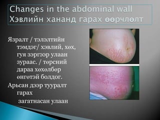
#3 1. Enlargement during pregnancy involves stretching and marked hypertrophy of existing muscle cells.2. In addition to an increase in the size of the uterine muscle cells, there is an increase in fibrous tissue, elastic tissue, blood vessels, and lymphatics.3. Enlargement and thickening of the uterine wall is most marked in the fundus.
#6 Pronounces softening and cyanosis – due to increased vascularity, edema, hypertrophy, and hyperplasia of the cervical glands.2. Clot of very thick mucus obstructs the cervical canal (cervical plug).3. Erosions of cervix, common during pregnancy, represent an extension of proliferating endocervical glands and columnar endocervical epithelium.
#7 1. Ovulation ceases during pregnancy; maturation of new follicles is suspended.2. One corpus luteum functions during early pregnancy (first 8 weeks), producing mainly progesterone.After 12weeks placenta produce estrogen and progesterone, these hormone suppress pituitary grand release FSH, LH.
#8 1. Increased vascularity, hyperemia, and softening of connective tissue in skin and muscles of perineum and vulva.2. Chadwick’s sign notes – characteristic violet color due to increased vascularity and hyperemia.3. Vaginal walls prepare for labor: mucosa increases in thickness, connective tissue loosens, and small-muscle cells hypertrophy.4. Vaginal secretions increase; pH is 3.5 t0 6 – because of increased production of lactic acid from glycogen in the vaginal epithelium by Lactobacillus acidophilus. (Acid pH probably aids in keeping vagina relatively free of pathogenic bacteria).
#9 Vulva area skin color is darkness due to melanin hormone, and purple color due to increased blood vessels.
#10 1. Tenderness and tingling occur in early weeks of pregnancy. 2. Increase in size by 2nd month – hypertrophy of mammary alveoli.3. Nipples become larger, more deeply pigmented, and more erectile early in pregnancy.4. Colostrum may be expressed by second trimester.5. Areolae become broader and more deeply pigmented. The depth of pigmentation varies with the individual’s complexion.6. Scattered through the areola are a number of small elevations (glands of Montgomery), which are hypertrophic sebaceous glands.
#33 Tone and motility of gastrointestinal tract decrease, leading to prolongation of gastric Stomach and intestines are displaced upward and laterally by the enlarging uterus. Heartburn is common, caused by reflux of acid secretions in the lower esophagus.
#34 Hemorrhoids are common because of elevated pressure in veins below the level of the large uterus and constipation. Liver function tests yield significantly different results during pregnancy. Distention of the gallbladder is common along with a decrease in emptying time and thickening of bile.
#35 The increasing mobility of sacroiliac, sacrococcygeal, and pelvic joints during pregnancy is a result of hormonal changes.2. This mobility contributes to alteration of maternal posture and to back pain. 3. Late in pregnancy, aching, numbness, and weakness in the upper extremities may occur because of lordosis, which ultimately produces traction on the ulnar and median nerves.4. Separation of the rectus muscles due to pressure of the growing uterus creates a diastasisrecti. If this is severe, a portion of the anterior uterine wall is covered by only a layer of skin, fascia, and peritoneum.