SlideShare a Scribd company logo
1 of 39
Download to read offline
7
Anatomy
Departmental Objectives
At the end of the Anatomy course, the students should be able to:
 mention, identify, show, draw and describe the anatomical structure of the human body responsible
for carrying out normal body functions.
 apply the acquired knowledge to understand and correlate the other pre-clinical, para-clinical and
clinical medical subjects.
 execute the acquired knowledge of Anatomy with the knowledge of other medical subjects to
provide optimum health services in the country and abroad.
List of Competencies to acquire:
 Ability to demonstrate knowledge & skill of understanding human anatomy, functions of different
components of human body, cell biology and human development in clinical perspective.
 Ability to utilize knowledge and skill of understanding of spatial relationship, course &
distributions of different components of human in real life situation.
 Ability to detect the deviation from normal state in the human body in relation to structure,
function and development.
 Ability to identify human body components and functional pattern by using internationally
accepted terminologies.
Distribution of teaching - learning hours
Lecture Tutorial Practical
(Histology)
Demons
tration
+Dissectio
n +Card
exam
Total
Teaching
hours
Integrat
ed
teaching
for
phase I
Formative Exam Summative exam
Preparato
ry leave+
post term
leave
Exam
time
Preparato
ry leave
Exam
time
115 hrs 53hrs 52 hrs 307 hrs 527 hrs 36 hrs 21+14=
35 days
42 days 30 days 30 days
Time for integrated teaching, examination, preparatory leave of formative & summative assessment is
common for all subjects of the phase
Related behavioral, professional & ethical issues will be discussed in all teaching learning sessions
Teaching - learning methods, teaching aids and evaluation
Teaching Methods
Teaching aids In course evaluation
Large group Small group Self learning
Lecture
Integrated
teaching
Tutorial
Practical
Demonstration
Dissection
Self-study &
self-assessment
Computer / Laptop & Multimedia
OHP, Transparency & Transparency marker
White board & different coloured white
board markers
Black board & white and coloured
chalks
Cadavers, prosected parts, bones, viscera
Histological slide, Microscope & Projection
microscope
Projection system and Virtual anatomy
dissection table
 Item
Examination
 Card
Examination
 Term Final
Examination
(written, oral+
practical)
Related Equipments: Flip Chart, Photograph, Model, X-ray films (CT scan and other imaging films), View
box, Diagram, Preserved specimens, Living body for surface marking, Simulator, various Projection system
and Virtual anatomy dissection table.
1st Professional Examination:
Marks distribution of Assessment of Anatomy
Total marks – 500
 Written=200 (Formative 20+MCQ (SBA+MTF) 40+(SAQ + SEQ) 140)
 SOE = 150
 Practical=150
8
Learning Objectives and Course Contents in Anatomy
Learning Objectives Contents
Teaching hours
Total : 12 hrs
General Anatomy
Student will be able to
 define anatomy and explain the subdivisions of anatomy.
 describe the anatomical terminology, planes & positions.
 define and classify bone. Describe the composition, blood supply,
functions, ossification of bones with clinical correlation.
 describe the composition, characteristics, location and functions
of different types of cartilages.
 define & classify joints. Describe the characters, stability &
movements of joints with clinical correlation.
 classify muscles. Describe their properties and functions.
 define & classify blood vessels.
 describe the different types of circulation.
 describe different types of vascular anastomosis with their
functional & clinical implications.
 describe components, functions & the general plan of lymphatic
drainage of the whole body.
 classify lymphoid organs. Describe the functions of lymphoid
organs with clinical significance.
CORE :
 Definition, subdivisions of anatomy and its importance in the
study of medicine.
 Anatomical terminologies, anatomical planes & positions.
 Skeletal system: Bones – classification, composition, functions,
parts of a developing long bone, blood supply, periosteum &
endosteum. Ossification – definition, centres, processes. Factors
affecting growth of bone.
 Cartilages: composition, types, characters, locations and
functions
 Joint: classification, characteristics of each type & movements,
stability of the joints. Clinical conditions associated with joints.
General plan of blood supply & nerve supply of joints.
 Muscular system: different ways of classification, characteristics
and functions different types.
Skeletal muscle – classification; Principle applied to innervation
& contraction.
 Blood vascular system: component parts, general plan, structure,
classification.
Differences between different types of vessel. Nutrition &
innervations of vessels.
Circulation – systemic, portal & pulmonary circulation and
characteristic features of each type.
Vascular anastomosis: types ,sites, characteristics ,functional and
clinical importance
 Lymph vascular system: components, characteristic features of
lymph capillaries. Comparison with blood capillary.
 Lymphoid organs: classification, distribution & functions,
TERM I
01 hr
01 hr
03 hrs
01 hr
02 hrs
01 hr
02 hrs
01 hr
9
Learning Objectives Contents Teaching hours
Total : 08 hrs
Cell Biology
Student will be able to:
 define and describe the human cell & its constituents, structure &
functions of all components of cell.
 describe the features of different types of cells.
Human Genetics
Students will be able to:
 describe the different basic features of chromosomes.
 define terms related to human genetics.
CORE:
 Human Cell – Basic organization, types of constituents, cell
membrane
 Nucleus – structure & functions
 Cytoplasm, organelles and inclusions – structure & functions
 Functional correlation of different types of cell (protein
secreting, ion transporting, steroid secreting, mucus secreting,
antibody producing cell) in respect of their nuclear,
cytoplasmic, membrane and surface feature
CORE:
 Chromosomes: Basic structure
 Terms & definitions: Gene, Gene locus, genome, genotype,
phenotype, genetic trait etc.
Total:06 hrs.
TERM I
02 hrs
01 hr
02 hrs
01hr
Total: 02 hrs
TERM I
01hr
01 hrs
10
Learning Objectives Contents
Teaching hours
Total: 14 hours
General Histology
Student will be able to:
 define and classify the basic tissues in the body
 identify under microscope different types of:
 Epithelial tissue
 Connective tissue
 Muscular tissue
 describe microscopic components and differentiate different
components of:
 Epithelial tissue
 Connective tissue
 Muscular tissue
 describe the histological structures of different types of muscle
tissue.
 describe the composition & functions of components of nervous
tissue
 correlate inter-relationship between structure and functions of each
tissue
General Histology
Basic tissues:
 Definition, classification, components, characters, distribution
and functions of
 Epithelial tissue and its subtypes
 Connective tissue and its subtypes
 Muscular tissue and its subtypes
 Structure and functions of
 Cell surface specialization
 Inter cellular junction
 Histological structure of
 Smooth muscle tissue
 Cardiac muscle tissue
 Skeletal muscle tissue
 Mechanism of muscle contraction
 Structure and function of Nervous tissue
 Neurons
 Neuroglia
TERM I
05hrs
05 hrs
TERM II
02 hrs
TERM III
02 hrs
11
Learning Objectives Contents
Teaching hours
Total: 18 hours
Systemic Histology: Students will be able to describe the histological
structures of organs of different systems of the body.
Systemic Histology : histological structures of
 Respiratory system : Respiratory tract & Lung
 Vascular system : Different types of artery, capillary &
vein
 Lymphoid organs: Thymus, spleen, lymph node & tonsil
 Digestive system & associated Glands : tongue,
oesophagus, stomach, intestine, Liver, gall bladder,
pancreas
 Exocrine glands : salivary glands
 Urinary system : kidney, ureter, urinary bladder
 Male reproductive system : testis, epididymis, vas deferens,
seminal vesicle
 Female reproductive system: ovary, uterus, uterine tube,
vagina
 Endocrine glands: pituitary, thyroid, parathyroid, adrenal
glands
 Skin and its appendages
TERM I
01 hr
01 hr
TERM II
02 hrs
04 hrs
01 hr
03 hr
02 hrs
02 hrs
TERM III
01 hr
01 hr
12
Learning Objectives Contents
Teaching hours
Total 18hrs
General Embryology
Students will be able to:
 define terms related to embryology
 explain the significance of study of embryology
 explain basic process of development
 describe different processes of cell division
 describe oogenesis and spermatogenesis
 describe the process of fertilization
 describe the events of 1st
week of development.
 describe the events2nd
week of development.
 describe the events 3rd
week of development.
 describe the development & derivatives of ectoderm, mesoderm &
endoderm.
 explain the development of foetal membranes
 explain the development of twins & their types.
 describe the causes & types of congenital anomalies
 explain the process of human evocation
 describe the Molecular regulation & cell signaling pathways
CORE:
 Introduction: terms and definition; Significance of study of
embryology.
 Basic process of development: proliferation, growth,
differentiation, inductors, evocators and organizer
 Cell division: Types
 chromosomal changes during cell division with anomalies
 Gametogenesis and maturation of Germ cells.
 Fertilization: Events, factors influencing the fertilization;
Progress in 1st
week of development
 Progress in 2nd
week of development.
 Progress in 3rd
week of development.
 Derivatives of germ layers: ectoderm, mesoderm & endoderm.
 Foetal membranes : Placenta, chorion, amnion, umbilical cord,
yolk sac etc.
 Twins
 Teratology
Additional:
 Human Evolution
 Concepts of medical biotechnology in relation to embryology
 Molecular regulation &cell signaling
TERM I
01 hr
01 hr
02 hrs
02 hrs
02 hrs
02 hr
02 hrs
01 hr
TERM II
03 hrs
01 hrs
01 hrs
13
Learning Objectives Contents
Teaching hours
Total: 24 hours
Systemic Developmental Anatomy
Student will be able to:
 describe the process of development of different systems of the
body
 describe the developmental anomalies of the organs of different
systems of the body
 mention general outline of development of: Thoracic duct,
Cisterna chyli, Inferior Vena Cava, Superior Vena Cava, Portal
Vein, Brachiocephalic veins & Renal veins.
CORE:
Development and their Anomalies of
 Skeletal system & vertebral column
Muscular system
 Upper and lower limb
 Digestive system with associated glands
 Respiratory system
 Cardiovascular System & aortic arches
 Coelomic cavity & the diaphragm
 Skin & mammary gland
 Urinary system
 Male and female Reproduction system
 Pituitary & suprarenal gland
 Face & neck & their associated organs
 Nervous System
 Eye & Ear
Additional:
Development of
 Lymphatic System
 Vascular System
TERM II
02 hrs
01 hr
03 hrs
01 hr
03 hrs
01 hr
01 hr
02 hrs
03 hrs
TERM III
01 hr
03 hrs
02 hrs
01 hr
14
Learning Objectives Contents
Teaching hours
Total: 21 hours
Neuroanatomy
Students will be able to:
 classify nervous system and describe each type.
 mention different parts of nervous system
 describe composition of nervous system
 describe autonomic nervous system,
 describe the coverings of central nervous system
 describe the ventricular system of CNS
 explain blood brain & blood CSF barrier
CORE:
 Introduction to Nervous system,
 Composition of grey matter and white matter
 Nerve fibers: structure classifications & functions,
myelination, degeneration, regeneration
 Receptors: definition, structure classifications location &
functions
 Synapse: definition, structure classifications & functions
 Autonomic nervous system: parts, autonomic nerve plexuses
& ganglia
 Coverings of brain and spinal cord : Pia, arachnoid and dura
mater, their extension, folds, spaces, nerve supply
& blood supply
 Ventricular system and Cerebrospinal fluid (CSF) : Location
of different ventricles of brain the formation, composition,
circulation, absorption & functions of CSF
 Blood-brain and Blood-CSF barriers: Composition & function
TERM I
01 hr
TERM III
01hr
01 hrs
TERM I & II
02 hrs
TERM III
01 hr
02 hr
15
Learning Objectives Contents Teaching hours
Neuroanatomy
Students will be able to:
 describe the anatomical aspects of motor and sensory parts of
nervous system with their functional and clinical significance
CORE:
Motor system
 Cerebrum (motor areas): Gyri, sulci and important functional
areas with effects of lesion; mode of blood supply
 Pyramidal & extrapyramidal system & effects of their lesion
 Cerebellum: parts, functional lobes, nuclei, peduncles &
functions, blood supply, clinical conditions
 Basal nuclei: locations, parts, functions artery supply &
clinical conditions
 Motor and mixed Cranial Nerves: Classification, functional
components, cranial nerve nuclei and course of cranial nerves
Sensory system
 Dermatome & axial line
 Cerebrum( sensory areas): Gyri, sulci and important
functional areas with effects of lesion; mode of blood supply
 Ascending tracts of spinal cord with effects of lesions
 Diencephalon: parts & functions
 Sensory cranial nerves & Smell, visual & auditory pathway
 Spinal Cord: Length, extension, enlargement, blood supply,
cross-sections at different level
 Brain stem: blood supply, cross sections at different levels
 Reticular formation
 Limbic system
TERM III
02 hrs
01 hr
01 hr
02 hr
01hr
01 hr
01 hr
01 hr
01 hr
01 hr
01hr
16
Learning Objectives Contents
Teaching hours
(Total 24 hrs)
Living (surface) Anatomy
Students will be able to:
 locate and count ribs & costal cartilages
 draw and demonstrate important anatomical points and structures
of Thorax on the surface of the body
Students will be able to:
 draw and demonstrate important anatomical points and structures
of Superior extremity on the surface of the body
Thorax
CORE:
 Counting of ribs and costal cartilages
 Heart- apex and borders
 Lung-borders and apex,
 Trachea & Bronchi
 Esophagus
 Triangle of auscultation
 Jugular notch
 Sternal angle
 Area of Superficial Cardiac dullness
Common carotid and subclavian artery
Internal thoracic artery
Superior extremity
CORE
 Nerves: Radial, Ulnar, Median nerve, Axillary nerve
 Arteries: Brachial, Radial, Ulnar artery, Superficial and deep
palmar arch
 Veins: cephalic, basilic & median cubital vein
 Flexor retinaculum
 Anatomical snuff box
 Medial humeral epicondyle
For Tutorial
06 hrs.
04 hrs.
17
Learning Objectives Contents Teaching hours
Living (surface) Anatomy
Students will be able to:
 locate, demonstrate the different anatomical planes and land
marks on the surface of the body
 draw, demonstrate the nine regions of the abdomen on the surface
of the body
 draw and indicate inguinal canal on the surface of the body
 draw and demonstrate important anatomical points, borders and
parts of important organs of abdomen on the surface of the body
Students will be able to:
 locate and demonstrate important points and structures of inferior
extremity on the surface of the body
CORE:
Abdomen
 Trans-pyloric plane, Trans tubercular plane, Subcostal plane,
mid clavicular line
 Regions of abdomen
 Superficial & deep inguinal ring, Inguinal canal
 Abdominal aorta & inferior vena cava
 Stomach, Duodenum, Pancreas, Liver, Gall bladder, Bile duct,
spleen, Kidney from back & Mac Burney’s point
 Transverse colon, ureter from front and back, celiac trunk,
splenic artery, Root of the mesentery
Inferior extremity
 Common peroneal nerve, Tibial nerve
 Popliteal artery
 Anterior & posterior tibial artery
 Arteria dorsalis pedis
 Great Saphenous vein
 Small Saphenous vein
 Adductor tubercle
 Lateral and Medial Malleolus
 Greater trochanter of femur
 Anterior superior iliac spine
Additional
 Femoral nerve, sural nerve, Medial and lateral plantar artery,
plantar arch.
For Tutorial
6 hrs.
4 hrs.
18
Learning Objectives Contents Teaching hours
Students will be able to:
 draw and demonstrate important anatomical points and structures
of Head and Neck on the surface of the body
Head and neck
 Facial artery, Facial vein
 Internal jugular vein, External jugular vein
 Common Carotid artery & its bifurcation
 Facial Nerve & their branches
 vagus nerve in the neck
 Parotid gland and its duct
 Frontal and maxillary air sinuses
 Thyroid gland
 Tip of the coracoid process
 Inferior angle of scapula
 Tip of the 7th
cervical spine
Additional:
 Pterion, lambda
 Middle meningeal artery
For Tutorial
04 hrs.
19
Learning Objectives Contents
Teaching hours
(Total 09 hrs)
Anatomy of Radiology & Images
Students will be able to:
 describe radio-opaque and radio-lucent structures
 identify and locate the normal structures in radiograph
CORE
Radio opaque structures
Radio-lucent structures
Plain X-ray of the
-chest PA view
-abdomen AP view
-pelvis AP view
-arm including proximal & distal
joints AP & lateral view
-forearm including proximal & distal
joints AP & lateral view
-hand including proximal & distal
joints
-thigh including proximal & distal
joints AP & lateral view
-leg including proximal & distal
joints AP & lateral view
-foot including proximal & distal
joints AP & lateral view
-head & neck (cervical spine) AP &
lateral view
-Paranasal sinuses OM view
Additional:
 Common normal Ultrasonographs, Isotope scan,
 Magnetic Resonance Images (MRI), CT Scan
 Coronary Angiograph
For Tutorial
09 hrs
20
Learning Objectives Contents
Teaching hours
(Total 20 hrs)
Clinical Anatomy
Students will be able to:
 describe the anatomical basis of clinical disorder of structures of the
thorax and the abdomen.
Thorax
• Pleurisy / Pleural effusion
• Pneumothorax
• Coronary artery disease
• Pericarditis/ pericardial effusion
• Flail chest
• Paralysis of the diaphragm
Abdomen
• Portal vein obstruction
• Hydrocele
• Hernia
• Peritonitis, ascites
• Gastric ulcer
• Duodenal ulcer
• Gall stone/cholecystitis
• appendicitis
• Benign hyperplasia of prostate, Prostatic cancer
• Cystocele
• Stress incontinence
• Rupture urethra
• Salpingitis
• Ectopic pregnancy
• Prolapse of uterus / vagina
• Haemorrhoids
• Undescended testis
• Psoas abscess
• Ischiorectal abscess
For Tutorial
03 hrs
06 hrs
21
Learning Objectives Contents
Teaching
hours
Clinical Anatomy
Students will be able to:
 describe the anatomical basis of clinical disorder of the structures
of head & neck, nervous system & extremities
Head & Neck
• Fracture of the skull bones
• Scalp injury
• Piriform fossa and foreign body
• Otitis media
• Sinusitis
• Epistaxis
• Tonsilitis
• Swelling of thyroid gland
• Mumps
• Cavernous vein thrombosis
• Cervical rib
CNS & Eyeball
• Injury to brain /eye ball / spinal cord/cranial nerves
• Meningitis
• Hydrocephalus
• Cerebral ischaemia, intracranial haemorrhage
(extradural,subarachnoid, cerebral)
• Papilledema
• Horner’s syndrome
Superior extremity
• Dislocation of shoulder joint
• Brachial plexus & injury to its nerves
• Carpal tunnel syndrome
• Colle’s fracture
• Breast abscess & breast cancer
Inferior extremity
• Varicose vein
• Deep vein thrombosis
• Nerve injury
• Dislocation of hip joint
• Rupture of menisci & cruciate ligament, Bursitis
• Deformities of foot
For Tutorial
03hr
.
03hr
03hr
02hr
22
Learning Objectives Contents
Teaching
hours
Clinical Anatomy
Students will be able to:
 describe the anatomical basis for selection of arteries, veins &
muscles of clinical importance.
 demonstrate the different auscultatory areas
 describe the anatomical basis for clinical procedure of Thorax,
Abdomen, Head & Neck , CNS & Eyeball.
 Arterial pulsation
 Intravenous injections
 Intramuscular injection
 Apex beat, mitral, tricuspid, aortic & pulmonary areas
• Sternal puncture
• Pleural effusion
• Pericardial effusion
• Coronary angiogram
• Bronchoscopy
• Laryngoscopy
• Paracentesis /peritoneal dialysis
• Ryle’s tube
• Endoscopy
• Liver abscess
• Vasectomy
• Tubal ligation
• Nasogastric intubation
• Palpation of Cervical lymph node
• Lumbar puncture
• Epidural/spinal anesthesia
• Pudendal block
• Fundoscopy
23
Regional Anatomy : THORAX CARD
(DISSECTION, DEMONSTRATION & TUTORIAL)
Learning Objectives Contents
Teaching
hours
Students will be able to:
 demonstrate the boundary & identify the contents of thoracic wall, thoracic cavity,
mediastinum & intercostal space
 identify & demonstrate the gross features of bones & joints of thorax
 describe the formation, course, branches & distribution of spinal nerve / intercostal nerve
 identify & demonstrate the surfaces, borders, parts, chambers- including structures within
the chambers of the heart
 explain blood supply & nerve supply of heart
 identify & demonstrate the layers of pericardium
 identify & demonstrate the surfaces, borders, fissures, lobes, hilus & bronchopulmonary
units of the lung
 identify & demonstrate the layers & parts of pleura.
 explain the blood supply, lymphatic drainage & nerve supply of lung & pleura.
 identify & demonstrate the trachea, bronchus & bronchial tree.
 explain blood supply & nerve supply of trachea & bronchial tree.
 explain the blood supply, nerve supply & lymphatic drainage of thoracic wall.
 identify & demonstrate the surfaces, parts openings, attachments of the diaphragm.
 explain the blood supply & nerve supply of the diaphragm.
 explain the significance of the orifices of the diaphragm.
 explain & demonstrate the extension, parts, relations & constrictions of oesophagus
 explain the blood supply, lymphatic drainage & nerve supply of the oesophagus.
 correlate clinical conditions associated with structures of thorax (Heart with its vessels,
lung, trachea, bronchus, bronchial tree & the diaphragm)
 Thoracic wall formation, thoracic cavity,
intercostal space and mediastinum.
 Bones and joints of the thorax
 Spinal nerve / intercostal nerve
 Heart with pericardium.
 Lung with pleura, trachea and bronchus.
 Blood vessels, nerves and lymphatics of the
thorax.
 The diaphragm.
 Oesophagus
 Clinical Anatomy
45 hrs.
NB: Previously mentioned 53 hours in pages 10-16 for Tutorial also have shown in this part (DISSECTION, DEMONSTRATION & TUTORIAL)
24
Regional Anatomy: SUPERIOR EXTREMITY CARD
(DISSECTION, DEMONSTRATION & TUTORIAL)
Learning Objectives Contents
Teaching
hours
Students will be able to:
 identify & demonstrate muscles, vessels, nerves of pectoral region including attachment
of muscles
 describe the parts of mammary gland & its blood supply, lymphatic drainage & nerve
supply
 demonstrate the boundary & identify the contents of axilla, quadrangular & triangular
spaces, & cubital fossa
 demonstrate the attachments of muscles, and identify vessels, nerves, lymphatics & lymph
nodes of different parts of superior extremity
 demonstrate the gross features of bones & joints of superior extremity and muscles acting
on joints
 correlate clinical conditions associated with structures (nerves, vessels, bones, joints) of
superior extremity
 Pectoral region with mammary gland
 Axilla
 Superficial dissection of the upper limb,
back and scapular region including
quadrangular & triangular space
Front of the arm, forearm and palm
 Back of the arm, forearm and dorsum of the
hand
 Blood supply, lymphatic drainage,
cutaneous innervation & dermatome of
superior extremity
 Bones & joints of the upper limb
 Clinical Anatomy
43 hrs.
25
Regional Anatomy: ABDOMEN CARD
(DISSECTION, DEMONSTRATION & TUTORIAL)
Learning Objectives Contents
Teaching
hours
Students will be able to:
 demonstrate the different layers of anterior abdominal wall & hernial region
 explain clinical types of hernia
 demonstrate the different parts of GI Tract & its peritoneum
 explain the mode of blood supply, lymphatic drainage & nerve supply of different organs
 demonstrate the features of liver, pancreas, supra renal gland & different parts of biliary
system
 explain blood supply, lymphatic drainage & nerve supply of them.
 demonstrate the features of kidney, suprarenal gland, ureter, urinary bladder, & urethra
 explain their blood supply, lymphatic drainage & nerve supply
 demonstrate the features of different parts of male & female reproductive system.
 explain their blood supply, lymphatic drainage & nerve supply.
 demonstrate the muscles and identify the vessels, nerves & lymphatics of posterior
abdominal wall
 demonstrate the parts and identify the contents of the pelvis
 differentiate between male & female pelvis
 demonstrate the gross features & joints of lumbar vertebra & bony pelvis and muscles
acting on joints
 correlate clinical conditions associated with different organs of the abdomen
 Anterior wall of the abdomen with hernial
region.
 Stomach, abdominal part of the oesophagus
 Duodenum, pancreas and spleen.
 The mesentery and mesenteric vessels,
jejunum and ileum.
 Large intestine. rectum & anal canal
 Liver with the biliary apparatus including
gall bladder; portal vein.
 Kidney, suprarenal gland, ureter, urinary
bladder & urethra.
 Ovary, uterus, uterine tube, female external
organs and perineum.
 Vas deferens, seminal vesicle, prostate and
male external genital organs.
 Muscles, blood vessels, lymphatics and
nerves of the posterior abdominal wall.
 Muscles, blood vessels lymphatics, nerves
of the pelvis.
 Lumbar vertebra, bony pelvis & joints
 Clinical Anatomy
103 hrs.
26
Regional Anatomy: INFERIOR EXTREMITY CARD
(DISSECTION, DEMONSTRATION & TUTORIAL)
Learning Objectives Contents
Teaching
hours
Students will be able to:
 demonstrate muscles attachments and identify vessels & nerves of different parts of
inferior extremity
 demonstrate the boundary and identify the contents of femoral triangle, adductor canal,
popliteal fossa & sole of the foot
 demonstrate the features of bones, joints, & muscles acting on joints
 explain the venous drainage, lymphatic drainage, & dermatome of inferior extremity
 correlate the clinical conditions associated with structures (nerves, vessels, bones, joints)
of inferior extremity
 Front and medial side of the thigh
 Gluteal region and back of the thigh
 Front of the leg and dorsum of the foot
 Lateral side, medial side and back of the leg
including the popliteal fossa sole of the foot
 Bones & joints of lower limb
 Arches of the foot
 Blood supply, lymphatic drainage,
cutaneous innervation & dermatome of
inferior extremity
 Clinical Anatomy
42 hrs.
27
Regional Anatomy: HEAD & NECK CARD
(DISSECTION, DEMONSTRATION & TUTORIAL)
Learning Objectives Contents
Teaching
hours
Students will be able to:
 identify and demonstrate the different parts of bones of head & neck, joints, & muscles
acting on joints
 state the gross features & attachments of skull bones including base of skull & cervical
vertebrae.
 demonstrate movements of joints of head & neck
 demonstrate the layers of scalp identify the contents of temporal region
 demonstrate the boundary of face and identify muscles and sensory supply of face
 identify parotid gland & duct & explain the structures within the parotid gland
 demonstrate the boundary and identify contents of different triangles of head & neck
region
 demonstrate the boundary and identify contents of mouth cavity
 demonstrate the gross features & nerve supply of tongue
 explain Auditory pathway (VIII – cranial nerve)
 demonstrate the different parts of pharynx with their extension & muscles of pharynx, the
walls of nose and paranasal air sinuses, the extension, cartilages & muscles of larynx
 identify structures present in the internal surface of the larynx
 demonstrate the region of vertebral column and attachments of muscles of the back
 demonstrate the different parts of ear
 correlate important clinical conditions associated with structures in Head & Neck
(Thyroid gland, parathyroid gland, air sinuses, larynx, scalp, ear, face etc.)
 Bones & joints of head and neck
 Scalp and temporal region
 Face and orbit
 Anterior triangle and its subdivisions,
submandibular region including thyroid
gland
 Posterior triangle
 Mouth and tongue
 Pharynx
 Nose and paranasal sinuses
 Larynx
 Vertebral column and deep dissection of the
back of the neck
 External, middle and internal ear.
 Clinical Anatomy
87 hrs.
28
Regional Anatomy: CENTRAL NERVOUS SYSTEM & EYEBALL CARD
(DISSECTION, DEMONSTRATION & TUTORIAL)
Learning Objectives Contents
Teaching
hours
Students will be able to:
 demonstrate
 the boundary & contents of cranial cavity & orbit
 the different parts of brain & cranial nerves attached to brain
 the layers of meninges- Pia, arachnoid, and dura mater
 explain the folds of dura & its contents
 explain the blood supply & nerve supply of the meninges
 demonstrate the boundary of different lobes of cerebrum, sulci, gyri & important
functional areas
 explain the blood supply of cerebrum including the formation of Circle Willis
 demonstrate the parts & describe the functions & connections of
 diencephalon, pituitary gland, basal nuclei,
 internal capsule, extra pyramidal system &
 limbic system, brain stem
 locate &describe
 the nuclei, course, functional components & distribution of cranial nerves
 the boundary & parts of ventricles circulation of CSF through ventricles
 gross features of spinal cord and its meninges and spinal nerves attached to it
 the coats of eyeball & the course of optic nerve
 explain refractive media
 explain the effects of lesion and loss of blood supply to different parts of nervous system.
 Introduction to the nervous system, cranial
cavity and orbit.
 General examination of the brain
 Superficial attachments of cranial
nerves
 meninges of the brain
 Cerebrum: lobes of cerebrum, sulci, gyri &
important functional areas, blood supply,
formation of Circle of Willis
 Diencephalon: thalamus, hypothalamus,
metathalamus, epithalmus and pituitary
gland
 Basal nuclei, internal capsule, extra
pyramidal system and limbic system
 Brain stem and reticular formation
 Cranial nerves
 Ventricles and cerebrospinal fluid
Spinal cord & spinal nerves
 eyeball
 Clinical Anatomy.

 40 hrs





















29
Cell Biology & Histology Tutorial & Practical (Card I)
Learning Objectives Contents
Teaching
hours
Students will be able to:
 demonstrate different parts of microscope & how to handle it
 state the principles of tissue preparation
 explain cell division
 identify different types of tissue on slide under microscope
 Microscope: Parts & how to handle
 Principles of different types of microscopy
 Principles of tissue preparation and staining:
 Fixation, embedding, sectioning & routine staining
 Cell and cell division
 Epithelium:
Simple squamous, cuboidal, columnar, Pseudo stratified
Stratified squamous, cuboidal, Stratified columnar
Transitional
 Connective tissue::
General, special, bone, cartilage
 Muscular tissue: smooth, skeletal & cardiac muscle
 Nervous tissue in general
17 hrs.
30
Cell Biology & Histology Tutorial & Practical (Card II)
Learning Objectives Contents
Teaching
hours
 Students will be able to identify different structures of the
following systems on slides under microscope:
Respiratory system.
Cardiovascular system
Digestive system and & associated
Glands.
Urinary system
Male reproductive system and
associated glands
female reproductive system and
associated glands
 Respiratory system
Larynx, trachea, bronchial tree and Lung
 Large artery, medium sized artery, large vein
 Digestive system & associated glands
Tongue, pharynx, oesophagus, stomach,
small intestine & large intestine
(including vermiform appendix)
Liver and gall bladder, Pancreas
 Urinary system
Kidney, ureter, urinary bladder, urethrae
 Male reproductive system and associated glands
Testis, epididymis, vas deferens,
seminal vesicle, prostate
 Female reproductive system and associated glands
Ovary, fallopian tube, uterus, vagina
 Mammary gland , placenta
17hrs.
31
Cell Biology & Histology Tutorial & Practical (Card III)
Learning Objectives Contents Teaching
hours
 Students will be able to identify following structures on slides under
microscope:
Lymphatic system
Salivary glands
Nervous system
Endocrine system
Special sense organs
Skin
 Lymphatic system
Lymph node, tonsil, spleen & thymus
 Exocrine glands (salivary glands}
 Nervous system
spinal cord, cerebrum, cerebellum, peripheral nerve
(including the optic nerve)
 Endocrine gland (Pituitary, Thyroid, Parathyroid, Adrenal
and Islet’s of Langerhans
 Special sense organs: Eyeball (cornea, retina), internal ear
 Thick skin & thin skin
18 hrs.
32
Teaching - Learning & Assessment Methods
Teaching / Learning
Method
Teaching Aid In Course Assessment Summative
Assessment
Lecture Computer & multimedia
Slide projector, overhead projector (OHP), black board white
and different colour chalk, white board and different colour
white board markers.
 Item Examination:
Oral,
Practical
 Card Completion
Examination
 Term
Examinations:
Written,
Oral,
Practical
 Preparation of
exercise book
• Written
• Oral
• Practical
Regional Anatomy:
Demonstration & Tutorial Cadavers, prosected parts, bones, viscera and other specimens
of body parts, models, charts, black board white and different
coloured chalk, white board and different coloured white
board markers, Illustration sheets/posters, OHP, video, slide
projector, computer with CD ROM, radiographs & other
images.
Projection system and Virtual anatomy dissection table
Regional Anatomy:
Dissection
Cadavers, prosected parts, specimens and bones, black board
white and different coloured chalk, white board and different
colour white board markers, Computer & multimedia.
Projection system and Virtual anatomy dissection table
Cell Biology & Histology
Tutorial & Practical
Histological slide, Microscope & Projection microscope
slide projector, black board white and different colour chalk,
white board and different coloured white board markers,
OHP, Illustration sheets (including photomicrographs &
drawings)/posters, video projector, computer with CD ROM
drive.
33
Assessment in Anatomy
Component Marks Total Marks
Formative assessment 10+10 20
WRITTEN EXAMINATION
paper-I- MCQ (SBA+MTF) 20
(SAQ+ SEQ) 70
paper-II- MCQ (SBA+MTF) 20 180
(SAQ+ SEQ) 70
ORAL EXAMINATION (Structured)
Board I 75 150
Board II 75
PRACTICAL EXAMINATION
Board I Board II
Objective structured practical Exam (OSPE)
Dissection
Anatomy of Radiology and imaging
Lucky slides
Living Anatomy
Practical Khata
30 30
10 15
10 10
10 10
10 10
05 ---
75 +75
Grand Total 500
 Topics: Board I: CNS & Eyeball, Head & Neck, Thorax (Gross anatomy, Clinical anatomy, Histology, Embryology).
Cell biology & Genetics. General Histology: Epithelial Tissue, Nervous Tissue. General Anatomy: Angiology, Neurology.
Board II: Abdomen, Inferior & Superior Extremity (Gross anatomy, Clinical anatomy, Histology, Embryology).
General Embryology. General Histology: Connective Tissue, Muscle Tissue General Anatomy: Osteology, Arthrology, Myology.
 Each student will appear in Board I & Board II in separate date/day for oral and practical examination
 Pass marks 60% in each of theoretical, oral and practical examination
34
Time allocation in Anatomy
Lecture & Review - 115 hours
Term General
Anatomy
Hours
Cell
Biology
Hours
General
Histology
Hours
Systemic
Histology
Hours
General
Embryology
Hours
Systemic
Embryology
Hours
Neuro
anatomy
Hours.
Human
Genetics
Hours.
Total
Hours
First Term 12 06 10 02 13 - 01 02 46
Second Term - - 02 14 05 17 02 - 40
Third Term - - 02 02 - 07 18 - 29
Grand Total
Hours (Class
+Exam)
12 06 14 18 18 24 21 02 115
Cell Biology & Histology - Tutorial & Practical – 52 hours
Term Class Hours (Including Item
Exam hrs)
Card Completion Exam Hours Total Hours
First Term (Card I) 15 2 17
Second Term (Card II) 15 2 17
Third Term (Card III) 16 2 18
Grand Total Hours 46 6 52
35
Term Cards Dissection
&
Demonstration
Tutorial Review Part Completion
Examination Hours
Total Hours
Living (surface)
Anatomy
Anatomy of
radiology &
Images
Clinical
Anatomy
First Term Thorax 34 6 1 3 01 45
Superior
Extremity
33 4 2 3 01 43
Second
Term
Abdomen 89 6 1 6 01 103
Inferior
Extremity
33 4 2 2 01 42
Third
Term
Head, Neck 77 4 2 3 01 87
Central
Nervous
system and
Eye ball
35 00 1 3 01 40
Grand
Total
Hours
301 24 9 20 06 360
36
ACADEMIC CALENDAR for ANATOMY
N.B.- Card completion examinations will be arranged on discussion with other departments
Class/Exam Hours(including
Class exams
hrs)
First Term
(14 working weeks)
Second Term
(15 working weeks)
Third Term
(14 working weeks)
Lecture and Review 115  General Anatomy-12 hrs
 Cell Biology -06 hrs
 Human Genetics - 02 hrs
 General Histology-10 hr
 Systemic Histology – 02
hrs
 General Embryology - 13
hrs
 Neuroanatomy – 01 hrs
 General Histology-02 hr
 Systemic Histology - 14 hrs
 General Embryology - 05 hrs
 Systemic Embryology- 17 hrs
 Neuroanatomy – 02 hrs
a) General histology - 02 hr
b) Systemic Histology -02 hrs
c) Systemic Embryology - 07 hrs
d) Neuroanatomy - 18hrs
Tutorial/ Review 53
Thorax Card – 10 hrs
Sup. Ext. Card – 09 hrs
Abdomen Card – 13 hrs
Inf. Ext. Card – 08 hrs
Head & Neck Card –09 hrs
C.N.S & Eyeball – 04 hrs
Dissection 301
Thorax Card - 34hrs
Sup Ext Card- 3hrs
Abdomen Card – 89hrs
Inf. Ext. Card – 33hrs
Head & Neck Card – 77 hrs
C.N.S & Eyeball Card - 35 hrs
Card Completion
Exam 06
Thorax Card- 01hrs
Sup Ext. Card- 01hrs
Abdomen Card– 01hrs
Inf. Ext. Card – 01hrs
Head & Neck Card –01 hrs
C.N.S & Eyeball Card - 01 hrs
Cell Biology &
Histology-Tutorial/
Practical
52 Card I – 17hrs Card II - 17hrs Card III – 18 hrs
Grand Total 527
(Physiology, Biochemistry)
Prerequisite for 1st
professional examination
1. A Student must pass all term exam before appearing 1st
professional exam.
2. Class attendance must be 75 %
Evaluation
&
leave
04
weeks
Evaluation
&
leave
04
weeks
2.Evaluation
&
preparatory
leave
for
first
prof–08
weeks
1.Evaluation
&
preparatory
leave
for
third
term;03
weeks
37
Year
Session
Roll No.
Batch
Card no.
Cadaver no.
Total marks
Pass marks
Name of the student
Period of placement From : To :
DEPARTMENT OF ANATOMY
………………………..MEDICAL COLLEGE
THORAX CARD
(ITEM EXAM FOLLOWING DISSECTION, DEMONSTRATION & TUTORIAL)
Part for dissection (item) Date of
beginning
Date of
examination
Marks
obtained
Remarks and
Signature of
the Lecturer
1.Thoracic wall, intercostal space, thoracic
cavity and mediastinum
2. Bones and joints of the thorax
3. Heart with pericardium
4. Lung, pleura, trachea and bronchial tree
5. The Diaphragm & oesophagus
6. Blood vessels, nerves and lymphatics
of the thorax
7. Living Anatomy
8. Anatomy of Radiology & Images
*Each item should cover related clinical & functional anatomy
No. of attendance in the practical classes
of the card
Out of
Mark obtained
Remarks
Signature of the Lecturer
Signature of Head of the Department
38
Year
Session
Roll No.
Batch
Card no.
Cadaver no.
Total marks
Pass marks
Name of the student
Period of placement From : To :
DEPARTMENT OF ANATOMY
………………………..MEDICAL COLLEGE
SUPERIOR EXTREMITY CARD
(ITEM EXAM FOLLOWING DISSECTION, DEMONSTRATION & TUTORIAL)
Part for dissection (item) Date of
beginning
Date of
examination
Marks
obtained
Remarks and
Signature of
the Lecturer
1. Bones and introduction to the joints of
the superior extremity
2. Pectoral region with mammary gland
3. Axilla
4. Superficial dissection of the upper limb,
back and scapular region.
5. Front of the arm, forearm & palm
6. Back of the arm, forearm & dorsum of
the hand
7. Blood vessels, nerves and lymphatics
of the superior extremity
8. Shoulder joint, acromioclavicular joint,
elbow joint, wrist joint, joints of hand
9. Living Anatomy
10. Anatomy of Radiology & Images
*Each item should cover related clinical & functional anatomy
No. of attendance in the practical
classes of the card
Out of
Mark obtained
Remarks
Signature of the Lecturer
Signature of Head of the Department
39
Year
Session
Roll No.
Batch
Card no.
Cadaver no.
Total marks
Pass marks
Name of the student
Period of placement From: To :
DEPARTMENT OF ANATOMY
………………………..MEDICAL COLLEGE
ABDOMEN CARD
(ITME EXAM FOLLOWING DISSECTION, DEMONSTRATION & TUTORIAL)
Part for dissection (item) Date of
beginning
Date of
examination
Mark
obtained
Remarks and
Signature of the
Lecturer
1.Bones and joints of abdomen & pelvis
2.Anterior wall of the abdomen with hernial
region
3.Stomach, abdominal part of the oesophagus
and coeliac trunk
4.Duodenum, pancreas and spleen
5.The mesentery and mesenteric vessels,
jejunum and ileum
6.Large intestine
7. Rectum and anal canal
8..Liver with the biliary apparatus including
gall bladder; portal vein
9. Kidneys, suprarenal gland, ureters. urinary
bladder, urethrae
10. Muscles, blood vessels, lymphatics and
nerves of the posterior abdominal wall
11. Muscles, blood vessels, lymphatics, nerves
of the pelvis
12. Ovaries, uterus, uterine tubes, vagina,
female external genital organs
13.Perineum, pelvic diaphragm, urogenital
diaphragm, perineal pouches, ischiorectal fossa
14.Vas deferens, seminal vesicles, prostate,
testes and male external genital organs
15. Living Anatomy
16.Anatomy of Radiology & Images
*Each item should cover related clinical & functional anatomy
No. of attendance in the practical classes of the
card
Out of
Mark obtained
Remarks
Signature of the Lecturer
Signature of Head of the Department
40
Year
Session
Roll No.
Batch
Card no.
Cadaver no.
Total marks
Pass marks
Name of the student
Period of placement From : To :
DEPARTMENT OF ANATOMY
………………………..MEDICAL COLLEGE
INFERIOR EXTREMITY CARD
(ITEM EXAM FOLLOWING DISSECTION, DEMONSTRATION & TUTORIAL)
Part for dissection (item) Date of
beginning
Date of
examination
Marks
obtained
Remarks and
Signature of the
Lecturer
1. Bones and introduction to the joints of the
inferior extremity
2. Front and medial side of the thigh
3. Gluteal region and back of the thigh
4. Front of the leg and dorsum of the foot
5. Lateral side, medial side and back of the leg
including the popliteal fossa., sole of the foot
6. Blood vessels, nerves and lymphatics of the
inferior extremity
7. Hip joint, knee joint, tibiofibular joints ,
ankle joint
8. Joints and arches of the foot
9. Living Anatomy
10. Anatomy of Radiology & Images
*Each item should cover related clinical & functional anatomy
No. of attendance in the practical classes of
the card
Out of
Mark obtained
Remarks
Signature of the Lecturer
Signature of Head of the Department
41
Year
Session
Roll No.
Batch
Card no.
Cadaver no.
Total marks
Pass marks
Name of the student
Period of placement From : To :
DEPARTMENT OF ANATOMY
………………………..MEDICAL COLLEGE
HEAD AND NECK CARD
(ITEM EXAM FOLLOWING DISSECTION, DEMONSTRATION & TUTORIAL)
Part for dissection (item) Date of
beginning
Date of
examination
Mark
obtained
Remarks and
Signature of
the Lecturer
1. Bones of head and neck
2. Joints of head and neck
3. Scalp and temporal region
4. Face and orbit
5. Anterior triangle and submandibular
region
6. Posterior triangle
7. Mouth and tongue
8. Pharynx
9. Nose and paranasal sinuses
10. Larynx
11. Vertebral column and deep dissection of the
back
12. Blood vessels, nerves and lymphatics
of the head & neck
13. Exocrine & endocrine glands of head &
neck
14. Organ of hearing and equilibrium (Ear)
15. Living Anatomy
16. Anatomy of Radiology & Images
*Each item should cover related clinical & functional anatomy
No. of attendance in the practical classes of the
card
Out of
Mark obtained
Remarks
Signature of the Lecturer
Signature of Head of the Department
42
Year
Session
Roll No.
Batch
Card no.
Cadaver no.
Total marks
Pass marks
Name of the student
Period of placement From : To :
DEPARTMENT OF ANATOMY
………………………..MEDICAL COLLEGE
CENTRAL NERVOUS SYSTEM AND EYEBALL CARD
(ITEM EXAM FOLLOWING DISSECTION, DEMONSTRATION & TUTORIAL)
Part for dissection (item) Date of
beginning
Date of
examination
Mark
obtained
Remarks and
Signature of
the Lecturer
1. General introduction to the nervous system,
cranial cavity and orbit
2. General examination of the brain with its
nerve attachments and meninges
3. Cranial nerve – nuclei, course. functional
components, supply & lesions
4. Cerebrum
5. Diencephalon
6. White matter of cerebrum, Basal ganglia,,
Pyramidal and extra -pyramidal system ,
limbic system
7. Brain stem, reticular formation &
Cerebellum
8. Ventricles and cerebrospinal fluid
9. Spinal cord & Spinal nerve
10. Eyeball
11. Living Anatomy
12. Anatomy of Radiology & Images
4. *Each item should cover related clinical & functional anatomy
No. of attendance in the practical
classes of the card
Out of
Mark obtained
Remarks
Signature of the Lecturer
Signature of Head of the Department
43
Year
Session
Roll No.
Batch
Total marks
Pass marks
Name of the student
Period of placement From : To :
DEPARTMENT OF ANATOMY
……………………MEDICAL COLLEGE
HISTOLOGY CARD NO. I
Item Date of
beginning
Date of
examination
Marks
obtained
Remarks
and
Signature
1. Study of microscope
2. Principles of tissue preparation and
staining (routine)
3. Cell and cell division
4. Epithelium
5. Connective tissue - General
6. Connective tissue – proper
7. Muscular tissue (skeletal, cardiac,
smooth)
8. Nervous tissue in general
Total No. of attendance Out of
Marks obtained
Remarks
Signature of the Lecturer
Signature of the Prof. of Anatomy
44
Year
Session
Roll No.
Batch
Total marks
Pass marks
Name of the student
Period of placement From : To :
DEPARTMENT OF ANATOMY
………………………..MEDICAL COLLEGE
HISTOLOGY CARD NO. II
Item
Date of
beginning
Date of
examination
Marks
obtained
Remarks
and
Signature
1. Vascular system
2. Respiratory system
3. Digestive system & associated glands
4. Urinary system
5. Male reproductive system
6. Female reproductive system
Total No. of attendance Out of
Marks obtained
Remarks
Signature of the Lecturer
Signature of the Prof. of Anatomy
45
Year
Session
Roll No.
Batch
Total marks
Pass marks
Name of the student
Period of placement From : To :
DEPARTMENT OF ANATOMY
………………………..MEDICAL COLLEGE
HISTOLOGY CARD NO. III
Item Date of
beginning
Date of
examination
Marks
obtained
Remarks
and
Signature
1. Lymphoid organ
2. Salivary glands
3. Endocrine Glands
4. Nervous system
5. Skin –thick skin & thin skin,
Special sense organ
(Cornea, Retina, Internal ear)
Total No. of attendance Out of
Marks obtained
Remarks
Signature of the Lecturer
Signature of the Prof. of Anatomy

More Related Content

Similar to 4. ANATOMY.pdf 2. PREFACES - MBBS COURSE CURRICULAM DOCS

Musculoskeletal system
Musculoskeletal system Musculoskeletal system
Musculoskeletal system Vamsi kumar
 
14_Human Anatomy & Physiology.pdf
14_Human Anatomy & Physiology.pdf14_Human Anatomy & Physiology.pdf
14_Human Anatomy & Physiology.pdfVamsi kumar
 
Introduction to Anatomy, Branches of Anatomy, Origin of Anatomy, BD Churasia ...
Introduction to Anatomy, Branches of Anatomy, Origin of Anatomy, BD Churasia ...Introduction to Anatomy, Branches of Anatomy, Origin of Anatomy, BD Churasia ...
Introduction to Anatomy, Branches of Anatomy, Origin of Anatomy, BD Churasia ...Dr Shahid Alam
 
Respiratory physiology
Respiratory physiologyRespiratory physiology
Respiratory physiologyVamsi kumar
 
INTRODUCTION TO ANATOMY AND PHYSIOLOGY FOR BEGINNERS
INTRODUCTION TO ANATOMY AND PHYSIOLOGY FOR BEGINNERSINTRODUCTION TO ANATOMY AND PHYSIOLOGY FOR BEGINNERS
INTRODUCTION TO ANATOMY AND PHYSIOLOGY FOR BEGINNERSissabbafoussam
 
HUMAN BODY AN OVERVIEW 1.pptx
HUMAN BODY AN OVERVIEW 1.pptxHUMAN BODY AN OVERVIEW 1.pptx
HUMAN BODY AN OVERVIEW 1.pptxRioRdd1
 
ANATOMY PHYSIOLOGY NOTES.pdf
ANATOMY PHYSIOLOGY NOTES.pdfANATOMY PHYSIOLOGY NOTES.pdf
ANATOMY PHYSIOLOGY NOTES.pdfJOEKALLVERXE
 
Unit 5 Assignment Brief
Unit 5 Assignment BriefUnit 5 Assignment Brief
Unit 5 Assignment Briefhscteacher04
 
ANATOMY AND PHYSIOLOGY NOTES FOR YEAR 1 slide one.pptx
ANATOMY AND PHYSIOLOGY NOTES FOR YEAR 1 slide one.pptxANATOMY AND PHYSIOLOGY NOTES FOR YEAR 1 slide one.pptx
ANATOMY AND PHYSIOLOGY NOTES FOR YEAR 1 slide one.pptxbarnabusmuriithi
 
scope of human anatomy and physiology
scope of human anatomy and physiologyscope of human anatomy and physiology
scope of human anatomy and physiologySindhoora Shetty
 
01 ch pg0001-0024_crivello(1)
01 ch pg0001-0024_crivello(1)01 ch pg0001-0024_crivello(1)
01 ch pg0001-0024_crivello(1)Hellen Kyakuwaire
 
Topic 1 lecture objectives
Topic 1 lecture objectivesTopic 1 lecture objectives
Topic 1 lecture objectivessalew1
 
introduction to anatomy fuad.pptx
introduction to anatomy fuad.pptxintroduction to anatomy fuad.pptx
introduction to anatomy fuad.pptxGemechisGudina1
 
B.Sc_Nursing_1st_Year_Syllabus_.pdf
B.Sc_Nursing_1st_Year_Syllabus_.pdfB.Sc_Nursing_1st_Year_Syllabus_.pdf
B.Sc_Nursing_1st_Year_Syllabus_.pdfNayanaHegde6
 
Human anatomy and physiology study course
Human anatomy and physiology study courseHuman anatomy and physiology study course
Human anatomy and physiology study courseSivanath N K
 
Blood properties, components and composition
Blood properties, components and compositionBlood properties, components and composition
Blood properties, components and compositionVamsi kumar
 

Similar to 4. ANATOMY.pdf 2. PREFACES - MBBS COURSE CURRICULAM DOCS (20)

Musculoskeletal system
Musculoskeletal system Musculoskeletal system
Musculoskeletal system
 
14_Human Anatomy & Physiology.pdf
14_Human Anatomy & Physiology.pdf14_Human Anatomy & Physiology.pdf
14_Human Anatomy & Physiology.pdf
 
Introduction to Anatomy, Branches of Anatomy, Origin of Anatomy, BD Churasia ...
Introduction to Anatomy, Branches of Anatomy, Origin of Anatomy, BD Churasia ...Introduction to Anatomy, Branches of Anatomy, Origin of Anatomy, BD Churasia ...
Introduction to Anatomy, Branches of Anatomy, Origin of Anatomy, BD Churasia ...
 
Respiratory physiology
Respiratory physiologyRespiratory physiology
Respiratory physiology
 
INTRODUCTION TO ANATOMY AND PHYSIOLOGY FOR BEGINNERS
INTRODUCTION TO ANATOMY AND PHYSIOLOGY FOR BEGINNERSINTRODUCTION TO ANATOMY AND PHYSIOLOGY FOR BEGINNERS
INTRODUCTION TO ANATOMY AND PHYSIOLOGY FOR BEGINNERS
 
Book-Anatomy.pdf
Book-Anatomy.pdfBook-Anatomy.pdf
Book-Anatomy.pdf
 
HUMAN BODY AN OVERVIEW 1.pptx
HUMAN BODY AN OVERVIEW 1.pptxHUMAN BODY AN OVERVIEW 1.pptx
HUMAN BODY AN OVERVIEW 1.pptx
 
ANATOMY PHYSIOLOGY NOTES.pdf
ANATOMY PHYSIOLOGY NOTES.pdfANATOMY PHYSIOLOGY NOTES.pdf
ANATOMY PHYSIOLOGY NOTES.pdf
 
Unit 5 Assignment Brief
Unit 5 Assignment BriefUnit 5 Assignment Brief
Unit 5 Assignment Brief
 
Human Anatomy and Physiology : Introduction
Human Anatomy and Physiology : IntroductionHuman Anatomy and Physiology : Introduction
Human Anatomy and Physiology : Introduction
 
ANATOMY AND PHYSIOLOGY NOTES FOR YEAR 1 slide one.pptx
ANATOMY AND PHYSIOLOGY NOTES FOR YEAR 1 slide one.pptxANATOMY AND PHYSIOLOGY NOTES FOR YEAR 1 slide one.pptx
ANATOMY AND PHYSIOLOGY NOTES FOR YEAR 1 slide one.pptx
 
scope of human anatomy and physiology
scope of human anatomy and physiologyscope of human anatomy and physiology
scope of human anatomy and physiology
 
01 ch pg0001-0024_crivello(1)
01 ch pg0001-0024_crivello(1)01 ch pg0001-0024_crivello(1)
01 ch pg0001-0024_crivello(1)
 
Topic 1 lecture objectives
Topic 1 lecture objectivesTopic 1 lecture objectives
Topic 1 lecture objectives
 
introduction to anatomy fuad.pptx
introduction to anatomy fuad.pptxintroduction to anatomy fuad.pptx
introduction to anatomy fuad.pptx
 
B.Sc_Nursing_1st_Year_Syllabus_.pdf
B.Sc_Nursing_1st_Year_Syllabus_.pdfB.Sc_Nursing_1st_Year_Syllabus_.pdf
B.Sc_Nursing_1st_Year_Syllabus_.pdf
 
0 module
0 module0 module
0 module
 
Lecture 1
Lecture 1Lecture 1
Lecture 1
 
Human anatomy and physiology study course
Human anatomy and physiology study courseHuman anatomy and physiology study course
Human anatomy and physiology study course
 
Blood properties, components and composition
Blood properties, components and compositionBlood properties, components and composition
Blood properties, components and composition
 

More from Lits IT

MEDICINE & ALIED SUBJECT.pdf MEDICAL SCIENCE RESEARCH & DEVELOPMENTS
MEDICINE & ALIED SUBJECT.pdf MEDICAL SCIENCE RESEARCH & DEVELOPMENTSMEDICINE & ALIED SUBJECT.pdf MEDICAL SCIENCE RESEARCH & DEVELOPMENTS
MEDICINE & ALIED SUBJECT.pdf MEDICAL SCIENCE RESEARCH & DEVELOPMENTSLits IT
 
CLIMATE CHANGE.pdf MEDICAL SCIENCE RESEARCH & DEVELOPMENTS
CLIMATE CHANGE.pdf MEDICAL SCIENCE RESEARCH & DEVELOPMENTSCLIMATE CHANGE.pdf MEDICAL SCIENCE RESEARCH & DEVELOPMENTS
CLIMATE CHANGE.pdf MEDICAL SCIENCE RESEARCH & DEVELOPMENTSLits IT
 
35. SURGERY & ALIED SUBJECT.pdf MEDICAL RESEARCH & DEVELOPMENTS
35. SURGERY & ALIED SUBJECT.pdf MEDICAL RESEARCH & DEVELOPMENTS35. SURGERY & ALIED SUBJECT.pdf MEDICAL RESEARCH & DEVELOPMENTS
35. SURGERY & ALIED SUBJECT.pdf MEDICAL RESEARCH & DEVELOPMENTSLits IT
 
34. MEDICINE & ALIED SUBJECT.pdf MEDICAL SCIENCE RESEARCH & DEVELOPMENTS
34. MEDICINE & ALIED SUBJECT.pdf MEDICAL SCIENCE RESEARCH & DEVELOPMENTS34. MEDICINE & ALIED SUBJECT.pdf MEDICAL SCIENCE RESEARCH & DEVELOPMENTS
34. MEDICINE & ALIED SUBJECT.pdf MEDICAL SCIENCE RESEARCH & DEVELOPMENTSLits IT
 
33. OBSTETRIC & GYNOCOLOGY.pdf MEDICAL SCIENCE R&D
33. OBSTETRIC & GYNOCOLOGY.pdf MEDICAL SCIENCE R&D33. OBSTETRIC & GYNOCOLOGY.pdf MEDICAL SCIENCE R&D
33. OBSTETRIC & GYNOCOLOGY.pdf MEDICAL SCIENCE R&DLits IT
 
33. MICRO BIOLOGY.pdf MEDICAL SCIENCE RESEARCH & DEVELOPMENTS
33. MICRO BIOLOGY.pdf MEDICAL SCIENCE RESEARCH & DEVELOPMENTS33. MICRO BIOLOGY.pdf MEDICAL SCIENCE RESEARCH & DEVELOPMENTS
33. MICRO BIOLOGY.pdf MEDICAL SCIENCE RESEARCH & DEVELOPMENTSLits IT
 
33. MICRO BIOLOGY.pdf MEDICAL SCIENCE RESEARCH & DEVELOPMENT
33. MICRO BIOLOGY.pdf MEDICAL SCIENCE RESEARCH & DEVELOPMENT33. MICRO BIOLOGY.pdf MEDICAL SCIENCE RESEARCH & DEVELOPMENT
33. MICRO BIOLOGY.pdf MEDICAL SCIENCE RESEARCH & DEVELOPMENTLits IT
 
32. PATHOLOGY.pdf MEDICAL SCIENCE RESEARCH & DEVELOPMENT
32. PATHOLOGY.pdf MEDICAL SCIENCE RESEARCH & DEVELOPMENT32. PATHOLOGY.pdf MEDICAL SCIENCE RESEARCH & DEVELOPMENT
32. PATHOLOGY.pdf MEDICAL SCIENCE RESEARCH & DEVELOPMENTLits IT
 
31. PHARMACOLOGY.pdf MEDICAL SCIENCE R&D
31. PHARMACOLOGY.pdf MEDICAL SCIENCE R&D31. PHARMACOLOGY.pdf MEDICAL SCIENCE R&D
31. PHARMACOLOGY.pdf MEDICAL SCIENCE R&DLits IT
 
30. COMMUNITY MEDICINE & PUBLIC HEALTH.pdf
30. COMMUNITY MEDICINE & PUBLIC HEALTH.pdf30. COMMUNITY MEDICINE & PUBLIC HEALTH.pdf
30. COMMUNITY MEDICINE & PUBLIC HEALTH.pdfLits IT
 
29. FORENSIC MEDICINE & TAXICOLOGY.pdf MEDICAL RESEARCH R&D
29. FORENSIC MEDICINE & TAXICOLOGY.pdf  MEDICAL RESEARCH R&D29. FORENSIC MEDICINE & TAXICOLOGY.pdf  MEDICAL RESEARCH R&D
29. FORENSIC MEDICINE & TAXICOLOGY.pdf MEDICAL RESEARCH R&DLits IT
 
28. PHARMACOLOGY & THERAPEUTICS.pdf MEDICAL R&D
28. PHARMACOLOGY & THERAPEUTICS.pdf MEDICAL R&D28. PHARMACOLOGY & THERAPEUTICS.pdf MEDICAL R&D
28. PHARMACOLOGY & THERAPEUTICS.pdf MEDICAL R&DLits IT
 
27. PHYSIOLOGY1.pdf MEDICAL SCIENCE RESEARCH & DEVELOPMENT
27. PHYSIOLOGY1.pdf MEDICAL SCIENCE RESEARCH & DEVELOPMENT27. PHYSIOLOGY1.pdf MEDICAL SCIENCE RESEARCH & DEVELOPMENT
27. PHYSIOLOGY1.pdf MEDICAL SCIENCE RESEARCH & DEVELOPMENTLits IT
 
26. PHYSIOLOGY.pdf MEDICAL SCIENCE RESEARCH & DEVELOPMENT
26. PHYSIOLOGY.pdf MEDICAL SCIENCE RESEARCH & DEVELOPMENT26. PHYSIOLOGY.pdf MEDICAL SCIENCE RESEARCH & DEVELOPMENT
26. PHYSIOLOGY.pdf MEDICAL SCIENCE RESEARCH & DEVELOPMENTLits IT
 
25. BIOCHEMISTRY 1.pdf MEDICAL SCIENCE R&D
25. BIOCHEMISTRY 1.pdf MEDICAL SCIENCE R&D25. BIOCHEMISTRY 1.pdf MEDICAL SCIENCE R&D
25. BIOCHEMISTRY 1.pdf MEDICAL SCIENCE R&DLits IT
 
24. BIOCHEMISTRY.pdf MEDICAL SCIENCE R&D
24. BIOCHEMISTRY.pdf MEDICAL SCIENCE R&D24. BIOCHEMISTRY.pdf MEDICAL SCIENCE R&D
24. BIOCHEMISTRY.pdf MEDICAL SCIENCE R&DLits IT
 
23. ANATOMY MANUAL2.pdf MEDICAL SCIENCE R&D
23.  ANATOMY MANUAL2.pdf MEDICAL SCIENCE R&D23.  ANATOMY MANUAL2.pdf MEDICAL SCIENCE R&D
23. ANATOMY MANUAL2.pdf MEDICAL SCIENCE R&DLits IT
 
22 ANATOMY MANUAL 1.pdf MEDICAL SCIENCE R&D
22 ANATOMY MANUAL 1.pdf MEDICAL SCIENCE R&D22 ANATOMY MANUAL 1.pdf MEDICAL SCIENCE R&D
22 ANATOMY MANUAL 1.pdf MEDICAL SCIENCE R&DLits IT
 
20. MBBS CURRICULUM.pdf MEDICAL SCIENCE RESEARCH & DEVELOPMENT
20. MBBS CURRICULUM.pdf MEDICAL SCIENCE RESEARCH & DEVELOPMENT20. MBBS CURRICULUM.pdf MEDICAL SCIENCE RESEARCH & DEVELOPMENT
20. MBBS CURRICULUM.pdf MEDICAL SCIENCE RESEARCH & DEVELOPMENTLits IT
 
18. MEDICAL HUMANATIES FOR INTERNSHIP PERIOD.pdf
18. MEDICAL HUMANATIES FOR INTERNSHIP PERIOD.pdf18. MEDICAL HUMANATIES FOR INTERNSHIP PERIOD.pdf
18. MEDICAL HUMANATIES FOR INTERNSHIP PERIOD.pdfLits IT
 

More from Lits IT (20)

MEDICINE & ALIED SUBJECT.pdf MEDICAL SCIENCE RESEARCH & DEVELOPMENTS
MEDICINE & ALIED SUBJECT.pdf MEDICAL SCIENCE RESEARCH & DEVELOPMENTSMEDICINE & ALIED SUBJECT.pdf MEDICAL SCIENCE RESEARCH & DEVELOPMENTS
MEDICINE & ALIED SUBJECT.pdf MEDICAL SCIENCE RESEARCH & DEVELOPMENTS
 
CLIMATE CHANGE.pdf MEDICAL SCIENCE RESEARCH & DEVELOPMENTS
CLIMATE CHANGE.pdf MEDICAL SCIENCE RESEARCH & DEVELOPMENTSCLIMATE CHANGE.pdf MEDICAL SCIENCE RESEARCH & DEVELOPMENTS
CLIMATE CHANGE.pdf MEDICAL SCIENCE RESEARCH & DEVELOPMENTS
 
35. SURGERY & ALIED SUBJECT.pdf MEDICAL RESEARCH & DEVELOPMENTS
35. SURGERY & ALIED SUBJECT.pdf MEDICAL RESEARCH & DEVELOPMENTS35. SURGERY & ALIED SUBJECT.pdf MEDICAL RESEARCH & DEVELOPMENTS
35. SURGERY & ALIED SUBJECT.pdf MEDICAL RESEARCH & DEVELOPMENTS
 
34. MEDICINE & ALIED SUBJECT.pdf MEDICAL SCIENCE RESEARCH & DEVELOPMENTS
34. MEDICINE & ALIED SUBJECT.pdf MEDICAL SCIENCE RESEARCH & DEVELOPMENTS34. MEDICINE & ALIED SUBJECT.pdf MEDICAL SCIENCE RESEARCH & DEVELOPMENTS
34. MEDICINE & ALIED SUBJECT.pdf MEDICAL SCIENCE RESEARCH & DEVELOPMENTS
 
33. OBSTETRIC & GYNOCOLOGY.pdf MEDICAL SCIENCE R&D
33. OBSTETRIC & GYNOCOLOGY.pdf MEDICAL SCIENCE R&D33. OBSTETRIC & GYNOCOLOGY.pdf MEDICAL SCIENCE R&D
33. OBSTETRIC & GYNOCOLOGY.pdf MEDICAL SCIENCE R&D
 
33. MICRO BIOLOGY.pdf MEDICAL SCIENCE RESEARCH & DEVELOPMENTS
33. MICRO BIOLOGY.pdf MEDICAL SCIENCE RESEARCH & DEVELOPMENTS33. MICRO BIOLOGY.pdf MEDICAL SCIENCE RESEARCH & DEVELOPMENTS
33. MICRO BIOLOGY.pdf MEDICAL SCIENCE RESEARCH & DEVELOPMENTS
 
33. MICRO BIOLOGY.pdf MEDICAL SCIENCE RESEARCH & DEVELOPMENT
33. MICRO BIOLOGY.pdf MEDICAL SCIENCE RESEARCH & DEVELOPMENT33. MICRO BIOLOGY.pdf MEDICAL SCIENCE RESEARCH & DEVELOPMENT
33. MICRO BIOLOGY.pdf MEDICAL SCIENCE RESEARCH & DEVELOPMENT
 
32. PATHOLOGY.pdf MEDICAL SCIENCE RESEARCH & DEVELOPMENT
32. PATHOLOGY.pdf MEDICAL SCIENCE RESEARCH & DEVELOPMENT32. PATHOLOGY.pdf MEDICAL SCIENCE RESEARCH & DEVELOPMENT
32. PATHOLOGY.pdf MEDICAL SCIENCE RESEARCH & DEVELOPMENT
 
31. PHARMACOLOGY.pdf MEDICAL SCIENCE R&D
31. PHARMACOLOGY.pdf MEDICAL SCIENCE R&D31. PHARMACOLOGY.pdf MEDICAL SCIENCE R&D
31. PHARMACOLOGY.pdf MEDICAL SCIENCE R&D
 
30. COMMUNITY MEDICINE & PUBLIC HEALTH.pdf
30. COMMUNITY MEDICINE & PUBLIC HEALTH.pdf30. COMMUNITY MEDICINE & PUBLIC HEALTH.pdf
30. COMMUNITY MEDICINE & PUBLIC HEALTH.pdf
 
29. FORENSIC MEDICINE & TAXICOLOGY.pdf MEDICAL RESEARCH R&D
29. FORENSIC MEDICINE & TAXICOLOGY.pdf  MEDICAL RESEARCH R&D29. FORENSIC MEDICINE & TAXICOLOGY.pdf  MEDICAL RESEARCH R&D
29. FORENSIC MEDICINE & TAXICOLOGY.pdf MEDICAL RESEARCH R&D
 
28. PHARMACOLOGY & THERAPEUTICS.pdf MEDICAL R&D
28. PHARMACOLOGY & THERAPEUTICS.pdf MEDICAL R&D28. PHARMACOLOGY & THERAPEUTICS.pdf MEDICAL R&D
28. PHARMACOLOGY & THERAPEUTICS.pdf MEDICAL R&D
 
27. PHYSIOLOGY1.pdf MEDICAL SCIENCE RESEARCH & DEVELOPMENT
27. PHYSIOLOGY1.pdf MEDICAL SCIENCE RESEARCH & DEVELOPMENT27. PHYSIOLOGY1.pdf MEDICAL SCIENCE RESEARCH & DEVELOPMENT
27. PHYSIOLOGY1.pdf MEDICAL SCIENCE RESEARCH & DEVELOPMENT
 
26. PHYSIOLOGY.pdf MEDICAL SCIENCE RESEARCH & DEVELOPMENT
26. PHYSIOLOGY.pdf MEDICAL SCIENCE RESEARCH & DEVELOPMENT26. PHYSIOLOGY.pdf MEDICAL SCIENCE RESEARCH & DEVELOPMENT
26. PHYSIOLOGY.pdf MEDICAL SCIENCE RESEARCH & DEVELOPMENT
 
25. BIOCHEMISTRY 1.pdf MEDICAL SCIENCE R&D
25. BIOCHEMISTRY 1.pdf MEDICAL SCIENCE R&D25. BIOCHEMISTRY 1.pdf MEDICAL SCIENCE R&D
25. BIOCHEMISTRY 1.pdf MEDICAL SCIENCE R&D
 
24. BIOCHEMISTRY.pdf MEDICAL SCIENCE R&D
24. BIOCHEMISTRY.pdf MEDICAL SCIENCE R&D24. BIOCHEMISTRY.pdf MEDICAL SCIENCE R&D
24. BIOCHEMISTRY.pdf MEDICAL SCIENCE R&D
 
23. ANATOMY MANUAL2.pdf MEDICAL SCIENCE R&D
23.  ANATOMY MANUAL2.pdf MEDICAL SCIENCE R&D23.  ANATOMY MANUAL2.pdf MEDICAL SCIENCE R&D
23. ANATOMY MANUAL2.pdf MEDICAL SCIENCE R&D
 
22 ANATOMY MANUAL 1.pdf MEDICAL SCIENCE R&D
22 ANATOMY MANUAL 1.pdf MEDICAL SCIENCE R&D22 ANATOMY MANUAL 1.pdf MEDICAL SCIENCE R&D
22 ANATOMY MANUAL 1.pdf MEDICAL SCIENCE R&D
 
20. MBBS CURRICULUM.pdf MEDICAL SCIENCE RESEARCH & DEVELOPMENT
20. MBBS CURRICULUM.pdf MEDICAL SCIENCE RESEARCH & DEVELOPMENT20. MBBS CURRICULUM.pdf MEDICAL SCIENCE RESEARCH & DEVELOPMENT
20. MBBS CURRICULUM.pdf MEDICAL SCIENCE RESEARCH & DEVELOPMENT
 
18. MEDICAL HUMANATIES FOR INTERNSHIP PERIOD.pdf
18. MEDICAL HUMANATIES FOR INTERNSHIP PERIOD.pdf18. MEDICAL HUMANATIES FOR INTERNSHIP PERIOD.pdf
18. MEDICAL HUMANATIES FOR INTERNSHIP PERIOD.pdf
 

Recently uploaded

All-domain Anomaly Resolution Office U.S. Department of Defense (U) Case: “Eg...
All-domain Anomaly Resolution Office U.S. Department of Defense (U) Case: “Eg...All-domain Anomaly Resolution Office U.S. Department of Defense (U) Case: “Eg...
All-domain Anomaly Resolution Office U.S. Department of Defense (U) Case: “Eg...Sérgio Sacani
 
Presentation Vikram Lander by Vedansh Gupta.pptx
Presentation Vikram Lander by Vedansh Gupta.pptxPresentation Vikram Lander by Vedansh Gupta.pptx
Presentation Vikram Lander by Vedansh Gupta.pptxgindu3009
 
Recombination DNA Technology (Nucleic Acid Hybridization )
Recombination DNA Technology (Nucleic Acid Hybridization )Recombination DNA Technology (Nucleic Acid Hybridization )
Recombination DNA Technology (Nucleic Acid Hybridization )aarthirajkumar25
 
Nanoparticles synthesis and characterization​ ​
Nanoparticles synthesis and characterization​  ​Nanoparticles synthesis and characterization​  ​
Nanoparticles synthesis and characterization​ ​kaibalyasahoo82800
 
Analytical Profile of Coleus Forskohlii | Forskolin .pdf
Analytical Profile of Coleus Forskohlii | Forskolin .pdfAnalytical Profile of Coleus Forskohlii | Forskolin .pdf
Analytical Profile of Coleus Forskohlii | Forskolin .pdfSwapnil Therkar
 
Artificial Intelligence In Microbiology by Dr. Prince C P
Artificial Intelligence In Microbiology by Dr. Prince C PArtificial Intelligence In Microbiology by Dr. Prince C P
Artificial Intelligence In Microbiology by Dr. Prince C PPRINCE C P
 
PossibleEoarcheanRecordsoftheGeomagneticFieldPreservedintheIsuaSupracrustalBe...
PossibleEoarcheanRecordsoftheGeomagneticFieldPreservedintheIsuaSupracrustalBe...PossibleEoarcheanRecordsoftheGeomagneticFieldPreservedintheIsuaSupracrustalBe...
PossibleEoarcheanRecordsoftheGeomagneticFieldPreservedintheIsuaSupracrustalBe...Sérgio Sacani
 
Call Girls in Mayapuri Delhi 💯Call Us 🔝9953322196🔝 💯Escort.
Call Girls in Mayapuri Delhi 💯Call Us 🔝9953322196🔝 💯Escort.Call Girls in Mayapuri Delhi 💯Call Us 🔝9953322196🔝 💯Escort.
Call Girls in Mayapuri Delhi 💯Call Us 🔝9953322196🔝 💯Escort.aasikanpl
 
Analytical Profile of Coleus Forskohlii | Forskolin .pptx
Analytical Profile of Coleus Forskohlii | Forskolin .pptxAnalytical Profile of Coleus Forskohlii | Forskolin .pptx
Analytical Profile of Coleus Forskohlii | Forskolin .pptxSwapnil Therkar
 
SOLUBLE PATTERN RECOGNITION RECEPTORS.pptx
SOLUBLE PATTERN RECOGNITION RECEPTORS.pptxSOLUBLE PATTERN RECOGNITION RECEPTORS.pptx
SOLUBLE PATTERN RECOGNITION RECEPTORS.pptxkessiyaTpeter
 
Recombinant DNA technology (Immunological screening)
Recombinant DNA technology (Immunological screening)Recombinant DNA technology (Immunological screening)
Recombinant DNA technology (Immunological screening)PraveenaKalaiselvan1
 
Cultivation of KODO MILLET . made by Ghanshyam pptx
Cultivation of KODO MILLET . made by Ghanshyam pptxCultivation of KODO MILLET . made by Ghanshyam pptx
Cultivation of KODO MILLET . made by Ghanshyam pptxpradhanghanshyam7136
 
Is RISC-V ready for HPC workload? Maybe?
Is RISC-V ready for HPC workload? Maybe?Is RISC-V ready for HPC workload? Maybe?
Is RISC-V ready for HPC workload? Maybe?Patrick Diehl
 
CALL ON ➥8923113531 🔝Call Girls Kesar Bagh Lucknow best Night Fun service 🪡
CALL ON ➥8923113531 🔝Call Girls Kesar Bagh Lucknow best Night Fun service  🪡CALL ON ➥8923113531 🔝Call Girls Kesar Bagh Lucknow best Night Fun service  🪡
CALL ON ➥8923113531 🔝Call Girls Kesar Bagh Lucknow best Night Fun service 🪡anilsa9823
 
Isotopic evidence of long-lived volcanism on Io
Isotopic evidence of long-lived volcanism on IoIsotopic evidence of long-lived volcanism on Io
Isotopic evidence of long-lived volcanism on IoSérgio Sacani
 
Orientation, design and principles of polyhouse
Orientation, design and principles of polyhouseOrientation, design and principles of polyhouse
Orientation, design and principles of polyhousejana861314
 
Biopesticide (2).pptx .This slides helps to know the different types of biop...
Biopesticide (2).pptx  .This slides helps to know the different types of biop...Biopesticide (2).pptx  .This slides helps to know the different types of biop...
Biopesticide (2).pptx .This slides helps to know the different types of biop...RohitNehra6
 
Physiochemical properties of nanomaterials and its nanotoxicity.pptx
Physiochemical properties of nanomaterials and its nanotoxicity.pptxPhysiochemical properties of nanomaterials and its nanotoxicity.pptx
Physiochemical properties of nanomaterials and its nanotoxicity.pptxAArockiyaNisha
 

Recently uploaded (20)

All-domain Anomaly Resolution Office U.S. Department of Defense (U) Case: “Eg...
All-domain Anomaly Resolution Office U.S. Department of Defense (U) Case: “Eg...All-domain Anomaly Resolution Office U.S. Department of Defense (U) Case: “Eg...
All-domain Anomaly Resolution Office U.S. Department of Defense (U) Case: “Eg...
 
Presentation Vikram Lander by Vedansh Gupta.pptx
Presentation Vikram Lander by Vedansh Gupta.pptxPresentation Vikram Lander by Vedansh Gupta.pptx
Presentation Vikram Lander by Vedansh Gupta.pptx
 
Recombination DNA Technology (Nucleic Acid Hybridization )
Recombination DNA Technology (Nucleic Acid Hybridization )Recombination DNA Technology (Nucleic Acid Hybridization )
Recombination DNA Technology (Nucleic Acid Hybridization )
 
Nanoparticles synthesis and characterization​ ​
Nanoparticles synthesis and characterization​  ​Nanoparticles synthesis and characterization​  ​
Nanoparticles synthesis and characterization​ ​
 
Analytical Profile of Coleus Forskohlii | Forskolin .pdf
Analytical Profile of Coleus Forskohlii | Forskolin .pdfAnalytical Profile of Coleus Forskohlii | Forskolin .pdf
Analytical Profile of Coleus Forskohlii | Forskolin .pdf
 
Artificial Intelligence In Microbiology by Dr. Prince C P
Artificial Intelligence In Microbiology by Dr. Prince C PArtificial Intelligence In Microbiology by Dr. Prince C P
Artificial Intelligence In Microbiology by Dr. Prince C P
 
PossibleEoarcheanRecordsoftheGeomagneticFieldPreservedintheIsuaSupracrustalBe...
PossibleEoarcheanRecordsoftheGeomagneticFieldPreservedintheIsuaSupracrustalBe...PossibleEoarcheanRecordsoftheGeomagneticFieldPreservedintheIsuaSupracrustalBe...
PossibleEoarcheanRecordsoftheGeomagneticFieldPreservedintheIsuaSupracrustalBe...
 
Call Girls in Mayapuri Delhi 💯Call Us 🔝9953322196🔝 💯Escort.
Call Girls in Mayapuri Delhi 💯Call Us 🔝9953322196🔝 💯Escort.Call Girls in Mayapuri Delhi 💯Call Us 🔝9953322196🔝 💯Escort.
Call Girls in Mayapuri Delhi 💯Call Us 🔝9953322196🔝 💯Escort.
 
Analytical Profile of Coleus Forskohlii | Forskolin .pptx
Analytical Profile of Coleus Forskohlii | Forskolin .pptxAnalytical Profile of Coleus Forskohlii | Forskolin .pptx
Analytical Profile of Coleus Forskohlii | Forskolin .pptx
 
SOLUBLE PATTERN RECOGNITION RECEPTORS.pptx
SOLUBLE PATTERN RECOGNITION RECEPTORS.pptxSOLUBLE PATTERN RECOGNITION RECEPTORS.pptx
SOLUBLE PATTERN RECOGNITION RECEPTORS.pptx
 
Recombinant DNA technology (Immunological screening)
Recombinant DNA technology (Immunological screening)Recombinant DNA technology (Immunological screening)
Recombinant DNA technology (Immunological screening)
 
Cultivation of KODO MILLET . made by Ghanshyam pptx
Cultivation of KODO MILLET . made by Ghanshyam pptxCultivation of KODO MILLET . made by Ghanshyam pptx
Cultivation of KODO MILLET . made by Ghanshyam pptx
 
Is RISC-V ready for HPC workload? Maybe?
Is RISC-V ready for HPC workload? Maybe?Is RISC-V ready for HPC workload? Maybe?
Is RISC-V ready for HPC workload? Maybe?
 
CALL ON ➥8923113531 🔝Call Girls Kesar Bagh Lucknow best Night Fun service 🪡
CALL ON ➥8923113531 🔝Call Girls Kesar Bagh Lucknow best Night Fun service  🪡CALL ON ➥8923113531 🔝Call Girls Kesar Bagh Lucknow best Night Fun service  🪡
CALL ON ➥8923113531 🔝Call Girls Kesar Bagh Lucknow best Night Fun service 🪡
 
Isotopic evidence of long-lived volcanism on Io
Isotopic evidence of long-lived volcanism on IoIsotopic evidence of long-lived volcanism on Io
Isotopic evidence of long-lived volcanism on Io
 
CELL -Structural and Functional unit of life.pdf
CELL -Structural and Functional unit of life.pdfCELL -Structural and Functional unit of life.pdf
CELL -Structural and Functional unit of life.pdf
 
9953056974 Young Call Girls In Mahavir enclave Indian Quality Escort service
9953056974 Young Call Girls In Mahavir enclave Indian Quality Escort service9953056974 Young Call Girls In Mahavir enclave Indian Quality Escort service
9953056974 Young Call Girls In Mahavir enclave Indian Quality Escort service
 
Orientation, design and principles of polyhouse
Orientation, design and principles of polyhouseOrientation, design and principles of polyhouse
Orientation, design and principles of polyhouse
 
Biopesticide (2).pptx .This slides helps to know the different types of biop...
Biopesticide (2).pptx  .This slides helps to know the different types of biop...Biopesticide (2).pptx  .This slides helps to know the different types of biop...
Biopesticide (2).pptx .This slides helps to know the different types of biop...
 
Physiochemical properties of nanomaterials and its nanotoxicity.pptx
Physiochemical properties of nanomaterials and its nanotoxicity.pptxPhysiochemical properties of nanomaterials and its nanotoxicity.pptx
Physiochemical properties of nanomaterials and its nanotoxicity.pptx
 

4. ANATOMY.pdf 2. PREFACES - MBBS COURSE CURRICULAM DOCS

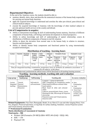
  • 1. 7 Anatomy Departmental Objectives At the end of the Anatomy course, the students should be able to:  mention, identify, show, draw and describe the anatomical structure of the human body responsible for carrying out normal body functions.  apply the acquired knowledge to understand and correlate the other pre-clinical, para-clinical and clinical medical subjects.  execute the acquired knowledge of Anatomy with the knowledge of other medical subjects to provide optimum health services in the country and abroad. List of Competencies to acquire:  Ability to demonstrate knowledge & skill of understanding human anatomy, functions of different components of human body, cell biology and human development in clinical perspective.  Ability to utilize knowledge and skill of understanding of spatial relationship, course & distributions of different components of human in real life situation.  Ability to detect the deviation from normal state in the human body in relation to structure, function and development.  Ability to identify human body components and functional pattern by using internationally accepted terminologies. Distribution of teaching - learning hours Lecture Tutorial Practical (Histology) Demons tration +Dissectio n +Card exam Total Teaching hours Integrat ed teaching for phase I Formative Exam Summative exam Preparato ry leave+ post term leave Exam time Preparato ry leave Exam time 115 hrs 53hrs 52 hrs 307 hrs 527 hrs 36 hrs 21+14= 35 days 42 days 30 days 30 days Time for integrated teaching, examination, preparatory leave of formative & summative assessment is common for all subjects of the phase Related behavioral, professional & ethical issues will be discussed in all teaching learning sessions Teaching - learning methods, teaching aids and evaluation Teaching Methods Teaching aids In course evaluation Large group Small group Self learning Lecture Integrated teaching Tutorial Practical Demonstration Dissection Self-study & self-assessment Computer / Laptop & Multimedia OHP, Transparency & Transparency marker White board & different coloured white board markers Black board & white and coloured chalks Cadavers, prosected parts, bones, viscera Histological slide, Microscope & Projection microscope Projection system and Virtual anatomy dissection table  Item Examination  Card Examination  Term Final Examination (written, oral+ practical) Related Equipments: Flip Chart, Photograph, Model, X-ray films (CT scan and other imaging films), View box, Diagram, Preserved specimens, Living body for surface marking, Simulator, various Projection system and Virtual anatomy dissection table. 1st Professional Examination: Marks distribution of Assessment of Anatomy Total marks – 500  Written=200 (Formative 20+MCQ (SBA+MTF) 40+(SAQ + SEQ) 140)  SOE = 150  Practical=150
  • 2. 8 Learning Objectives and Course Contents in Anatomy Learning Objectives Contents Teaching hours Total : 12 hrs General Anatomy Student will be able to  define anatomy and explain the subdivisions of anatomy.  describe the anatomical terminology, planes & positions.  define and classify bone. Describe the composition, blood supply, functions, ossification of bones with clinical correlation.  describe the composition, characteristics, location and functions of different types of cartilages.  define & classify joints. Describe the characters, stability & movements of joints with clinical correlation.  classify muscles. Describe their properties and functions.  define & classify blood vessels.  describe the different types of circulation.  describe different types of vascular anastomosis with their functional & clinical implications.  describe components, functions & the general plan of lymphatic drainage of the whole body.  classify lymphoid organs. Describe the functions of lymphoid organs with clinical significance. CORE :  Definition, subdivisions of anatomy and its importance in the study of medicine.  Anatomical terminologies, anatomical planes & positions.  Skeletal system: Bones – classification, composition, functions, parts of a developing long bone, blood supply, periosteum & endosteum. Ossification – definition, centres, processes. Factors affecting growth of bone.  Cartilages: composition, types, characters, locations and functions  Joint: classification, characteristics of each type & movements, stability of the joints. Clinical conditions associated with joints. General plan of blood supply & nerve supply of joints.  Muscular system: different ways of classification, characteristics and functions different types. Skeletal muscle – classification; Principle applied to innervation & contraction.  Blood vascular system: component parts, general plan, structure, classification. Differences between different types of vessel. Nutrition & innervations of vessels. Circulation – systemic, portal & pulmonary circulation and characteristic features of each type. Vascular anastomosis: types ,sites, characteristics ,functional and clinical importance  Lymph vascular system: components, characteristic features of lymph capillaries. Comparison with blood capillary.  Lymphoid organs: classification, distribution & functions, TERM I 01 hr 01 hr 03 hrs 01 hr 02 hrs 01 hr 02 hrs 01 hr
  • 3. 9 Learning Objectives Contents Teaching hours Total : 08 hrs Cell Biology Student will be able to:  define and describe the human cell & its constituents, structure & functions of all components of cell.  describe the features of different types of cells. Human Genetics Students will be able to:  describe the different basic features of chromosomes.  define terms related to human genetics. CORE:  Human Cell – Basic organization, types of constituents, cell membrane  Nucleus – structure & functions  Cytoplasm, organelles and inclusions – structure & functions  Functional correlation of different types of cell (protein secreting, ion transporting, steroid secreting, mucus secreting, antibody producing cell) in respect of their nuclear, cytoplasmic, membrane and surface feature CORE:  Chromosomes: Basic structure  Terms & definitions: Gene, Gene locus, genome, genotype, phenotype, genetic trait etc. Total:06 hrs. TERM I 02 hrs 01 hr 02 hrs 01hr Total: 02 hrs TERM I 01hr 01 hrs
  • 4. 10 Learning Objectives Contents Teaching hours Total: 14 hours General Histology Student will be able to:  define and classify the basic tissues in the body  identify under microscope different types of:  Epithelial tissue  Connective tissue  Muscular tissue  describe microscopic components and differentiate different components of:  Epithelial tissue  Connective tissue  Muscular tissue  describe the histological structures of different types of muscle tissue.  describe the composition & functions of components of nervous tissue  correlate inter-relationship between structure and functions of each tissue General Histology Basic tissues:  Definition, classification, components, characters, distribution and functions of  Epithelial tissue and its subtypes  Connective tissue and its subtypes  Muscular tissue and its subtypes  Structure and functions of  Cell surface specialization  Inter cellular junction  Histological structure of  Smooth muscle tissue  Cardiac muscle tissue  Skeletal muscle tissue  Mechanism of muscle contraction  Structure and function of Nervous tissue  Neurons  Neuroglia TERM I 05hrs 05 hrs TERM II 02 hrs TERM III 02 hrs
  • 5. 11 Learning Objectives Contents Teaching hours Total: 18 hours Systemic Histology: Students will be able to describe the histological structures of organs of different systems of the body. Systemic Histology : histological structures of  Respiratory system : Respiratory tract & Lung  Vascular system : Different types of artery, capillary & vein  Lymphoid organs: Thymus, spleen, lymph node & tonsil  Digestive system & associated Glands : tongue, oesophagus, stomach, intestine, Liver, gall bladder, pancreas  Exocrine glands : salivary glands  Urinary system : kidney, ureter, urinary bladder  Male reproductive system : testis, epididymis, vas deferens, seminal vesicle  Female reproductive system: ovary, uterus, uterine tube, vagina  Endocrine glands: pituitary, thyroid, parathyroid, adrenal glands  Skin and its appendages TERM I 01 hr 01 hr TERM II 02 hrs 04 hrs 01 hr 03 hr 02 hrs 02 hrs TERM III 01 hr 01 hr
  • 6. 12 Learning Objectives Contents Teaching hours Total 18hrs General Embryology Students will be able to:  define terms related to embryology  explain the significance of study of embryology  explain basic process of development  describe different processes of cell division  describe oogenesis and spermatogenesis  describe the process of fertilization  describe the events of 1st week of development.  describe the events2nd week of development.  describe the events 3rd week of development.  describe the development & derivatives of ectoderm, mesoderm & endoderm.  explain the development of foetal membranes  explain the development of twins & their types.  describe the causes & types of congenital anomalies  explain the process of human evocation  describe the Molecular regulation & cell signaling pathways CORE:  Introduction: terms and definition; Significance of study of embryology.  Basic process of development: proliferation, growth, differentiation, inductors, evocators and organizer  Cell division: Types  chromosomal changes during cell division with anomalies  Gametogenesis and maturation of Germ cells.  Fertilization: Events, factors influencing the fertilization; Progress in 1st week of development  Progress in 2nd week of development.  Progress in 3rd week of development.  Derivatives of germ layers: ectoderm, mesoderm & endoderm.  Foetal membranes : Placenta, chorion, amnion, umbilical cord, yolk sac etc.  Twins  Teratology Additional:  Human Evolution  Concepts of medical biotechnology in relation to embryology  Molecular regulation &cell signaling TERM I 01 hr 01 hr 02 hrs 02 hrs 02 hrs 02 hr 02 hrs 01 hr TERM II 03 hrs 01 hrs 01 hrs
  • 7. 13 Learning Objectives Contents Teaching hours Total: 24 hours Systemic Developmental Anatomy Student will be able to:  describe the process of development of different systems of the body  describe the developmental anomalies of the organs of different systems of the body  mention general outline of development of: Thoracic duct, Cisterna chyli, Inferior Vena Cava, Superior Vena Cava, Portal Vein, Brachiocephalic veins & Renal veins. CORE: Development and their Anomalies of  Skeletal system & vertebral column Muscular system  Upper and lower limb  Digestive system with associated glands  Respiratory system  Cardiovascular System & aortic arches  Coelomic cavity & the diaphragm  Skin & mammary gland  Urinary system  Male and female Reproduction system  Pituitary & suprarenal gland  Face & neck & their associated organs  Nervous System  Eye & Ear Additional: Development of  Lymphatic System  Vascular System TERM II 02 hrs 01 hr 03 hrs 01 hr 03 hrs 01 hr 01 hr 02 hrs 03 hrs TERM III 01 hr 03 hrs 02 hrs 01 hr
  • 8. 14 Learning Objectives Contents Teaching hours Total: 21 hours Neuroanatomy Students will be able to:  classify nervous system and describe each type.  mention different parts of nervous system  describe composition of nervous system  describe autonomic nervous system,  describe the coverings of central nervous system  describe the ventricular system of CNS  explain blood brain & blood CSF barrier CORE:  Introduction to Nervous system,  Composition of grey matter and white matter  Nerve fibers: structure classifications & functions, myelination, degeneration, regeneration  Receptors: definition, structure classifications location & functions  Synapse: definition, structure classifications & functions  Autonomic nervous system: parts, autonomic nerve plexuses & ganglia  Coverings of brain and spinal cord : Pia, arachnoid and dura mater, their extension, folds, spaces, nerve supply & blood supply  Ventricular system and Cerebrospinal fluid (CSF) : Location of different ventricles of brain the formation, composition, circulation, absorption & functions of CSF  Blood-brain and Blood-CSF barriers: Composition & function TERM I 01 hr TERM III 01hr 01 hrs TERM I & II 02 hrs TERM III 01 hr 02 hr
  • 9. 15 Learning Objectives Contents Teaching hours Neuroanatomy Students will be able to:  describe the anatomical aspects of motor and sensory parts of nervous system with their functional and clinical significance CORE: Motor system  Cerebrum (motor areas): Gyri, sulci and important functional areas with effects of lesion; mode of blood supply  Pyramidal & extrapyramidal system & effects of their lesion  Cerebellum: parts, functional lobes, nuclei, peduncles & functions, blood supply, clinical conditions  Basal nuclei: locations, parts, functions artery supply & clinical conditions  Motor and mixed Cranial Nerves: Classification, functional components, cranial nerve nuclei and course of cranial nerves Sensory system  Dermatome & axial line  Cerebrum( sensory areas): Gyri, sulci and important functional areas with effects of lesion; mode of blood supply  Ascending tracts of spinal cord with effects of lesions  Diencephalon: parts & functions  Sensory cranial nerves & Smell, visual & auditory pathway  Spinal Cord: Length, extension, enlargement, blood supply, cross-sections at different level  Brain stem: blood supply, cross sections at different levels  Reticular formation  Limbic system TERM III 02 hrs 01 hr 01 hr 02 hr 01hr 01 hr 01 hr 01 hr 01 hr 01 hr 01hr
  • 10. 16 Learning Objectives Contents Teaching hours (Total 24 hrs) Living (surface) Anatomy Students will be able to:  locate and count ribs & costal cartilages  draw and demonstrate important anatomical points and structures of Thorax on the surface of the body Students will be able to:  draw and demonstrate important anatomical points and structures of Superior extremity on the surface of the body Thorax CORE:  Counting of ribs and costal cartilages  Heart- apex and borders  Lung-borders and apex,  Trachea & Bronchi  Esophagus  Triangle of auscultation  Jugular notch  Sternal angle  Area of Superficial Cardiac dullness Common carotid and subclavian artery Internal thoracic artery Superior extremity CORE  Nerves: Radial, Ulnar, Median nerve, Axillary nerve  Arteries: Brachial, Radial, Ulnar artery, Superficial and deep palmar arch  Veins: cephalic, basilic & median cubital vein  Flexor retinaculum  Anatomical snuff box  Medial humeral epicondyle For Tutorial 06 hrs. 04 hrs.
  • 11. 17 Learning Objectives Contents Teaching hours Living (surface) Anatomy Students will be able to:  locate, demonstrate the different anatomical planes and land marks on the surface of the body  draw, demonstrate the nine regions of the abdomen on the surface of the body  draw and indicate inguinal canal on the surface of the body  draw and demonstrate important anatomical points, borders and parts of important organs of abdomen on the surface of the body Students will be able to:  locate and demonstrate important points and structures of inferior extremity on the surface of the body CORE: Abdomen  Trans-pyloric plane, Trans tubercular plane, Subcostal plane, mid clavicular line  Regions of abdomen  Superficial & deep inguinal ring, Inguinal canal  Abdominal aorta & inferior vena cava  Stomach, Duodenum, Pancreas, Liver, Gall bladder, Bile duct, spleen, Kidney from back & Mac Burney’s point  Transverse colon, ureter from front and back, celiac trunk, splenic artery, Root of the mesentery Inferior extremity  Common peroneal nerve, Tibial nerve  Popliteal artery  Anterior & posterior tibial artery  Arteria dorsalis pedis  Great Saphenous vein  Small Saphenous vein  Adductor tubercle  Lateral and Medial Malleolus  Greater trochanter of femur  Anterior superior iliac spine Additional  Femoral nerve, sural nerve, Medial and lateral plantar artery, plantar arch. For Tutorial 6 hrs. 4 hrs.
  • 12. 18 Learning Objectives Contents Teaching hours Students will be able to:  draw and demonstrate important anatomical points and structures of Head and Neck on the surface of the body Head and neck  Facial artery, Facial vein  Internal jugular vein, External jugular vein  Common Carotid artery & its bifurcation  Facial Nerve & their branches  vagus nerve in the neck  Parotid gland and its duct  Frontal and maxillary air sinuses  Thyroid gland  Tip of the coracoid process  Inferior angle of scapula  Tip of the 7th cervical spine Additional:  Pterion, lambda  Middle meningeal artery For Tutorial 04 hrs.
  • 13. 19 Learning Objectives Contents Teaching hours (Total 09 hrs) Anatomy of Radiology & Images Students will be able to:  describe radio-opaque and radio-lucent structures  identify and locate the normal structures in radiograph CORE Radio opaque structures Radio-lucent structures Plain X-ray of the -chest PA view -abdomen AP view -pelvis AP view -arm including proximal & distal joints AP & lateral view -forearm including proximal & distal joints AP & lateral view -hand including proximal & distal joints -thigh including proximal & distal joints AP & lateral view -leg including proximal & distal joints AP & lateral view -foot including proximal & distal joints AP & lateral view -head & neck (cervical spine) AP & lateral view -Paranasal sinuses OM view Additional:  Common normal Ultrasonographs, Isotope scan,  Magnetic Resonance Images (MRI), CT Scan  Coronary Angiograph For Tutorial 09 hrs
  • 14. 20 Learning Objectives Contents Teaching hours (Total 20 hrs) Clinical Anatomy Students will be able to:  describe the anatomical basis of clinical disorder of structures of the thorax and the abdomen. Thorax • Pleurisy / Pleural effusion • Pneumothorax • Coronary artery disease • Pericarditis/ pericardial effusion • Flail chest • Paralysis of the diaphragm Abdomen • Portal vein obstruction • Hydrocele • Hernia • Peritonitis, ascites • Gastric ulcer • Duodenal ulcer • Gall stone/cholecystitis • appendicitis • Benign hyperplasia of prostate, Prostatic cancer • Cystocele • Stress incontinence • Rupture urethra • Salpingitis • Ectopic pregnancy • Prolapse of uterus / vagina • Haemorrhoids • Undescended testis • Psoas abscess • Ischiorectal abscess For Tutorial 03 hrs 06 hrs
  • 15. 21 Learning Objectives Contents Teaching hours Clinical Anatomy Students will be able to:  describe the anatomical basis of clinical disorder of the structures of head & neck, nervous system & extremities Head & Neck • Fracture of the skull bones • Scalp injury • Piriform fossa and foreign body • Otitis media • Sinusitis • Epistaxis • Tonsilitis • Swelling of thyroid gland • Mumps • Cavernous vein thrombosis • Cervical rib CNS & Eyeball • Injury to brain /eye ball / spinal cord/cranial nerves • Meningitis • Hydrocephalus • Cerebral ischaemia, intracranial haemorrhage (extradural,subarachnoid, cerebral) • Papilledema • Horner’s syndrome Superior extremity • Dislocation of shoulder joint • Brachial plexus & injury to its nerves • Carpal tunnel syndrome • Colle’s fracture • Breast abscess & breast cancer Inferior extremity • Varicose vein • Deep vein thrombosis • Nerve injury • Dislocation of hip joint • Rupture of menisci & cruciate ligament, Bursitis • Deformities of foot For Tutorial 03hr . 03hr 03hr 02hr
  • 16. 22 Learning Objectives Contents Teaching hours Clinical Anatomy Students will be able to:  describe the anatomical basis for selection of arteries, veins & muscles of clinical importance.  demonstrate the different auscultatory areas  describe the anatomical basis for clinical procedure of Thorax, Abdomen, Head & Neck , CNS & Eyeball.  Arterial pulsation  Intravenous injections  Intramuscular injection  Apex beat, mitral, tricuspid, aortic & pulmonary areas • Sternal puncture • Pleural effusion • Pericardial effusion • Coronary angiogram • Bronchoscopy • Laryngoscopy • Paracentesis /peritoneal dialysis • Ryle’s tube • Endoscopy • Liver abscess • Vasectomy • Tubal ligation • Nasogastric intubation • Palpation of Cervical lymph node • Lumbar puncture • Epidural/spinal anesthesia • Pudendal block • Fundoscopy
  • 17. 23 Regional Anatomy : THORAX CARD (DISSECTION, DEMONSTRATION & TUTORIAL) Learning Objectives Contents Teaching hours Students will be able to:  demonstrate the boundary & identify the contents of thoracic wall, thoracic cavity, mediastinum & intercostal space  identify & demonstrate the gross features of bones & joints of thorax  describe the formation, course, branches & distribution of spinal nerve / intercostal nerve  identify & demonstrate the surfaces, borders, parts, chambers- including structures within the chambers of the heart  explain blood supply & nerve supply of heart  identify & demonstrate the layers of pericardium  identify & demonstrate the surfaces, borders, fissures, lobes, hilus & bronchopulmonary units of the lung  identify & demonstrate the layers & parts of pleura.  explain the blood supply, lymphatic drainage & nerve supply of lung & pleura.  identify & demonstrate the trachea, bronchus & bronchial tree.  explain blood supply & nerve supply of trachea & bronchial tree.  explain the blood supply, nerve supply & lymphatic drainage of thoracic wall.  identify & demonstrate the surfaces, parts openings, attachments of the diaphragm.  explain the blood supply & nerve supply of the diaphragm.  explain the significance of the orifices of the diaphragm.  explain & demonstrate the extension, parts, relations & constrictions of oesophagus  explain the blood supply, lymphatic drainage & nerve supply of the oesophagus.  correlate clinical conditions associated with structures of thorax (Heart with its vessels, lung, trachea, bronchus, bronchial tree & the diaphragm)  Thoracic wall formation, thoracic cavity, intercostal space and mediastinum.  Bones and joints of the thorax  Spinal nerve / intercostal nerve  Heart with pericardium.  Lung with pleura, trachea and bronchus.  Blood vessels, nerves and lymphatics of the thorax.  The diaphragm.  Oesophagus  Clinical Anatomy 45 hrs. NB: Previously mentioned 53 hours in pages 10-16 for Tutorial also have shown in this part (DISSECTION, DEMONSTRATION & TUTORIAL)
  • 18. 24 Regional Anatomy: SUPERIOR EXTREMITY CARD (DISSECTION, DEMONSTRATION & TUTORIAL) Learning Objectives Contents Teaching hours Students will be able to:  identify & demonstrate muscles, vessels, nerves of pectoral region including attachment of muscles  describe the parts of mammary gland & its blood supply, lymphatic drainage & nerve supply  demonstrate the boundary & identify the contents of axilla, quadrangular & triangular spaces, & cubital fossa  demonstrate the attachments of muscles, and identify vessels, nerves, lymphatics & lymph nodes of different parts of superior extremity  demonstrate the gross features of bones & joints of superior extremity and muscles acting on joints  correlate clinical conditions associated with structures (nerves, vessels, bones, joints) of superior extremity  Pectoral region with mammary gland  Axilla  Superficial dissection of the upper limb, back and scapular region including quadrangular & triangular space Front of the arm, forearm and palm  Back of the arm, forearm and dorsum of the hand  Blood supply, lymphatic drainage, cutaneous innervation & dermatome of superior extremity  Bones & joints of the upper limb  Clinical Anatomy 43 hrs.
  • 19. 25 Regional Anatomy: ABDOMEN CARD (DISSECTION, DEMONSTRATION & TUTORIAL) Learning Objectives Contents Teaching hours Students will be able to:  demonstrate the different layers of anterior abdominal wall & hernial region  explain clinical types of hernia  demonstrate the different parts of GI Tract & its peritoneum  explain the mode of blood supply, lymphatic drainage & nerve supply of different organs  demonstrate the features of liver, pancreas, supra renal gland & different parts of biliary system  explain blood supply, lymphatic drainage & nerve supply of them.  demonstrate the features of kidney, suprarenal gland, ureter, urinary bladder, & urethra  explain their blood supply, lymphatic drainage & nerve supply  demonstrate the features of different parts of male & female reproductive system.  explain their blood supply, lymphatic drainage & nerve supply.  demonstrate the muscles and identify the vessels, nerves & lymphatics of posterior abdominal wall  demonstrate the parts and identify the contents of the pelvis  differentiate between male & female pelvis  demonstrate the gross features & joints of lumbar vertebra & bony pelvis and muscles acting on joints  correlate clinical conditions associated with different organs of the abdomen  Anterior wall of the abdomen with hernial region.  Stomach, abdominal part of the oesophagus  Duodenum, pancreas and spleen.  The mesentery and mesenteric vessels, jejunum and ileum.  Large intestine. rectum & anal canal  Liver with the biliary apparatus including gall bladder; portal vein.  Kidney, suprarenal gland, ureter, urinary bladder & urethra.  Ovary, uterus, uterine tube, female external organs and perineum.  Vas deferens, seminal vesicle, prostate and male external genital organs.  Muscles, blood vessels, lymphatics and nerves of the posterior abdominal wall.  Muscles, blood vessels lymphatics, nerves of the pelvis.  Lumbar vertebra, bony pelvis & joints  Clinical Anatomy 103 hrs.
  • 20. 26 Regional Anatomy: INFERIOR EXTREMITY CARD (DISSECTION, DEMONSTRATION & TUTORIAL) Learning Objectives Contents Teaching hours Students will be able to:  demonstrate muscles attachments and identify vessels & nerves of different parts of inferior extremity  demonstrate the boundary and identify the contents of femoral triangle, adductor canal, popliteal fossa & sole of the foot  demonstrate the features of bones, joints, & muscles acting on joints  explain the venous drainage, lymphatic drainage, & dermatome of inferior extremity  correlate the clinical conditions associated with structures (nerves, vessels, bones, joints) of inferior extremity  Front and medial side of the thigh  Gluteal region and back of the thigh  Front of the leg and dorsum of the foot  Lateral side, medial side and back of the leg including the popliteal fossa sole of the foot  Bones & joints of lower limb  Arches of the foot  Blood supply, lymphatic drainage, cutaneous innervation & dermatome of inferior extremity  Clinical Anatomy 42 hrs.
  • 21. 27 Regional Anatomy: HEAD & NECK CARD (DISSECTION, DEMONSTRATION & TUTORIAL) Learning Objectives Contents Teaching hours Students will be able to:  identify and demonstrate the different parts of bones of head & neck, joints, & muscles acting on joints  state the gross features & attachments of skull bones including base of skull & cervical vertebrae.  demonstrate movements of joints of head & neck  demonstrate the layers of scalp identify the contents of temporal region  demonstrate the boundary of face and identify muscles and sensory supply of face  identify parotid gland & duct & explain the structures within the parotid gland  demonstrate the boundary and identify contents of different triangles of head & neck region  demonstrate the boundary and identify contents of mouth cavity  demonstrate the gross features & nerve supply of tongue  explain Auditory pathway (VIII – cranial nerve)  demonstrate the different parts of pharynx with their extension & muscles of pharynx, the walls of nose and paranasal air sinuses, the extension, cartilages & muscles of larynx  identify structures present in the internal surface of the larynx  demonstrate the region of vertebral column and attachments of muscles of the back  demonstrate the different parts of ear  correlate important clinical conditions associated with structures in Head & Neck (Thyroid gland, parathyroid gland, air sinuses, larynx, scalp, ear, face etc.)  Bones & joints of head and neck  Scalp and temporal region  Face and orbit  Anterior triangle and its subdivisions, submandibular region including thyroid gland  Posterior triangle  Mouth and tongue  Pharynx  Nose and paranasal sinuses  Larynx  Vertebral column and deep dissection of the back of the neck  External, middle and internal ear.  Clinical Anatomy 87 hrs.
  • 22. 28 Regional Anatomy: CENTRAL NERVOUS SYSTEM & EYEBALL CARD (DISSECTION, DEMONSTRATION & TUTORIAL) Learning Objectives Contents Teaching hours Students will be able to:  demonstrate  the boundary & contents of cranial cavity & orbit  the different parts of brain & cranial nerves attached to brain  the layers of meninges- Pia, arachnoid, and dura mater  explain the folds of dura & its contents  explain the blood supply & nerve supply of the meninges  demonstrate the boundary of different lobes of cerebrum, sulci, gyri & important functional areas  explain the blood supply of cerebrum including the formation of Circle Willis  demonstrate the parts & describe the functions & connections of  diencephalon, pituitary gland, basal nuclei,  internal capsule, extra pyramidal system &  limbic system, brain stem  locate &describe  the nuclei, course, functional components & distribution of cranial nerves  the boundary & parts of ventricles circulation of CSF through ventricles  gross features of spinal cord and its meninges and spinal nerves attached to it  the coats of eyeball & the course of optic nerve  explain refractive media  explain the effects of lesion and loss of blood supply to different parts of nervous system.  Introduction to the nervous system, cranial cavity and orbit.  General examination of the brain  Superficial attachments of cranial nerves  meninges of the brain  Cerebrum: lobes of cerebrum, sulci, gyri & important functional areas, blood supply, formation of Circle of Willis  Diencephalon: thalamus, hypothalamus, metathalamus, epithalmus and pituitary gland  Basal nuclei, internal capsule, extra pyramidal system and limbic system  Brain stem and reticular formation  Cranial nerves  Ventricles and cerebrospinal fluid Spinal cord & spinal nerves  eyeball  Clinical Anatomy.   40 hrs                     
  • 23. 29 Cell Biology & Histology Tutorial & Practical (Card I) Learning Objectives Contents Teaching hours Students will be able to:  demonstrate different parts of microscope & how to handle it  state the principles of tissue preparation  explain cell division  identify different types of tissue on slide under microscope  Microscope: Parts & how to handle  Principles of different types of microscopy  Principles of tissue preparation and staining:  Fixation, embedding, sectioning & routine staining  Cell and cell division  Epithelium: Simple squamous, cuboidal, columnar, Pseudo stratified Stratified squamous, cuboidal, Stratified columnar Transitional  Connective tissue:: General, special, bone, cartilage  Muscular tissue: smooth, skeletal & cardiac muscle  Nervous tissue in general 17 hrs.
  • 24. 30 Cell Biology & Histology Tutorial & Practical (Card II) Learning Objectives Contents Teaching hours  Students will be able to identify different structures of the following systems on slides under microscope: Respiratory system. Cardiovascular system Digestive system and & associated Glands. Urinary system Male reproductive system and associated glands female reproductive system and associated glands  Respiratory system Larynx, trachea, bronchial tree and Lung  Large artery, medium sized artery, large vein  Digestive system & associated glands Tongue, pharynx, oesophagus, stomach, small intestine & large intestine (including vermiform appendix) Liver and gall bladder, Pancreas  Urinary system Kidney, ureter, urinary bladder, urethrae  Male reproductive system and associated glands Testis, epididymis, vas deferens, seminal vesicle, prostate  Female reproductive system and associated glands Ovary, fallopian tube, uterus, vagina  Mammary gland , placenta 17hrs.
  • 25. 31 Cell Biology & Histology Tutorial & Practical (Card III) Learning Objectives Contents Teaching hours  Students will be able to identify following structures on slides under microscope: Lymphatic system Salivary glands Nervous system Endocrine system Special sense organs Skin  Lymphatic system Lymph node, tonsil, spleen & thymus  Exocrine glands (salivary glands}  Nervous system spinal cord, cerebrum, cerebellum, peripheral nerve (including the optic nerve)  Endocrine gland (Pituitary, Thyroid, Parathyroid, Adrenal and Islet’s of Langerhans  Special sense organs: Eyeball (cornea, retina), internal ear  Thick skin & thin skin 18 hrs.
  • 26. 32 Teaching - Learning & Assessment Methods Teaching / Learning Method Teaching Aid In Course Assessment Summative Assessment Lecture Computer & multimedia Slide projector, overhead projector (OHP), black board white and different colour chalk, white board and different colour white board markers.  Item Examination: Oral, Practical  Card Completion Examination  Term Examinations: Written, Oral, Practical  Preparation of exercise book • Written • Oral • Practical Regional Anatomy: Demonstration & Tutorial Cadavers, prosected parts, bones, viscera and other specimens of body parts, models, charts, black board white and different coloured chalk, white board and different coloured white board markers, Illustration sheets/posters, OHP, video, slide projector, computer with CD ROM, radiographs & other images. Projection system and Virtual anatomy dissection table Regional Anatomy: Dissection Cadavers, prosected parts, specimens and bones, black board white and different coloured chalk, white board and different colour white board markers, Computer & multimedia. Projection system and Virtual anatomy dissection table Cell Biology & Histology Tutorial & Practical Histological slide, Microscope & Projection microscope slide projector, black board white and different colour chalk, white board and different coloured white board markers, OHP, Illustration sheets (including photomicrographs & drawings)/posters, video projector, computer with CD ROM drive.
  • 27. 33 Assessment in Anatomy Component Marks Total Marks Formative assessment 10+10 20 WRITTEN EXAMINATION paper-I- MCQ (SBA+MTF) 20 (SAQ+ SEQ) 70 paper-II- MCQ (SBA+MTF) 20 180 (SAQ+ SEQ) 70 ORAL EXAMINATION (Structured) Board I 75 150 Board II 75 PRACTICAL EXAMINATION Board I Board II Objective structured practical Exam (OSPE) Dissection Anatomy of Radiology and imaging Lucky slides Living Anatomy Practical Khata 30 30 10 15 10 10 10 10 10 10 05 --- 75 +75 Grand Total 500  Topics: Board I: CNS & Eyeball, Head & Neck, Thorax (Gross anatomy, Clinical anatomy, Histology, Embryology). Cell biology & Genetics. General Histology: Epithelial Tissue, Nervous Tissue. General Anatomy: Angiology, Neurology. Board II: Abdomen, Inferior & Superior Extremity (Gross anatomy, Clinical anatomy, Histology, Embryology). General Embryology. General Histology: Connective Tissue, Muscle Tissue General Anatomy: Osteology, Arthrology, Myology.  Each student will appear in Board I & Board II in separate date/day for oral and practical examination  Pass marks 60% in each of theoretical, oral and practical examination
  • 28. 34 Time allocation in Anatomy Lecture & Review - 115 hours Term General Anatomy Hours Cell Biology Hours General Histology Hours Systemic Histology Hours General Embryology Hours Systemic Embryology Hours Neuro anatomy Hours. Human Genetics Hours. Total Hours First Term 12 06 10 02 13 - 01 02 46 Second Term - - 02 14 05 17 02 - 40 Third Term - - 02 02 - 07 18 - 29 Grand Total Hours (Class +Exam) 12 06 14 18 18 24 21 02 115 Cell Biology & Histology - Tutorial & Practical – 52 hours Term Class Hours (Including Item Exam hrs) Card Completion Exam Hours Total Hours First Term (Card I) 15 2 17 Second Term (Card II) 15 2 17 Third Term (Card III) 16 2 18 Grand Total Hours 46 6 52
  • 29. 35 Term Cards Dissection & Demonstration Tutorial Review Part Completion Examination Hours Total Hours Living (surface) Anatomy Anatomy of radiology & Images Clinical Anatomy First Term Thorax 34 6 1 3 01 45 Superior Extremity 33 4 2 3 01 43 Second Term Abdomen 89 6 1 6 01 103 Inferior Extremity 33 4 2 2 01 42 Third Term Head, Neck 77 4 2 3 01 87 Central Nervous system and Eye ball 35 00 1 3 01 40 Grand Total Hours 301 24 9 20 06 360
  • 30. 36 ACADEMIC CALENDAR for ANATOMY N.B.- Card completion examinations will be arranged on discussion with other departments Class/Exam Hours(including Class exams hrs) First Term (14 working weeks) Second Term (15 working weeks) Third Term (14 working weeks) Lecture and Review 115  General Anatomy-12 hrs  Cell Biology -06 hrs  Human Genetics - 02 hrs  General Histology-10 hr  Systemic Histology – 02 hrs  General Embryology - 13 hrs  Neuroanatomy – 01 hrs  General Histology-02 hr  Systemic Histology - 14 hrs  General Embryology - 05 hrs  Systemic Embryology- 17 hrs  Neuroanatomy – 02 hrs a) General histology - 02 hr b) Systemic Histology -02 hrs c) Systemic Embryology - 07 hrs d) Neuroanatomy - 18hrs Tutorial/ Review 53 Thorax Card – 10 hrs Sup. Ext. Card – 09 hrs Abdomen Card – 13 hrs Inf. Ext. Card – 08 hrs Head & Neck Card –09 hrs C.N.S & Eyeball – 04 hrs Dissection 301 Thorax Card - 34hrs Sup Ext Card- 3hrs Abdomen Card – 89hrs Inf. Ext. Card – 33hrs Head & Neck Card – 77 hrs C.N.S & Eyeball Card - 35 hrs Card Completion Exam 06 Thorax Card- 01hrs Sup Ext. Card- 01hrs Abdomen Card– 01hrs Inf. Ext. Card – 01hrs Head & Neck Card –01 hrs C.N.S & Eyeball Card - 01 hrs Cell Biology & Histology-Tutorial/ Practical 52 Card I – 17hrs Card II - 17hrs Card III – 18 hrs Grand Total 527 (Physiology, Biochemistry) Prerequisite for 1st professional examination 1. A Student must pass all term exam before appearing 1st professional exam. 2. Class attendance must be 75 % Evaluation & leave 04 weeks Evaluation & leave 04 weeks 2.Evaluation & preparatory leave for first prof–08 weeks 1.Evaluation & preparatory leave for third term;03 weeks
  • 31. 37 Year Session Roll No. Batch Card no. Cadaver no. Total marks Pass marks Name of the student Period of placement From : To : DEPARTMENT OF ANATOMY ………………………..MEDICAL COLLEGE THORAX CARD (ITEM EXAM FOLLOWING DISSECTION, DEMONSTRATION & TUTORIAL) Part for dissection (item) Date of beginning Date of examination Marks obtained Remarks and Signature of the Lecturer 1.Thoracic wall, intercostal space, thoracic cavity and mediastinum 2. Bones and joints of the thorax 3. Heart with pericardium 4. Lung, pleura, trachea and bronchial tree 5. The Diaphragm & oesophagus 6. Blood vessels, nerves and lymphatics of the thorax 7. Living Anatomy 8. Anatomy of Radiology & Images *Each item should cover related clinical & functional anatomy No. of attendance in the practical classes of the card Out of Mark obtained Remarks Signature of the Lecturer Signature of Head of the Department
  • 32. 38 Year Session Roll No. Batch Card no. Cadaver no. Total marks Pass marks Name of the student Period of placement From : To : DEPARTMENT OF ANATOMY ………………………..MEDICAL COLLEGE SUPERIOR EXTREMITY CARD (ITEM EXAM FOLLOWING DISSECTION, DEMONSTRATION & TUTORIAL) Part for dissection (item) Date of beginning Date of examination Marks obtained Remarks and Signature of the Lecturer 1. Bones and introduction to the joints of the superior extremity 2. Pectoral region with mammary gland 3. Axilla 4. Superficial dissection of the upper limb, back and scapular region. 5. Front of the arm, forearm & palm 6. Back of the arm, forearm & dorsum of the hand 7. Blood vessels, nerves and lymphatics of the superior extremity 8. Shoulder joint, acromioclavicular joint, elbow joint, wrist joint, joints of hand 9. Living Anatomy 10. Anatomy of Radiology & Images *Each item should cover related clinical & functional anatomy No. of attendance in the practical classes of the card Out of Mark obtained Remarks Signature of the Lecturer Signature of Head of the Department
  • 33. 39 Year Session Roll No. Batch Card no. Cadaver no. Total marks Pass marks Name of the student Period of placement From: To : DEPARTMENT OF ANATOMY ………………………..MEDICAL COLLEGE ABDOMEN CARD (ITME EXAM FOLLOWING DISSECTION, DEMONSTRATION & TUTORIAL) Part for dissection (item) Date of beginning Date of examination Mark obtained Remarks and Signature of the Lecturer 1.Bones and joints of abdomen & pelvis 2.Anterior wall of the abdomen with hernial region 3.Stomach, abdominal part of the oesophagus and coeliac trunk 4.Duodenum, pancreas and spleen 5.The mesentery and mesenteric vessels, jejunum and ileum 6.Large intestine 7. Rectum and anal canal 8..Liver with the biliary apparatus including gall bladder; portal vein 9. Kidneys, suprarenal gland, ureters. urinary bladder, urethrae 10. Muscles, blood vessels, lymphatics and nerves of the posterior abdominal wall 11. Muscles, blood vessels, lymphatics, nerves of the pelvis 12. Ovaries, uterus, uterine tubes, vagina, female external genital organs 13.Perineum, pelvic diaphragm, urogenital diaphragm, perineal pouches, ischiorectal fossa 14.Vas deferens, seminal vesicles, prostate, testes and male external genital organs 15. Living Anatomy 16.Anatomy of Radiology & Images *Each item should cover related clinical & functional anatomy No. of attendance in the practical classes of the card Out of Mark obtained Remarks Signature of the Lecturer Signature of Head of the Department
  • 34. 40 Year Session Roll No. Batch Card no. Cadaver no. Total marks Pass marks Name of the student Period of placement From : To : DEPARTMENT OF ANATOMY ………………………..MEDICAL COLLEGE INFERIOR EXTREMITY CARD (ITEM EXAM FOLLOWING DISSECTION, DEMONSTRATION & TUTORIAL) Part for dissection (item) Date of beginning Date of examination Marks obtained Remarks and Signature of the Lecturer 1. Bones and introduction to the joints of the inferior extremity 2. Front and medial side of the thigh 3. Gluteal region and back of the thigh 4. Front of the leg and dorsum of the foot 5. Lateral side, medial side and back of the leg including the popliteal fossa., sole of the foot 6. Blood vessels, nerves and lymphatics of the inferior extremity 7. Hip joint, knee joint, tibiofibular joints , ankle joint 8. Joints and arches of the foot 9. Living Anatomy 10. Anatomy of Radiology & Images *Each item should cover related clinical & functional anatomy No. of attendance in the practical classes of the card Out of Mark obtained Remarks Signature of the Lecturer Signature of Head of the Department
  • 35. 41 Year Session Roll No. Batch Card no. Cadaver no. Total marks Pass marks Name of the student Period of placement From : To : DEPARTMENT OF ANATOMY ………………………..MEDICAL COLLEGE HEAD AND NECK CARD (ITEM EXAM FOLLOWING DISSECTION, DEMONSTRATION & TUTORIAL) Part for dissection (item) Date of beginning Date of examination Mark obtained Remarks and Signature of the Lecturer 1. Bones of head and neck 2. Joints of head and neck 3. Scalp and temporal region 4. Face and orbit 5. Anterior triangle and submandibular region 6. Posterior triangle 7. Mouth and tongue 8. Pharynx 9. Nose and paranasal sinuses 10. Larynx 11. Vertebral column and deep dissection of the back 12. Blood vessels, nerves and lymphatics of the head & neck 13. Exocrine & endocrine glands of head & neck 14. Organ of hearing and equilibrium (Ear) 15. Living Anatomy 16. Anatomy of Radiology & Images *Each item should cover related clinical & functional anatomy No. of attendance in the practical classes of the card Out of Mark obtained Remarks Signature of the Lecturer Signature of Head of the Department
  • 36. 42 Year Session Roll No. Batch Card no. Cadaver no. Total marks Pass marks Name of the student Period of placement From : To : DEPARTMENT OF ANATOMY ………………………..MEDICAL COLLEGE CENTRAL NERVOUS SYSTEM AND EYEBALL CARD (ITEM EXAM FOLLOWING DISSECTION, DEMONSTRATION & TUTORIAL) Part for dissection (item) Date of beginning Date of examination Mark obtained Remarks and Signature of the Lecturer 1. General introduction to the nervous system, cranial cavity and orbit 2. General examination of the brain with its nerve attachments and meninges 3. Cranial nerve – nuclei, course. functional components, supply & lesions 4. Cerebrum 5. Diencephalon 6. White matter of cerebrum, Basal ganglia,, Pyramidal and extra -pyramidal system , limbic system 7. Brain stem, reticular formation & Cerebellum 8. Ventricles and cerebrospinal fluid 9. Spinal cord & Spinal nerve 10. Eyeball 11. Living Anatomy 12. Anatomy of Radiology & Images 4. *Each item should cover related clinical & functional anatomy No. of attendance in the practical classes of the card Out of Mark obtained Remarks Signature of the Lecturer Signature of Head of the Department
  • 37. 43 Year Session Roll No. Batch Total marks Pass marks Name of the student Period of placement From : To : DEPARTMENT OF ANATOMY ……………………MEDICAL COLLEGE HISTOLOGY CARD NO. I Item Date of beginning Date of examination Marks obtained Remarks and Signature 1. Study of microscope 2. Principles of tissue preparation and staining (routine) 3. Cell and cell division 4. Epithelium 5. Connective tissue - General 6. Connective tissue – proper 7. Muscular tissue (skeletal, cardiac, smooth) 8. Nervous tissue in general Total No. of attendance Out of Marks obtained Remarks Signature of the Lecturer Signature of the Prof. of Anatomy
  • 38. 44 Year Session Roll No. Batch Total marks Pass marks Name of the student Period of placement From : To : DEPARTMENT OF ANATOMY ………………………..MEDICAL COLLEGE HISTOLOGY CARD NO. II Item Date of beginning Date of examination Marks obtained Remarks and Signature 1. Vascular system 2. Respiratory system 3. Digestive system & associated glands 4. Urinary system 5. Male reproductive system 6. Female reproductive system Total No. of attendance Out of Marks obtained Remarks Signature of the Lecturer Signature of the Prof. of Anatomy
  • 39. 45 Year Session Roll No. Batch Total marks Pass marks Name of the student Period of placement From : To : DEPARTMENT OF ANATOMY ………………………..MEDICAL COLLEGE HISTOLOGY CARD NO. III Item Date of beginning Date of examination Marks obtained Remarks and Signature 1. Lymphoid organ 2. Salivary glands 3. Endocrine Glands 4. Nervous system 5. Skin –thick skin & thin skin, Special sense organ (Cornea, Retina, Internal ear) Total No. of attendance Out of Marks obtained Remarks Signature of the Lecturer Signature of the Prof. of Anatomy