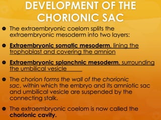
Downloaded 23 times

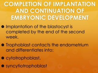
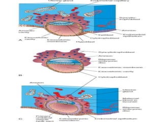
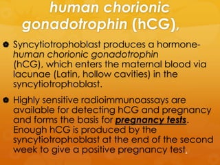
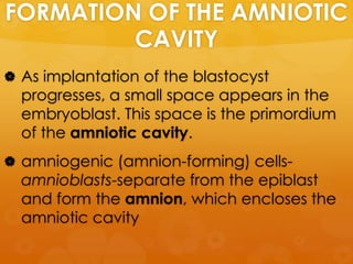
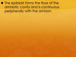

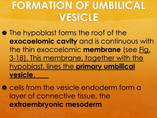
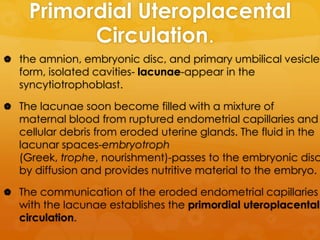
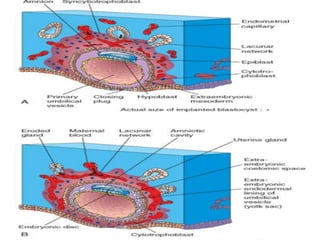


Implantation of the blastocyst is completed by the end of the second week of development. The trophoblast differentiates and the syncytiotrophoblast produces human chorionic gonadotropin (hCG) which enters maternal blood. HCG can be detected via pregnancy tests by the end of the second week. The amniotic cavity and embryonic disc form from the embryoblast, and the hypoblast forms the umbilical vesicle and extraembryonic mesoderm. Changes in the trophoblast and endometrium result in the formation of the extraembryonic coelom and chorionic sac, within which the embryo develops.


![PERI-PROSTHETIC FRACTURE NAIL-PLATE CONSTRUCT [NPC].pptx](https://cdn.slidesharecdn.com/ss_thumbnails/drarunkumardrmohamedashrafperiprostheticfrasturenail-plateconstructnpc-260209164459-7e9d15a1-thumbnail.jpg?width=640&height=640&fit=bounds)

