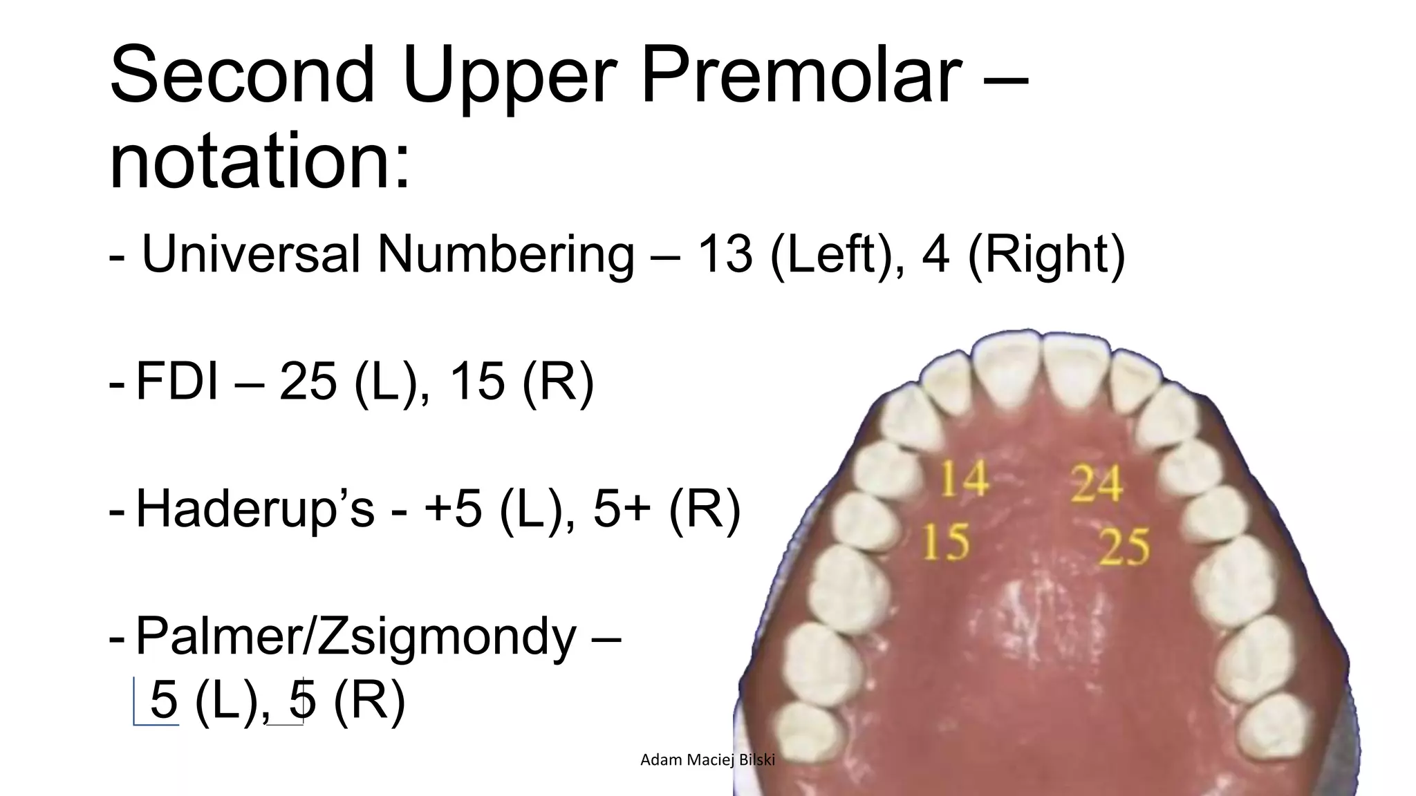
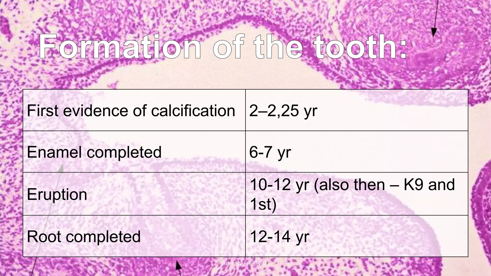
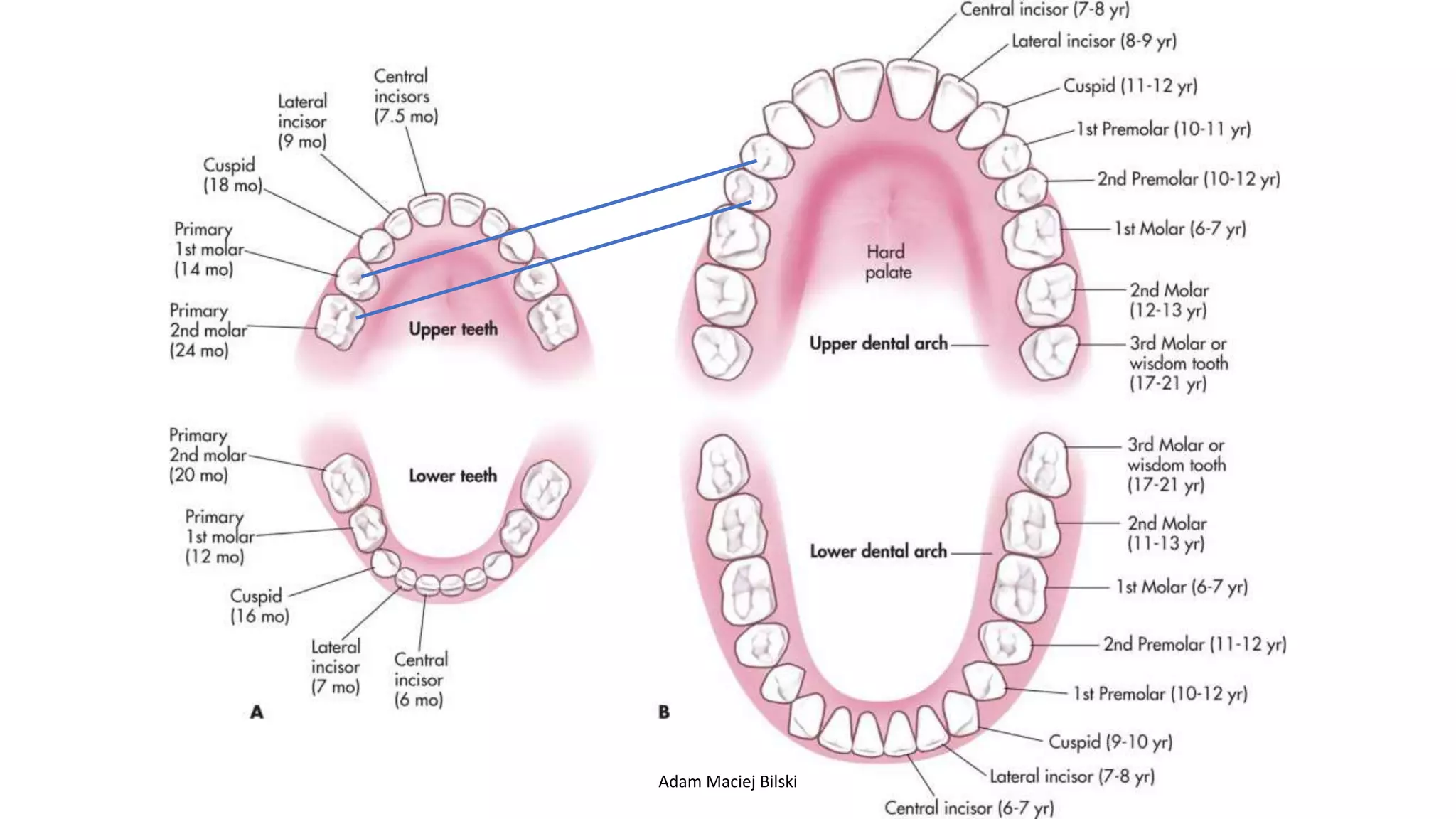
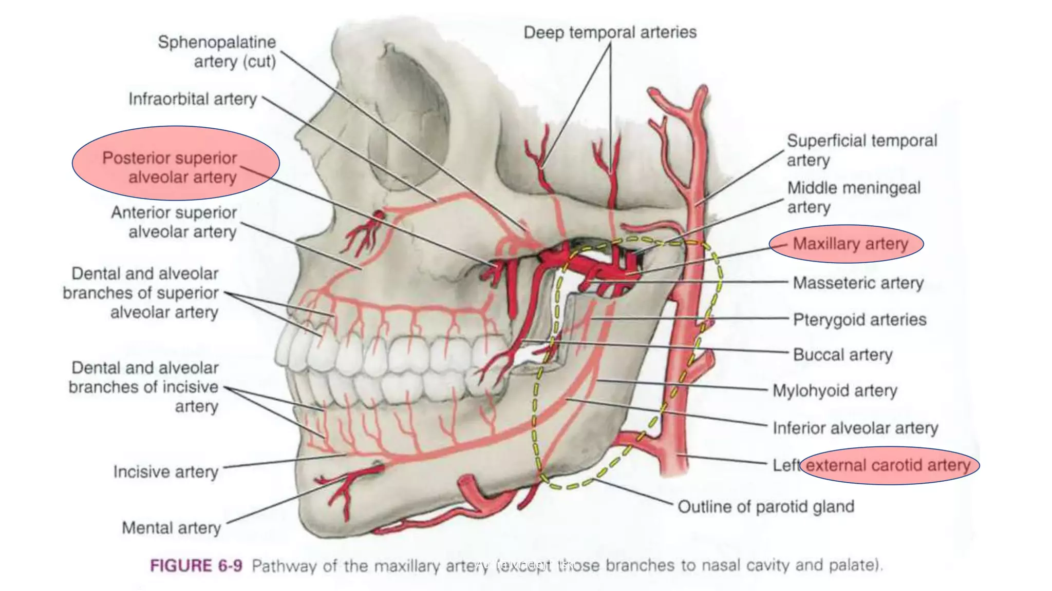
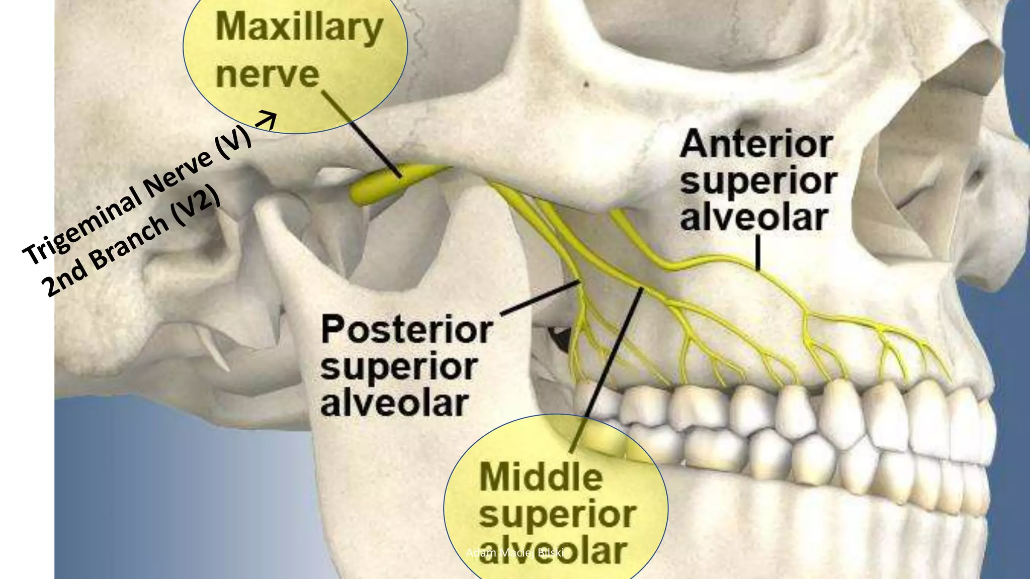
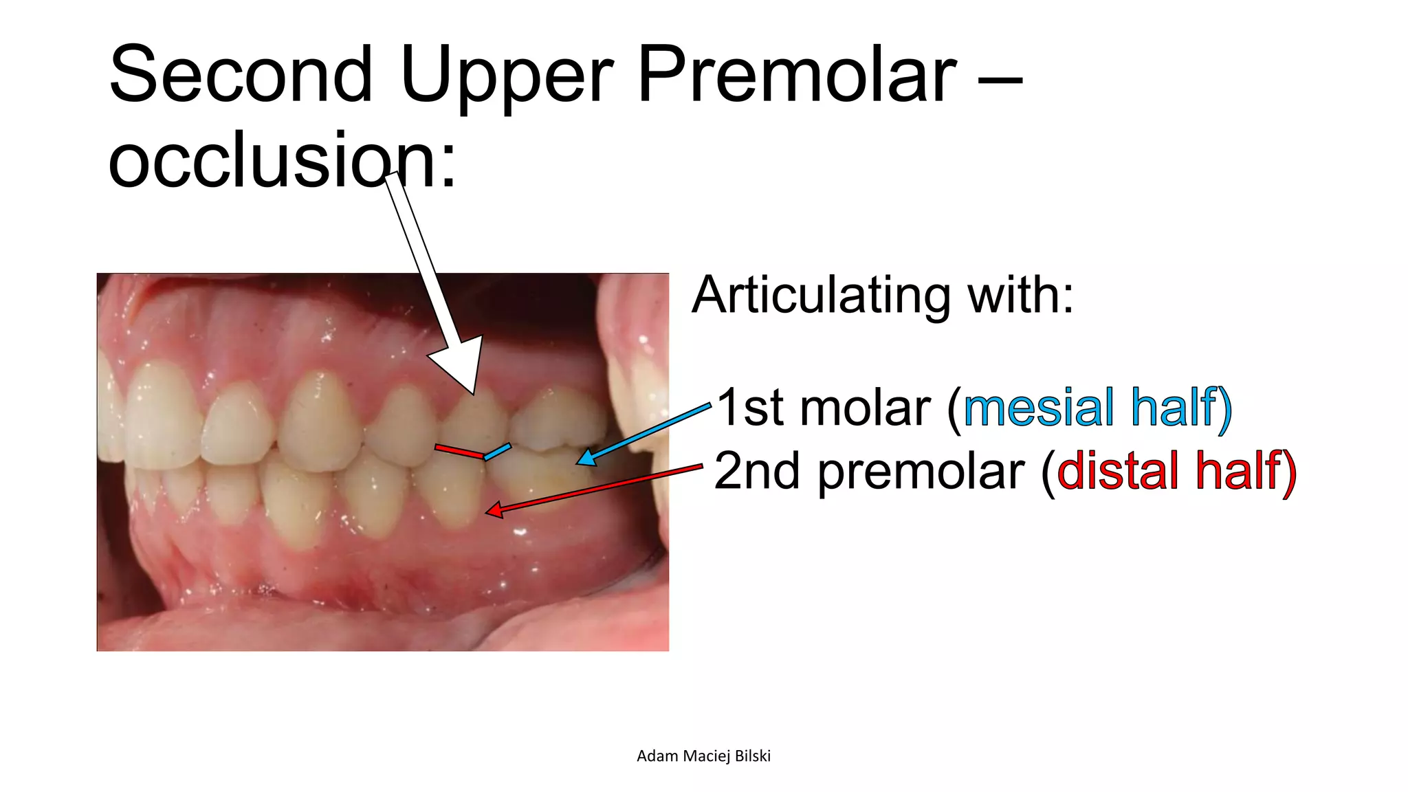
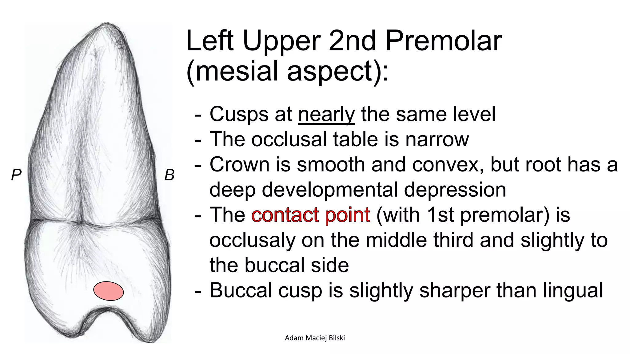
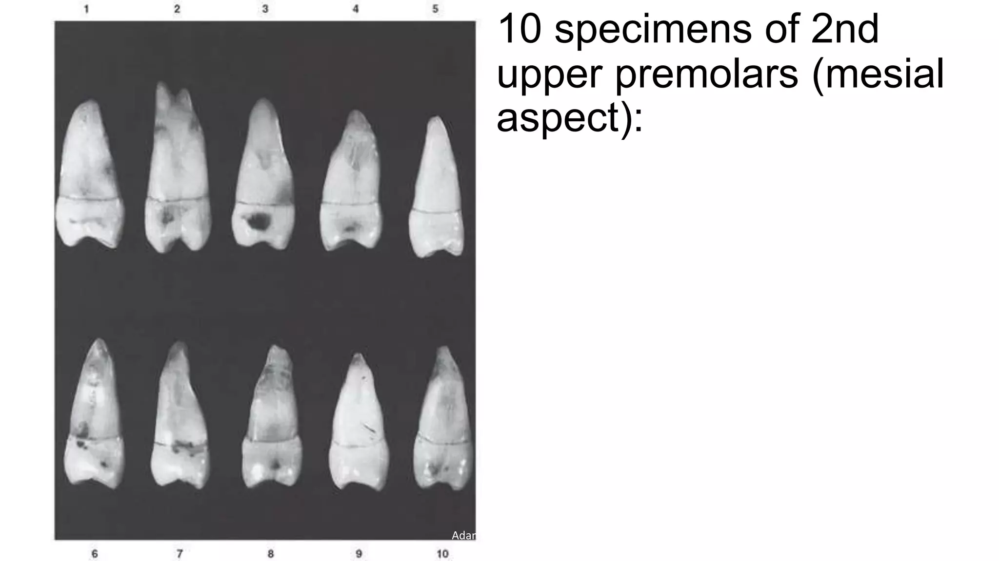
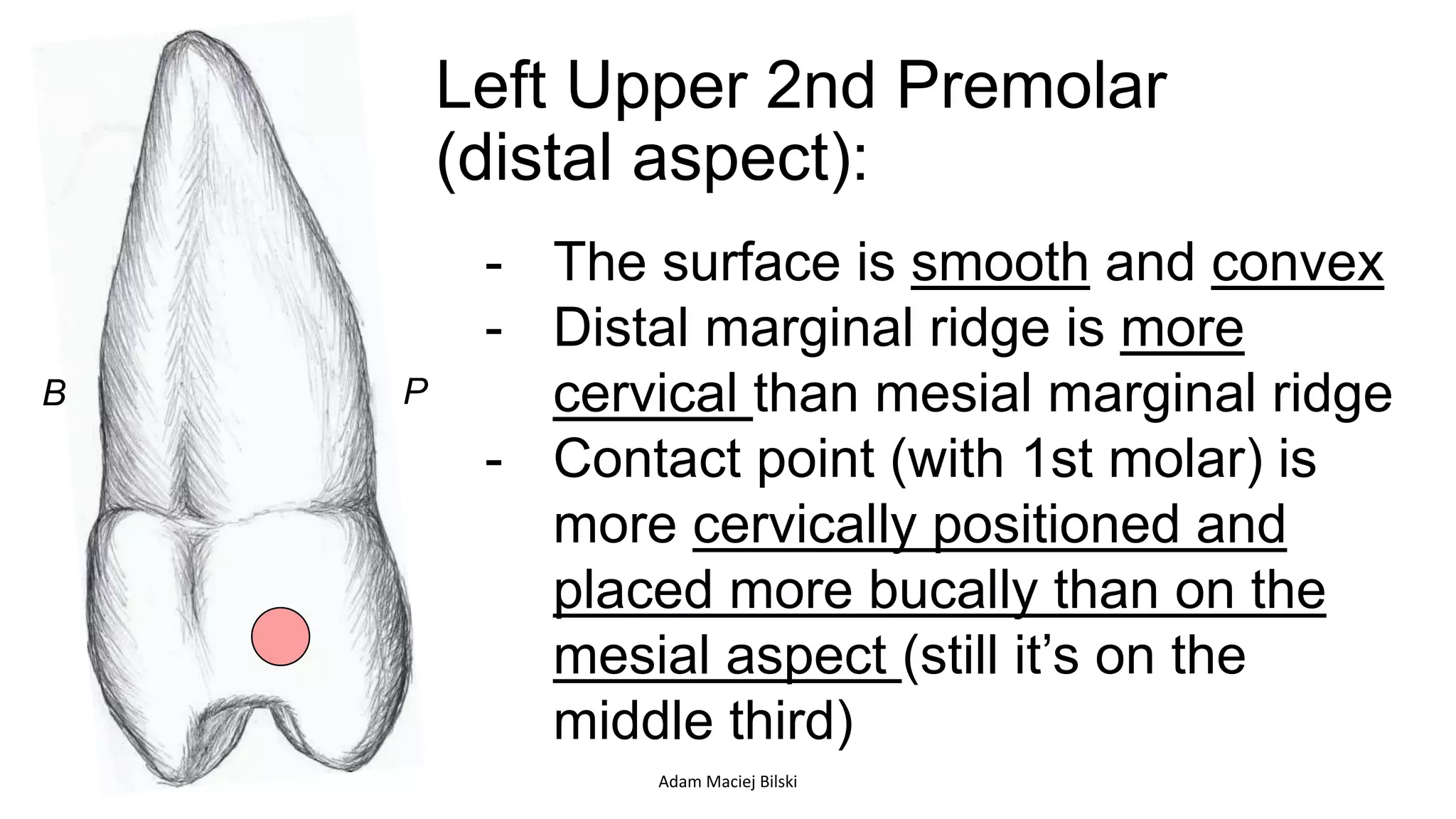
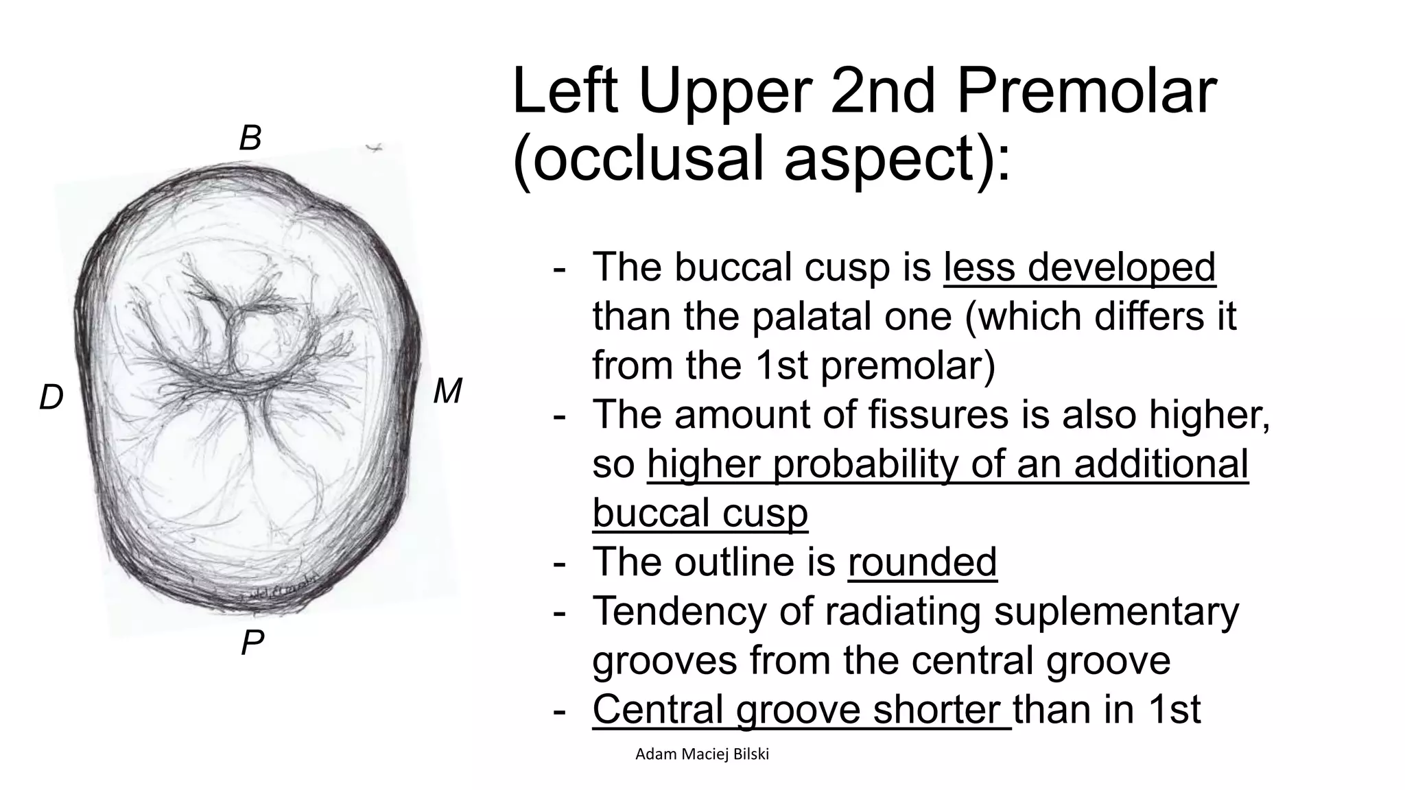
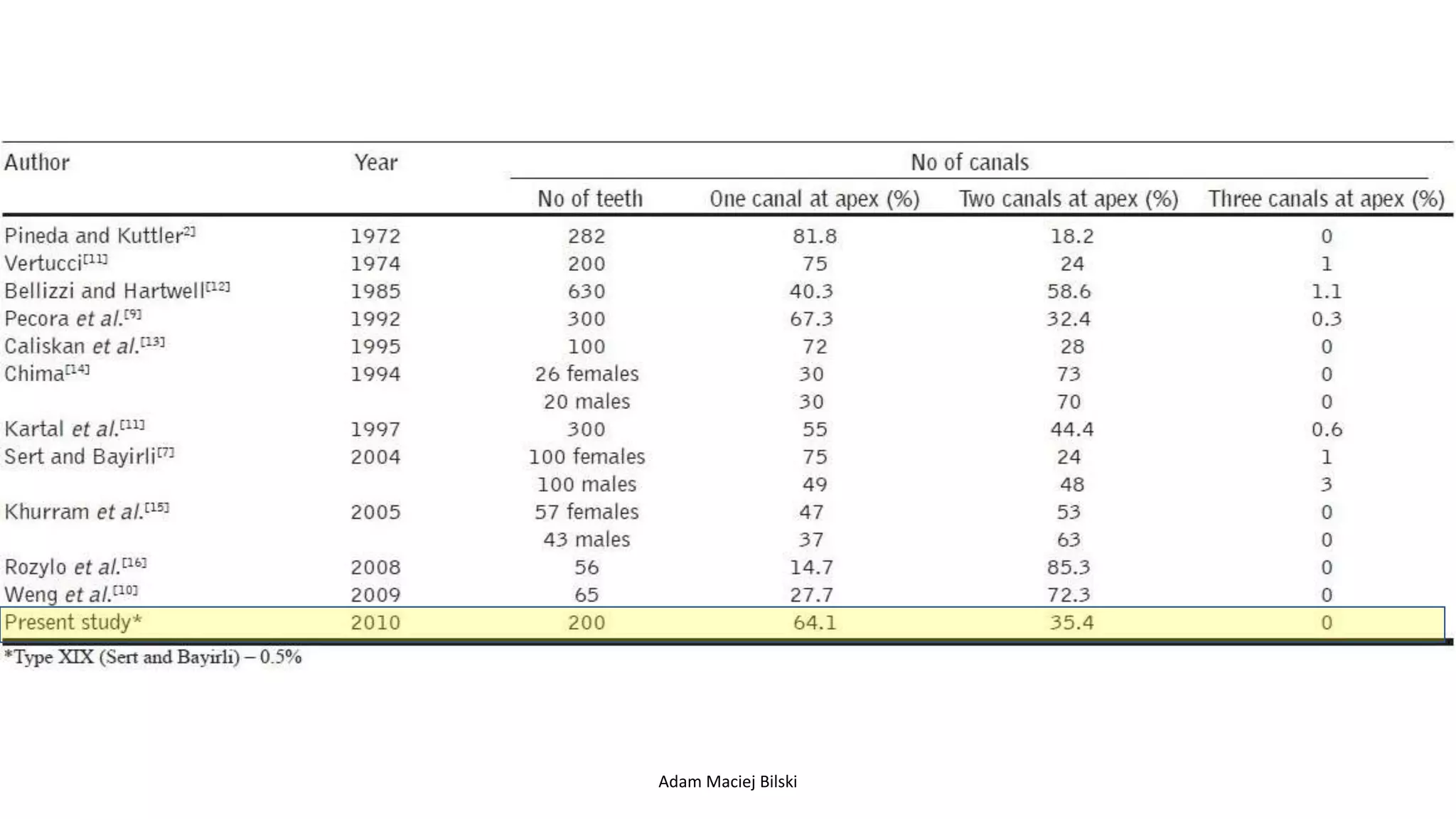
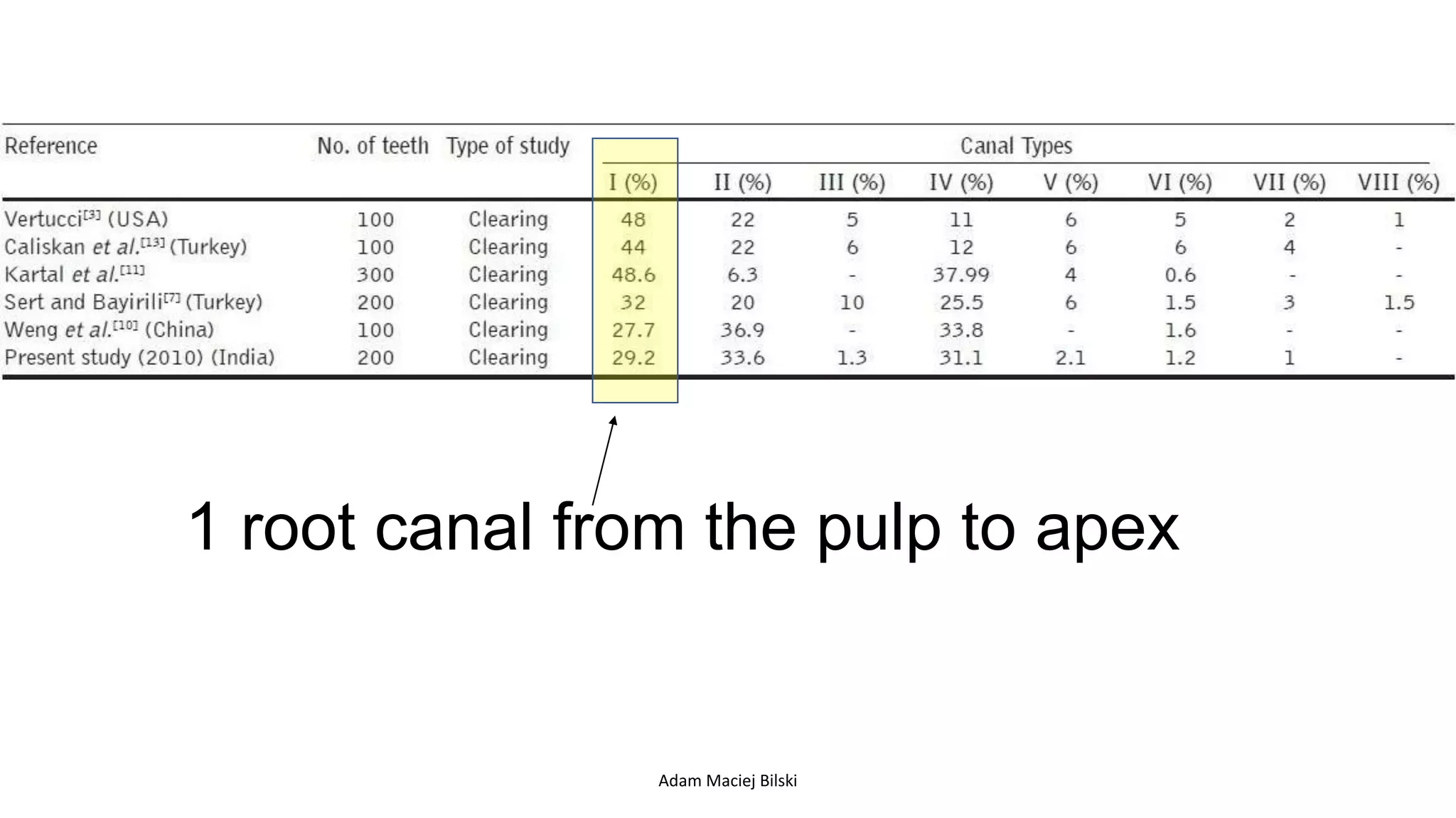
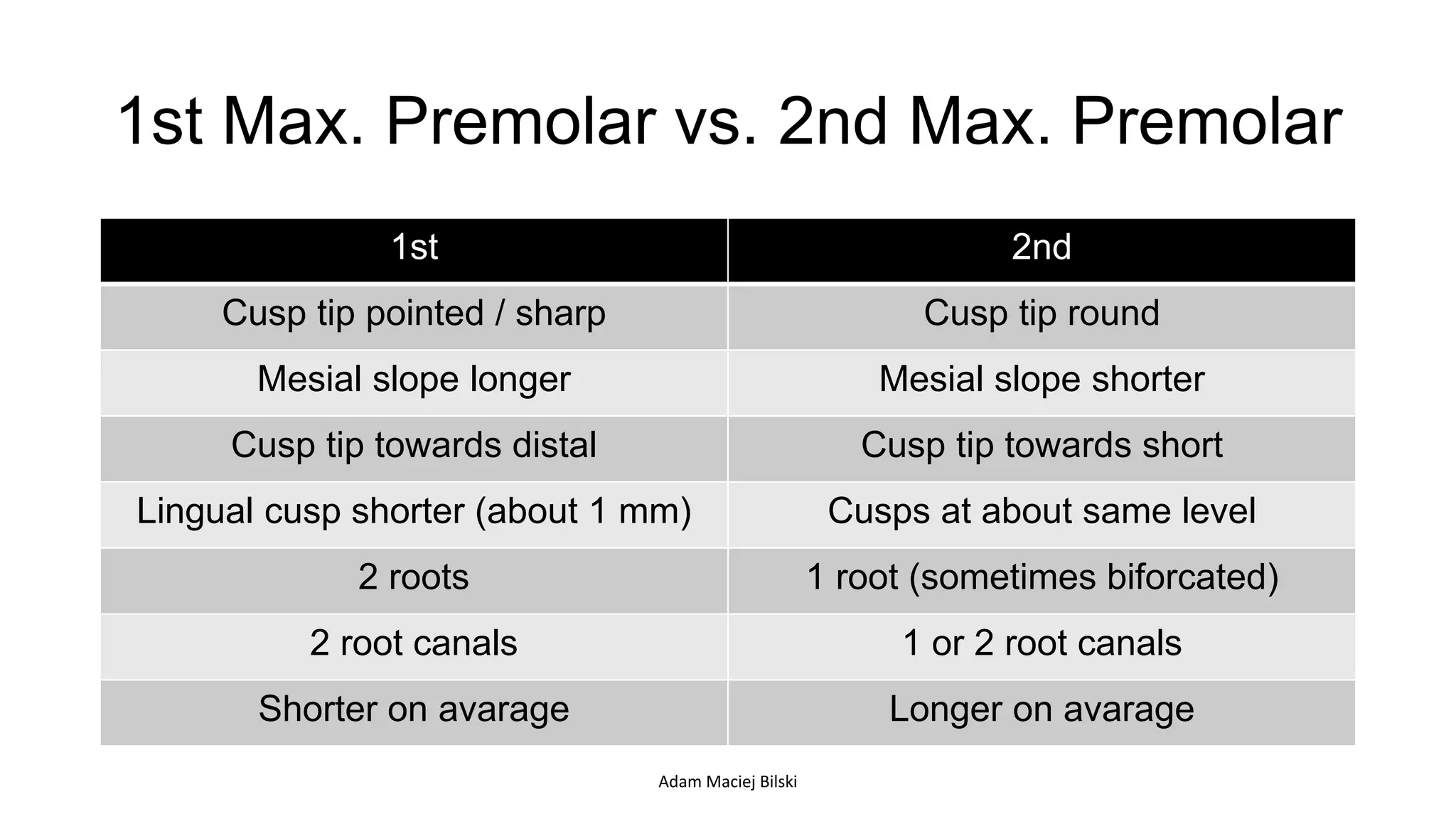
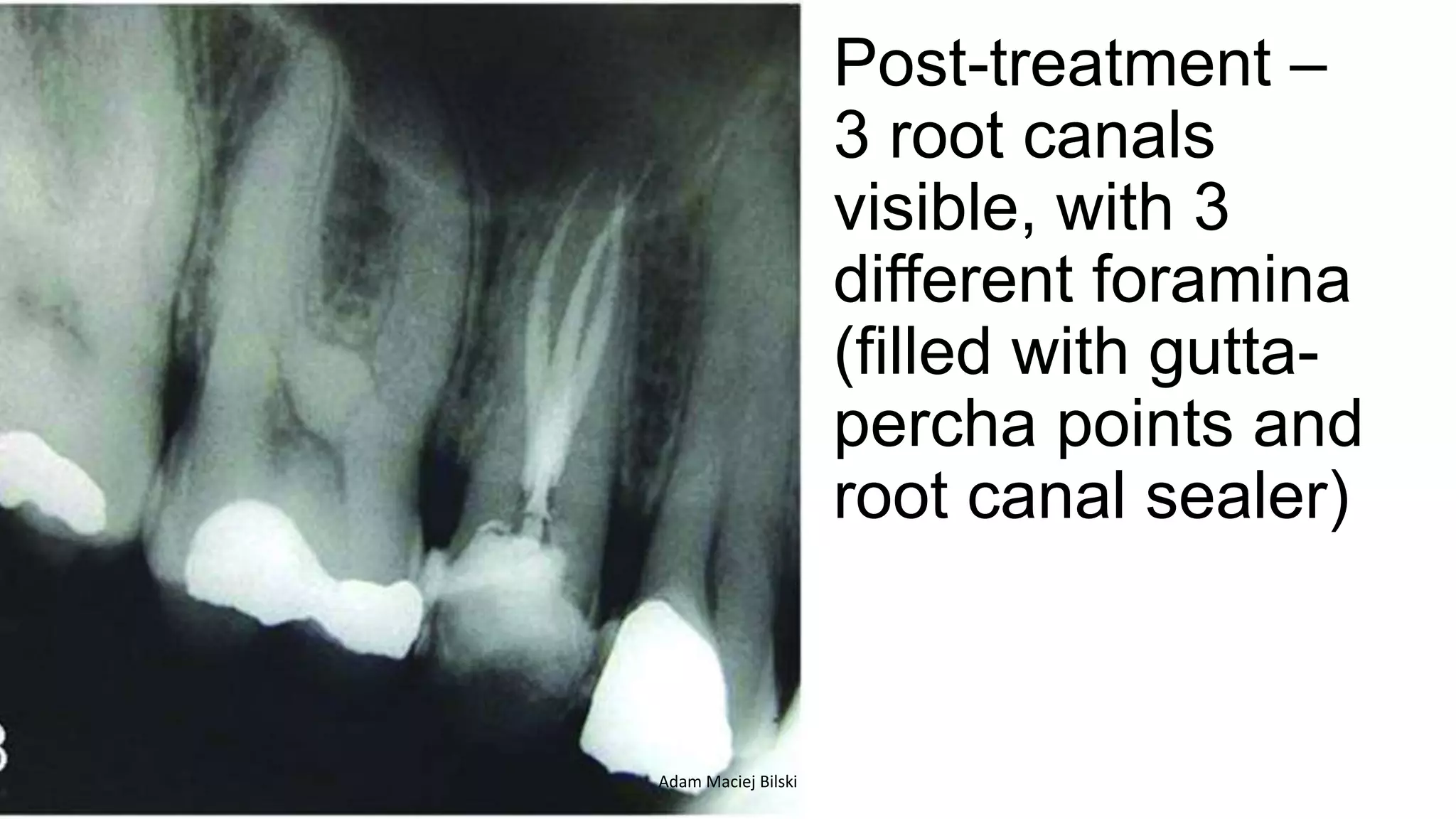
Second Upper
Premolar
Adam Bilski
Second Upper Premolar –
notation:
- Universal Numbering – 13 (Left), 4 (Right)
- FDI – 25 (L), 15 (R)
- Haderup’s - +5 (L), 5+ (R)
- Palmer/Zsigmondy –
5 (L), 5 (R)
Adam Maciej Bilski
Adam Maciej Bilski
v
Adam Maciej Bilski
Adam Maciej Bilski
2nd upper premolar – blood supply:
Adam Maciej Bilski
Adam Maciej Bilski
Second Upper Premolar –
occlusion:
Articulating with:
1st molar (
2nd premolar (
Adam Maciej Bilski
Their function is to masticate, and maintain
the vertical dimention of occlusion (*OVD).
*Vertical Dimention of Occlusion -
is a term used in dentistry to
indicate the superior-inferior
relationship of the maxilla and
the mandible when the teeth
are occluded in maximum
intercuspation.
*RVD – Rest Vertical Dimention
*
Adam Maciej Bilski
Aesthetics
Adam Maciej Bilski
Adam Maciej Bilski
Left Upper 2nd Premolar
(buccal/facial aspect):
M D
- Cusp tip is less pointed, and towards
mesial side
slope is shorter than
(opposite to 1st premolar)
is not as prominent as in
maxillary 1st premolar
- Cervical line is less curvy than in 1st
premolar
Adam Maciej Bilski
10 specimens of 2nd
upper premolars (buccal
aspect):
outline of the
crown visible (smallest line
– cervical part of the tooth)
but it’s more oval than 1st
MD
Adam Maciej Bilski
Left Upper 2nd Premolar
(palatal/lingual aspect):
D M
- Lingual cusp the same height
as buccal one
- Crown is 7,5 - 8,5 mm long
- Mesial slope shorter
Adam Maciej Bilski
Left Upper 2nd Premolar
(mesial aspect):
P B
- Cusps at nearly the same level
- The occlusal table is narrow
- Crown is smooth and convex, but root has a
deep developmental depression
- The (with 1st premolar) is
occlusaly on the middle third and slightly to
the buccal side
- Buccal cusp is slightly sharper than lingual
Adam Maciej Bilski
10 specimens of 2nd
upper premolars (mesial
aspect):
Adam Maciej Bilski
Left Upper 2nd Premolar
(distal aspect):
PB
- The surface is smooth and convex
- Distal marginal ridge is more
cervical than mesial marginal ridge
- Contact point (with 1st molar) is
more cervically positioned and
placed more bucally than on the
mesial aspect (still it’s on the
middle third)
Adam Maciej Bilski
Left Upper 2nd Premolar
(occlusal aspect):B
P
MD
- The buccal cusp is less developed
than the palatal one (which differs it
from the 1st premolar)
- The amount of fissures is also higher,
so higher probability of an additional
buccal cusp
- The outline is rounded
- Tendency of radiating suplementary
grooves from the central groove
- Central groove shorter than in 1st
Adam Maciej Bilski
10 specimens of 2nd
upper premolars
(occlusal aspect):
Adam Maciej Bilski
Root:
• Lenght of root – 13 - 14 mm (longer then the 1st one’s)
• Usually 1 (very rarely 2 roots present)
• In case of 2 the lingual is shorter and its apex is more blunt
• From lateral view – usually cervical part of root and crown is the
thickest
• Often pointed distally at the apical part (not a rule though)
Adam Maciej Bilski
Root Canal:
21,5 mm
- Not well-defined border
of pulp-root canal
- Pulp horns often visible
- Pulp cavity narrows
usually at the apical
half of the tooth
Adam Maciej Bilski
Dentinal
islands
pulpal projections
or fins are
sometimes
present at the
level of the CEJ
OR constrictions
at the same level
Adam Maciej Bilski
Adam Maciej Bilski
1 root canal from the pulp to apex
Adam Maciej Bilski
1st Max. Premolar vs. 2nd Max. Premolar
1st 2nd
Cusp tip pointed / sharp Cusp tip round
Mesial slope longer Mesial slope shorter
Cusp tip towards distal Cusp tip towards short
Lingual cusp shorter (about 1 mm) Cusps at about same level
2 roots 1 root (sometimes biforcated)
2 root canals 1 or 2 root canals
Shorter on avarage Longer on avarage
Adam Maciej Bilski
Pre-
treatment
radiograph:
- 32 year old male
patient
- Pain
- There has to be
endodontic
treatment under
the temporary
restoration
Adam Maciej Bilski
Post-treatment –
3 root canals
visible, with 3
different foramina
(filled with gutta-
percha points and
root canal sealer)
Adam Maciej Bilski
2nd premolars
were blocked
and couldn’t
erupt correctly
they have to be
removed
Adam Maciej Bilski
Adam Maciej Bilski
Adam Maciej Bilski
Adam Maciej Bilski
Adam Maciej Bilski
Nicely
erupted
Adam Maciej Bilski
Adam Maciej Bilski

2nd Upper Premolar Anatomy

  • 1.
  • 2.
    Second Upper Premolar– notation: - Universal Numbering – 13 (Left), 4 (Right) - FDI – 25 (L), 15 (R) - Haderup’s - +5 (L), 5+ (R) - Palmer/Zsigmondy – 5 (L), 5 (R) Adam Maciej Bilski
  • 3.
  • 4.
  • 5.
  • 6.
    2nd upper premolar– blood supply: Adam Maciej Bilski
  • 7.
  • 8.
    Second Upper Premolar– occlusion: Articulating with: 1st molar ( 2nd premolar ( Adam Maciej Bilski
  • 9.
    Their function isto masticate, and maintain the vertical dimention of occlusion (*OVD). *Vertical Dimention of Occlusion - is a term used in dentistry to indicate the superior-inferior relationship of the maxilla and the mandible when the teeth are occluded in maximum intercuspation. *RVD – Rest Vertical Dimention * Adam Maciej Bilski
  • 10.
  • 11.
  • 12.
    Left Upper 2ndPremolar (buccal/facial aspect): M D - Cusp tip is less pointed, and towards mesial side slope is shorter than (opposite to 1st premolar) is not as prominent as in maxillary 1st premolar - Cervical line is less curvy than in 1st premolar Adam Maciej Bilski
  • 13.
    10 specimens of2nd upper premolars (buccal aspect): outline of the crown visible (smallest line – cervical part of the tooth) but it’s more oval than 1st MD Adam Maciej Bilski
  • 14.
    Left Upper 2ndPremolar (palatal/lingual aspect): D M - Lingual cusp the same height as buccal one - Crown is 7,5 - 8,5 mm long - Mesial slope shorter Adam Maciej Bilski
  • 15.
    Left Upper 2ndPremolar (mesial aspect): P B - Cusps at nearly the same level - The occlusal table is narrow - Crown is smooth and convex, but root has a deep developmental depression - The (with 1st premolar) is occlusaly on the middle third and slightly to the buccal side - Buccal cusp is slightly sharper than lingual Adam Maciej Bilski
  • 16.
    10 specimens of2nd upper premolars (mesial aspect): Adam Maciej Bilski
  • 17.
    Left Upper 2ndPremolar (distal aspect): PB - The surface is smooth and convex - Distal marginal ridge is more cervical than mesial marginal ridge - Contact point (with 1st molar) is more cervically positioned and placed more bucally than on the mesial aspect (still it’s on the middle third) Adam Maciej Bilski
  • 18.
    Left Upper 2ndPremolar (occlusal aspect):B P MD - The buccal cusp is less developed than the palatal one (which differs it from the 1st premolar) - The amount of fissures is also higher, so higher probability of an additional buccal cusp - The outline is rounded - Tendency of radiating suplementary grooves from the central groove - Central groove shorter than in 1st Adam Maciej Bilski
  • 19.
    10 specimens of2nd upper premolars (occlusal aspect): Adam Maciej Bilski
  • 20.
    Root: • Lenght ofroot – 13 - 14 mm (longer then the 1st one’s) • Usually 1 (very rarely 2 roots present) • In case of 2 the lingual is shorter and its apex is more blunt • From lateral view – usually cervical part of root and crown is the thickest • Often pointed distally at the apical part (not a rule though) Adam Maciej Bilski
  • 21.
    Root Canal: 21,5 mm -Not well-defined border of pulp-root canal - Pulp horns often visible - Pulp cavity narrows usually at the apical half of the tooth Adam Maciej Bilski
  • 22.
    Dentinal islands pulpal projections or finsare sometimes present at the level of the CEJ OR constrictions at the same level Adam Maciej Bilski
  • 23.
  • 24.
    1 root canalfrom the pulp to apex Adam Maciej Bilski
  • 25.
    1st Max. Premolarvs. 2nd Max. Premolar 1st 2nd Cusp tip pointed / sharp Cusp tip round Mesial slope longer Mesial slope shorter Cusp tip towards distal Cusp tip towards short Lingual cusp shorter (about 1 mm) Cusps at about same level 2 roots 1 root (sometimes biforcated) 2 root canals 1 or 2 root canals Shorter on avarage Longer on avarage Adam Maciej Bilski
  • 26.
    Pre- treatment radiograph: - 32 yearold male patient - Pain - There has to be endodontic treatment under the temporary restoration Adam Maciej Bilski
  • 27.
    Post-treatment – 3 rootcanals visible, with 3 different foramina (filled with gutta- percha points and root canal sealer) Adam Maciej Bilski
  • 28.
    2nd premolars were blocked andcouldn’t erupt correctly they have to be removed Adam Maciej Bilski
  • 29.
  • 30.
  • 31.
  • 32.
  • 33.
  • 34.