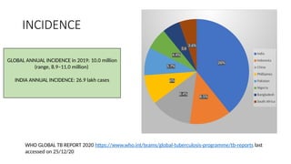
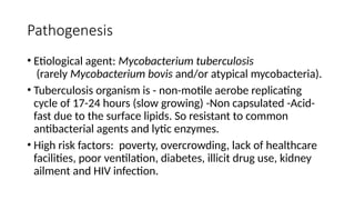
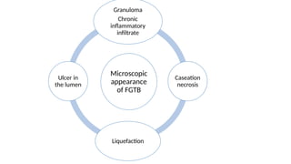
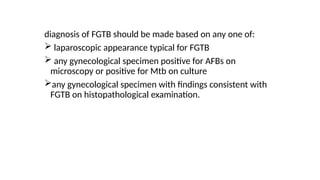
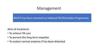
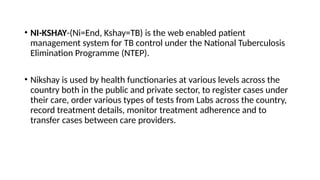
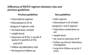
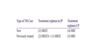
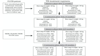
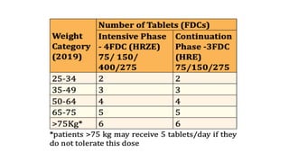
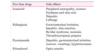
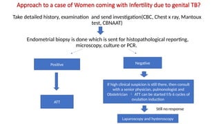
GENITAL TUBERCULOSIS
PRESENTER: Dr. BHAVYA GUPTA (JR-1)
MODERATOR: Dr. AMRITA SINGH
Learning Objectives
•Disease burden of Genital tuberculosis.
•Pathophysiology of FGTB
•Clinical symptoms
•Diagnosis
•Management
•Treatment
INCIDENCE
GLOBAL ANNUAL INCIDENCE in 2019: 10.0 million
(range, 8.9–11.0 million)
INDIA ANNUAL INCIDENCE: 26.9 lakh cases
WHO GLOBAL TB REPORT 2020 https://www.who.int/teams/global-tuberculosis-programme/tb-reports last
accessed on 25/12/20
26%
8.5%
8.4%
6%
5.7%
4.4%
3.6
3.6%
India
Indonesia
China
Phillipines
Pakistan
Nigeria
Bangladesh
South Africa
Pathogenesis
• Etiological agent: Mycobacterium tuberculosis
(rarely Mycobacterium bovis and/or atypical mycobacteria).
• Tuberculosis organism is - non-motile aerobe replicating
cycle of 17-24 hours (slow growing) -Non capsulated -Acid-
fast due to the surface lipids. So resistant to common
antibacterial agents and lytic enzymes.
• High risk factors: poverty, overcrowding, lack of healthcare
facilities, poor ventilation, diabetes, illicit drug use, kidney
ailment and HIV infection.
PATHOPHYSIOLOGY of FTGB
• Always secondary to primary focus elsewhere in the body.
• Mode of spread : Hematogenous (90%)
Lymphatic
Direct (less commonly, direct contiguous spread from
nearby organs like intestines or abdominal lymph node)
Ascending
• Age: Developed countries: pre-menopausal women over 40 years.
Developing countries: 20-40 years (due to early marriages/child bearing
at younger age)
• Less common in postmenopausal women because atropic endometrium
offers a poor mileu for the the growth of bacillus.
Epidemiology of female genital tuberculosis
• Female genital TB (FGTB) was first reported by Morgagni in 1744 on
the autopsy of a young woman who died of TB peritonitis.
• Genitourinary TB is responsible for 27.1 per cent cases of EPTB with
genital TB being seen in 9 % cases .
• The exact incidence of FGTB is not known due to underreporting of
cases, asymptomatic cases, vague symptomatology and the lack of
reliable diagnostics with high sensitivity
Characteristics of Mycobacteirum: Granuloma
formation
Frequency of tuberculosis in Genital organs
Genital Organ Frequency
Fallopian tube 90-100% cases
Uterus/Endometrium 50-80% cases
Ovaries 20-30% cases
Cervix 5-15% cases
Vagina and vulva 1-2% cases
Sharma JB, Sharma E, Sharma S, Dharmendra S. Female genital tuberculosis: Revisited. The Indian journal of medical
research. 2018 Dec;148(Suppl 1):S71.
Fallopian tubes
• MC site to be affected: Ampulla
• First site of infection:
Submucosal layer (interstitial salpingitis) of ampulla
ENDOSALPINGITIS EXOSALPINGITIS INTERSTITIAL
SALPINGITS
• Hematogenous
• Mucosa and
muscle layer
• Edematous and
thick tubes
• Granulamatous
caseation in tubal
walls
• Pyosalpinx or tubal
distention
• Direct spread
• Serosa
• Miliary Tubercles
on tubal surface
• Dense peritubal
adhesions
• Hematogenous
• Tubal wall
• Thickened tubes +-
• Peritubal
adhesions
Microscopic
appearance
of FGTB
Granuloma
Chronic
inflammatory
infiltrate
Caseation
necrosis
Liquefaction
Ulcer in
the lumen
Uterus- tuberculosis of endometrium
• Grossly: endometrium appears unremarkable in majority of cases
because of cyclical menstrual shedding (majority of cases).
• Initially, no macroscopic disease but with progression of disease
caseation and ulceration may occur.
• In advanced stages, distortion of cavity may occur, varying from slight
distortion to complete obliteration due to adhesions
Ashermann’s syndrome Secondary
ammenorhea
• Microscopically: TB granuloma may be seen especially in
premenstrual phase and close to the surface of endometrium
( may not always be seen due to repeated shedding)
• Some authors suggest that even in the absence of typical
granuloma, endometrial TB maybe diagnosed in the presence
of focal collection of lymphocytes with or without the presence
of dilated glands and destruction of epithelium.
• Post-menopausal women may present with pyometra.
Ovaries TB
• Isolated TB oophirits is rare
• Seen as a part of TB peritonitis/TB of genital organs (tubes or
endometrium) usually by DIRECT extension
• Depending on severity and stage of disease, there may be
 Tubercles on the ovary
 Adhesions
Caseation
Tubo-ovarian cyst or mass
Sometimes ovary can be completely destroyed by the
disease
Cervix
• Usually secondary to tubal or endometrial TB, but may rarely be
primary disease transmitted by the partner through infected semen.
• It manifests as polypoidal growth or ulceration simulating cervical
cancer necessitating biopsy and histopathological demonstration of
granulomatous inflammation
Vagina and vulva
• Usually secondary to endometrial or cervical TB, but may rarely be
primary disease transmitted by the partner through infected semen.
• There may be a hypertrophic ulcer or growth on the vulva or vagina
requiring biopsy and histopathological demonstration of granuloma
and to rule out cancer and other diseases such as syphilis and
lymphogranuloma venereum
Genital tuberculosis and Infertility
Tubal factor:
•Blockage of fallopian tubes.
•Loss of tubal function due to ciliary damage causing
infertility and ectopic gestation
•Perisalpingitis causing adhesions and Tubo-ovarian
mass formation
•Tubercular hydrosaplpinx with or without obstruction.
Defective ovarian function:
• Due to tubo-ovarian mass formation, adhesions, anovulation
and poor ovarian reserve.
• Endocrine disruption, chronic anovulation , Mycobacterium
tuberculosis has anti-gonadotropic effect.
• Poor oocyte quality (intrinsic oocyte defect)  poor quality
of embryo.
• The basal production of progesterone is inhibited by
mycobacteria and the stimulatory effect of hCG being corpus
luteum causing LPD, implantation failure, lower pregnancy
rate and higher miscarriage rates.
Uterine (endometrial) factors:
• Adverse effects on endometrial markers .
• Defective vascularization of endometrium
• Endometrial atrophy
• Synechiae formation
Endometrial factor
Adverse Effect on Endometrial
marker
• Which are essential to make the
endometrium receptive for
embryonic implantation
Defective vascularization of
endometrium
• Reduced sub-endometrial flow
due to involvement of basal
layer of endometrium
• Vascular thrombus formation
a) Release of harmful cytokines and growth factor (IL-2, IL-8 and TNF alpha) in the
endometrium
b) Natural killer cells
c) Lymphocytes activated killer cell
d) T helper -1 cell response instead of Th-2 cell response
General symptoms :
Fever, weight loss, anorexia, poor
general health
Abdomen:
Lump, abdominal pain, chronic
pelvic pain, acute abdomen-
rupture of the abscess, abnormal
vaginal discharge
Unusual symptoms:
Ulcers in vagina or vulva, labial
swelling, retention of urine, urinary
incontinence and fecal
incontinence
Menstrual symptoms:
Puberty mennorhagia, Post
menopausal bleeding,
Oligomennorhea,
hypomennorhea, amenorrhea
(primary/secondary)
CLINICAL FEATURES
Diagnosis of TB
Clinical suspicion
Past h/o TB
Family history of
TB
Hematology
Microbiology
Chest x-ray
USG
HSG
Laparoscopy
Histopathology
Physical
examination
Lymphadenopathy
Bones, joints and
skin
Chest TB
Abdominal TB
Speculum/
Bimanual
examination
1. Blood test: may show anemia, raised leucocytes and lymphocytes
and raised ESR in TB.
Not considered sensitive and specific, hence banned by WHO
and Government of India
2. X-ray Chest (PA view): may show active pulmonary TB or old healed
lesions of past.
SMEAR MICROSCOPY
• Ziehl-Neelsen
• Fluorochrome - Auramine-rhodamine (direct fluorescence)
• Simple & rapid procedure
• faster screening , low sensitivity = 55 %
• Requires 10,000 mycobacteria/ml for positive smear
• negative smear does not rule out GTB
TUBERCULIN (MANTOUX) TESTS
• 0.1 ML PPD IS INJECTED
INTRADERMALLY
• Delayed hypersensitivity reaction
• Read between 48 and 72 hours after
the injection
• The tuberculin skin test has a sensitivity
of 55% and specificity of 80% in
patients with genital tuberculosis
IGRA
• Interferon-gamma release assays (IGRAs) is to diagnostic test
to screen for active or latent infection.
• In vitro assay that measure interferon (IFN-γ) released by
sensitized T cells after stimulation by M. tuberculosis
antigens.
• Not affected by Bacille Calmette-Guérin (BCG) vaccination
status, IGRAs are useful for evaluation of LTBI in BCG-
vaccinated individuals, particularly in settings where BCG
vaccination is administered after infancy or multiple
(booster) BCG vaccinations are given.
QUANTIFERON-GOLD
•Active Tuberculosis disease OR latent tuberculosis infection
(LTBI)- cannot distinguish between them
•Intended for use in conjunction with risk assessment,
radiography, and other medical and diagnostic evaluations.
•Sensitivity- 89% and specificity- 98%
PRINCIPLE OF QUANTIFERON GOLD
Fresh heparinised whole blood from sensitised persons
Incubated with mixtures of synthetic peptides (two proteins present in M.
tuberculosis)
ESAT-6 (early secretory antigenic target-6)
CFP-10 (culture filtrate protein-10)
Lymphocytes recognize these mycobacterial antigens
Generation and secretion of interferon-γ (IFN-γ)
Detection and subsequent quantification of IFN-γ by ELISA
QFT-Gold Plus /TB sure test
• CD4 (Helper) cells– if they recognize TB antigens- will secrete γ
interferon. This response does not tell us anything about disease
activity- just a Yes or No answer. A positive response still needs a
clinical evaluation to determine infection versus active disease.
• The new Quantiferon Gold Plus test also tests CD8 cell response to TB
antigens – these killer cells only get activated and secrete γ
interferon in the presence of recent infection or active disease.
Unlike the persistent positive CD4 cell response, if the infection is old
and not active- CD8 cells shut off and do not secrete γ interferon.
CULTURE
• Provides definite diagnosis – Gold Standard
• For isolation of bacteria – a surest test
• More sensitive requiring 1000 organism/ml of sputum.
AFB culture
SOLID MEDIA
Egg based: LJ media
LIQUID BASED
Middlebrook 7H12
BACTEC
Sensitivity 80-93 percent
Specificity: 98%
LJ media
Radiometric Culture Method
•BACTEC -- is based on measurement of carbon dioxide
released by bacteria during growth in liquid medium
•Radioactive carbon labelled substrate like palmitic acid
or formic acid is used as marker for bacterial growth
• Diagnosis is made in 1-2 weeks
•Strong suspicion of TB is seen when the growth index
is greater than 10.
Polymerase Chain Reaction
• PCR based sequencing
- Amplification of DNA using specific primers
-- Few bacterial cells used for amplification
- Detectable with even 1-10 organisms / ml
- Results available in 2-3 days
- Cumbersome and expensive
- Disadvantage: cannot distinguish between live or dead
bacilli, hence may give false positive.
CB-NAAT
• The CBNAAT/ Xpert MTB/Rif test is a cartridge- based fully
automated NAAT (nucleic acid amplification test) for TB case
detection and Rifampicin resistance testing.
• Purifies, concentrates, amplifies (by rapid, real-time PCR) and
identifies targeted nucleic acid. It sequences in the TB
genome), and provides results from unprocessed sputum
samples in less than 2 hours, with minimal hands-on
technical time.
Imaging methods
• These are of use only in tubo-ovarian masses.
• USG:
may show tubo-obarian masses with calcification and fluid in
pouch of douglas.
Endometrial changes like thin endometrium, calcification of
bands and intrauterine synechiae
Hydrosalpix with cog wheel sign may be demonstrated
Hysterosalpingraphy
Tubal findings Endometrial findings
• Occlusion
• Hydrosaplinx
• Beading
• Tobacco pouch
• Maltese cross appearance
• calcification
• Synechiae
• Irregular contour
In known cases or in circumstances in
which genital TB is highly suspected,
HSG should be avoided because of
risk of activation.
Tobacco- pouch appearance
Tube is enlarged and distended,
ostium remain patent with
recognizable everted fimbriae
Beaded appearance
• Multiple
constrictions along
the course of
fallopian tube on
HSG due to fibrotic
strictures.
Pipe stem appearance/ Rigid pipe
• Scarring fallopian
tubes.
• Irregular and
rigid.
• Filling defect in
uterine cavity –
adhesion.
Hysteroscopy
• It is endoscopic visualization of endometrial cavity and may be normal
in the absence of endometrial TB.
• Usually, there is a pale looking cavity, the presence of tubercles, small
white caseous nodules, varying grades of intrauterine synechaie and
Asherman's syndrome
Diagnostic laproscopy
• A laparoscopy under general anaesthesia is the most reliable modality
for the detection of FGTB and abdomino-pelvic TB, particularly
affection of oviducts, ovaries and peritoneum.
• Questionable lesions maybe biopsied in an attempt to obtain
histopathological diagnosis at the same time.
• Most common findings are pelvic adhesions followed by tubal
pathology, occlusions, tubo-ovarian mass, ascites and caseous and
granulomatous nodules.
Sharma JB, Sharma E, Sharma S, Dharmendra S. Female genital tuberculosis: Revisited. The Indian
journal of medical research. 2018 Dec;148(Suppl 1):S71.
Sharma JB, Sharma E, Sharma S, Dharmendra S. Female genital tuberculosis: Revisited. The Indian
journal of medical research. 2018 Dec;148(Suppl 1):S71.
Sharma JB, Sharma E, Sharma S, Dharmendra S. Female genital tuberculosis: Revisited.
The Indian journal of medical research. 2018 Dec;148(Suppl 1):S71.
Sharma JB, Sharma E, Sharma S, Dharmendra S. Female genital tuberculosis: Revisited. The Indian journal of
medical research. 2018 Dec;148(Suppl 1):S71.
Sharma JB, Sharma E, Sharma S, Dharmendra S. Female genital tuberculosis: Revisited. The Indian
journal of medical research. 2018 Dec;148(Suppl 1):S71.
Diagnosis
Test Patients Comments
X-ray of chest All All patients presenting with
symptoms consistent with TB
should have a chest X-ray to look
for evidence of previous or active
pulmonary TB.
HIV test All EPTB is associated with HIV
infection. All patients should be
offered integrated counseling and
testing.
Pelvic ultrasound All Part of the initial assessment of
most patients presenting with
gynaecological symptoms
Endometrial aspirate Selected Where facilities exist, endometrial
aspirate can be obtained and sent
for: a) staining and microscopy for
AFB; b) culture and drug
susceptibility testing. Sensitivity is
low, and negative results cannot
rule out FGTB.
CT pelvis or MRI
pelvis
Selected To further characterize lesions and plan surgical
intervention in selected patients. Disadvantage
of CT is exposure to ionising radiation, which is
particularly a concern in women of childbearing
age
Laparoscopy Selected Laparoscopy with biopsy of lesions is required
when - other less invasive tests are inconclusive
- malignancy is also suspected - as part of
infertility investigations when less invasive tests
are inconclusive Laparoscopy offers the dual
advantage of pelvic organ visualization and
specimen collection from otherwise inaccessible
sites. Specimens should be subject to: a)
staining and microscopy for AFBs; b) culture and
drug susceptibility testing; and c)
histopathology.
diagnosis of FGTB should be made based on any one of:
 laparoscopic appearance typical for FGTB
 any gynecological specimen positive for AFBs on
microscopy or positive for Mtb on culture
any gynecological specimen with findings consistent with
FGTB on histopathological examination.
Management
Aims of treatment:
• To achieve TB cure
• To prevent the long term sequelae
• To restore normal anatomy if has been distorted
RNTCP has been renamed as National TB Elimination Programme.
• NI-KSHAY-(Ni=End, Kshay=TB) is the web enabled patient
management system for TB control under the National Tuberculosis
Elimination Programme (NTEP).
• Nikshay is used by health functionaries at various levels across the
country both in the public and private sector, to register cases under
their care, order various types of tests from Labs across the country,
record treatment details, monitor treatment adherence and to
transfer cases between care providers.
Difference of RNTCP regimen between new and
previous guidelines
Previous guidelines
• Intermittent regimen
• Ethambutol in CP of
category II regimen only
• No fixed dose, limited
• weight band
• Extension of IP for 1 month if
sputum is positive at the
end of IP
• Follow up-laboratory only
‑
• No long term follow up
‑ ‑
New guidelines
• Daily regimen
• Ethambutol in CP of both
categories I and II regimen
• Fixed dose combination as
per
• weight band
• No need of extension of IP
• Follow up-clinical, laboratory
‑
Investigation
• Long term follow up up to 2
‑ ‑
years
• Pregnant and breast-feeding women may be treated with
RHZE with pyridoxine 10 mg daily, as for other patients.
• There is no need to cease breast feeding. Some drugs
used in secondary regimens such as streptomycin,
ethionamide and the quinolones are contraindicated due
to teratogenicity.
• Women who need contraception should be counselled on
the use of oral contraceptives while receiving rifampicin.
• Women should be offered an oral contraceptive pill
containing a higher dose of estrogen (50 μg) after
consultation with a clinician, or a non-hormonal method
of contraception while taking rifampicin and for 1 month
after the end of treatment.
NEWER ANTI TB DRUGS
Bedaquiline:
• Bedaquiline works by blocking an enzyme inside the Mycobacterium
tuberculosis bacteria called ATP synthase.
• The recommended dose is 400 mg a day for two weeks and then 200
mg taken three times a week (with at least 48 hours between doses).
• ADVERSE EFFECTS: The most common side effects are headache,
dizziness, malaise, joint pain, QT prolongation and increases in liver
enzymes
Delamanid
• Delamanid is a dihydro-nitroimidazooxazole derivative.
• It acts by inhibiting the synthesis of mycobacterial cell wall
components, methoxy mycolic acid and ketomycolic acid.
• Dose: 100 mg/day
• ADVERSE EFFECTS: The most common side effects are
nausea, vomiting and dizziness. Anxiety, and shaking are
other important side effects QT prolongation
Surgical treatment
Approach to a case of Women coming with Infertility due to genital TB?
Take detailed history, examination and send investigation(CBC, Chest x ray, Mantoux
test, CBNAAT)
Endometrial biopsy is done which is sent for histopathological reporting,
microscopy, culture or PCR.
Positive Negative
ATT
If high clinical suspicion is still there, then consult
with a senior physician, pulmonologist and
Obstetrician  ATT can be started f/b 6 cycles of
ovulation induction
Laparoscopy and hysteroscopy
Still no response
Thank you

tb [Autosaved]. PPT by doc maney....pptx

  • 1.
    GENITAL TUBERCULOSIS PRESENTER: Dr.BHAVYA GUPTA (JR-1) MODERATOR: Dr. AMRITA SINGH
  • 2.
    Learning Objectives •Disease burdenof Genital tuberculosis. •Pathophysiology of FGTB •Clinical symptoms •Diagnosis •Management •Treatment
  • 3.
    INCIDENCE GLOBAL ANNUAL INCIDENCEin 2019: 10.0 million (range, 8.9–11.0 million) INDIA ANNUAL INCIDENCE: 26.9 lakh cases WHO GLOBAL TB REPORT 2020 https://www.who.int/teams/global-tuberculosis-programme/tb-reports last accessed on 25/12/20 26% 8.5% 8.4% 6% 5.7% 4.4% 3.6 3.6% India Indonesia China Phillipines Pakistan Nigeria Bangladesh South Africa
  • 4.
    Pathogenesis • Etiological agent:Mycobacterium tuberculosis (rarely Mycobacterium bovis and/or atypical mycobacteria). • Tuberculosis organism is - non-motile aerobe replicating cycle of 17-24 hours (slow growing) -Non capsulated -Acid- fast due to the surface lipids. So resistant to common antibacterial agents and lytic enzymes. • High risk factors: poverty, overcrowding, lack of healthcare facilities, poor ventilation, diabetes, illicit drug use, kidney ailment and HIV infection.
  • 6.
    PATHOPHYSIOLOGY of FTGB •Always secondary to primary focus elsewhere in the body. • Mode of spread : Hematogenous (90%) Lymphatic Direct (less commonly, direct contiguous spread from nearby organs like intestines or abdominal lymph node) Ascending • Age: Developed countries: pre-menopausal women over 40 years. Developing countries: 20-40 years (due to early marriages/child bearing at younger age) • Less common in postmenopausal women because atropic endometrium offers a poor mileu for the the growth of bacillus.
  • 7.
    Epidemiology of femalegenital tuberculosis • Female genital TB (FGTB) was first reported by Morgagni in 1744 on the autopsy of a young woman who died of TB peritonitis. • Genitourinary TB is responsible for 27.1 per cent cases of EPTB with genital TB being seen in 9 % cases . • The exact incidence of FGTB is not known due to underreporting of cases, asymptomatic cases, vague symptomatology and the lack of reliable diagnostics with high sensitivity
  • 9.
  • 10.
    Frequency of tuberculosisin Genital organs Genital Organ Frequency Fallopian tube 90-100% cases Uterus/Endometrium 50-80% cases Ovaries 20-30% cases Cervix 5-15% cases Vagina and vulva 1-2% cases Sharma JB, Sharma E, Sharma S, Dharmendra S. Female genital tuberculosis: Revisited. The Indian journal of medical research. 2018 Dec;148(Suppl 1):S71.
  • 11.
    Fallopian tubes • MCsite to be affected: Ampulla • First site of infection: Submucosal layer (interstitial salpingitis) of ampulla
  • 12.
    ENDOSALPINGITIS EXOSALPINGITIS INTERSTITIAL SALPINGITS •Hematogenous • Mucosa and muscle layer • Edematous and thick tubes • Granulamatous caseation in tubal walls • Pyosalpinx or tubal distention • Direct spread • Serosa • Miliary Tubercles on tubal surface • Dense peritubal adhesions • Hematogenous • Tubal wall • Thickened tubes +- • Peritubal adhesions
  • 13.
  • 14.
    Uterus- tuberculosis ofendometrium • Grossly: endometrium appears unremarkable in majority of cases because of cyclical menstrual shedding (majority of cases). • Initially, no macroscopic disease but with progression of disease caseation and ulceration may occur. • In advanced stages, distortion of cavity may occur, varying from slight distortion to complete obliteration due to adhesions Ashermann’s syndrome Secondary ammenorhea
  • 15.
    • Microscopically: TBgranuloma may be seen especially in premenstrual phase and close to the surface of endometrium ( may not always be seen due to repeated shedding) • Some authors suggest that even in the absence of typical granuloma, endometrial TB maybe diagnosed in the presence of focal collection of lymphocytes with or without the presence of dilated glands and destruction of epithelium. • Post-menopausal women may present with pyometra.
  • 16.
    Ovaries TB • IsolatedTB oophirits is rare • Seen as a part of TB peritonitis/TB of genital organs (tubes or endometrium) usually by DIRECT extension • Depending on severity and stage of disease, there may be  Tubercles on the ovary  Adhesions Caseation Tubo-ovarian cyst or mass Sometimes ovary can be completely destroyed by the disease
  • 17.
    Cervix • Usually secondaryto tubal or endometrial TB, but may rarely be primary disease transmitted by the partner through infected semen. • It manifests as polypoidal growth or ulceration simulating cervical cancer necessitating biopsy and histopathological demonstration of granulomatous inflammation
  • 18.
    Vagina and vulva •Usually secondary to endometrial or cervical TB, but may rarely be primary disease transmitted by the partner through infected semen. • There may be a hypertrophic ulcer or growth on the vulva or vagina requiring biopsy and histopathological demonstration of granuloma and to rule out cancer and other diseases such as syphilis and lymphogranuloma venereum
  • 19.
    Genital tuberculosis andInfertility Tubal factor: •Blockage of fallopian tubes. •Loss of tubal function due to ciliary damage causing infertility and ectopic gestation •Perisalpingitis causing adhesions and Tubo-ovarian mass formation •Tubercular hydrosaplpinx with or without obstruction.
  • 20.
    Defective ovarian function: •Due to tubo-ovarian mass formation, adhesions, anovulation and poor ovarian reserve. • Endocrine disruption, chronic anovulation , Mycobacterium tuberculosis has anti-gonadotropic effect. • Poor oocyte quality (intrinsic oocyte defect)  poor quality of embryo. • The basal production of progesterone is inhibited by mycobacteria and the stimulatory effect of hCG being corpus luteum causing LPD, implantation failure, lower pregnancy rate and higher miscarriage rates.
  • 21.
    Uterine (endometrial) factors: •Adverse effects on endometrial markers . • Defective vascularization of endometrium • Endometrial atrophy • Synechiae formation
  • 22.
    Endometrial factor Adverse Effecton Endometrial marker • Which are essential to make the endometrium receptive for embryonic implantation Defective vascularization of endometrium • Reduced sub-endometrial flow due to involvement of basal layer of endometrium • Vascular thrombus formation a) Release of harmful cytokines and growth factor (IL-2, IL-8 and TNF alpha) in the endometrium b) Natural killer cells c) Lymphocytes activated killer cell d) T helper -1 cell response instead of Th-2 cell response
  • 23.
    General symptoms : Fever,weight loss, anorexia, poor general health Abdomen: Lump, abdominal pain, chronic pelvic pain, acute abdomen- rupture of the abscess, abnormal vaginal discharge Unusual symptoms: Ulcers in vagina or vulva, labial swelling, retention of urine, urinary incontinence and fecal incontinence Menstrual symptoms: Puberty mennorhagia, Post menopausal bleeding, Oligomennorhea, hypomennorhea, amenorrhea (primary/secondary) CLINICAL FEATURES
  • 24.
    Diagnosis of TB Clinicalsuspicion Past h/o TB Family history of TB Hematology Microbiology Chest x-ray USG HSG Laparoscopy Histopathology Physical examination Lymphadenopathy Bones, joints and skin Chest TB Abdominal TB Speculum/ Bimanual examination
  • 25.
    1. Blood test:may show anemia, raised leucocytes and lymphocytes and raised ESR in TB. Not considered sensitive and specific, hence banned by WHO and Government of India 2. X-ray Chest (PA view): may show active pulmonary TB or old healed lesions of past.
  • 26.
    SMEAR MICROSCOPY • Ziehl-Neelsen •Fluorochrome - Auramine-rhodamine (direct fluorescence) • Simple & rapid procedure • faster screening , low sensitivity = 55 % • Requires 10,000 mycobacteria/ml for positive smear • negative smear does not rule out GTB
  • 27.
    TUBERCULIN (MANTOUX) TESTS •0.1 ML PPD IS INJECTED INTRADERMALLY • Delayed hypersensitivity reaction • Read between 48 and 72 hours after the injection • The tuberculin skin test has a sensitivity of 55% and specificity of 80% in patients with genital tuberculosis
  • 29.
    IGRA • Interferon-gamma releaseassays (IGRAs) is to diagnostic test to screen for active or latent infection. • In vitro assay that measure interferon (IFN-γ) released by sensitized T cells after stimulation by M. tuberculosis antigens. • Not affected by Bacille Calmette-Guérin (BCG) vaccination status, IGRAs are useful for evaluation of LTBI in BCG- vaccinated individuals, particularly in settings where BCG vaccination is administered after infancy or multiple (booster) BCG vaccinations are given.
  • 30.
    QUANTIFERON-GOLD •Active Tuberculosis diseaseOR latent tuberculosis infection (LTBI)- cannot distinguish between them •Intended for use in conjunction with risk assessment, radiography, and other medical and diagnostic evaluations. •Sensitivity- 89% and specificity- 98%
  • 31.
    PRINCIPLE OF QUANTIFERONGOLD Fresh heparinised whole blood from sensitised persons Incubated with mixtures of synthetic peptides (two proteins present in M. tuberculosis) ESAT-6 (early secretory antigenic target-6) CFP-10 (culture filtrate protein-10) Lymphocytes recognize these mycobacterial antigens Generation and secretion of interferon-γ (IFN-γ) Detection and subsequent quantification of IFN-γ by ELISA
  • 32.
    QFT-Gold Plus /TBsure test • CD4 (Helper) cells– if they recognize TB antigens- will secrete γ interferon. This response does not tell us anything about disease activity- just a Yes or No answer. A positive response still needs a clinical evaluation to determine infection versus active disease. • The new Quantiferon Gold Plus test also tests CD8 cell response to TB antigens – these killer cells only get activated and secrete γ interferon in the presence of recent infection or active disease. Unlike the persistent positive CD4 cell response, if the infection is old and not active- CD8 cells shut off and do not secrete γ interferon.
  • 33.
    CULTURE • Provides definitediagnosis – Gold Standard • For isolation of bacteria – a surest test • More sensitive requiring 1000 organism/ml of sputum. AFB culture SOLID MEDIA Egg based: LJ media LIQUID BASED Middlebrook 7H12 BACTEC Sensitivity 80-93 percent Specificity: 98%
  • 34.
  • 35.
    Radiometric Culture Method •BACTEC-- is based on measurement of carbon dioxide released by bacteria during growth in liquid medium •Radioactive carbon labelled substrate like palmitic acid or formic acid is used as marker for bacterial growth • Diagnosis is made in 1-2 weeks •Strong suspicion of TB is seen when the growth index is greater than 10.
  • 36.
    Polymerase Chain Reaction •PCR based sequencing - Amplification of DNA using specific primers -- Few bacterial cells used for amplification - Detectable with even 1-10 organisms / ml - Results available in 2-3 days - Cumbersome and expensive - Disadvantage: cannot distinguish between live or dead bacilli, hence may give false positive.
  • 38.
    CB-NAAT • The CBNAAT/Xpert MTB/Rif test is a cartridge- based fully automated NAAT (nucleic acid amplification test) for TB case detection and Rifampicin resistance testing. • Purifies, concentrates, amplifies (by rapid, real-time PCR) and identifies targeted nucleic acid. It sequences in the TB genome), and provides results from unprocessed sputum samples in less than 2 hours, with minimal hands-on technical time.
  • 39.
    Imaging methods • Theseare of use only in tubo-ovarian masses. • USG: may show tubo-obarian masses with calcification and fluid in pouch of douglas. Endometrial changes like thin endometrium, calcification of bands and intrauterine synechiae Hydrosalpix with cog wheel sign may be demonstrated
  • 40.
    Hysterosalpingraphy Tubal findings Endometrialfindings • Occlusion • Hydrosaplinx • Beading • Tobacco pouch • Maltese cross appearance • calcification • Synechiae • Irregular contour In known cases or in circumstances in which genital TB is highly suspected, HSG should be avoided because of risk of activation.
  • 42.
    Tobacco- pouch appearance Tubeis enlarged and distended, ostium remain patent with recognizable everted fimbriae
  • 43.
    Beaded appearance • Multiple constrictionsalong the course of fallopian tube on HSG due to fibrotic strictures.
  • 44.
    Pipe stem appearance/Rigid pipe • Scarring fallopian tubes. • Irregular and rigid. • Filling defect in uterine cavity – adhesion.
  • 48.
    Hysteroscopy • It isendoscopic visualization of endometrial cavity and may be normal in the absence of endometrial TB. • Usually, there is a pale looking cavity, the presence of tubercles, small white caseous nodules, varying grades of intrauterine synechaie and Asherman's syndrome
  • 49.
    Diagnostic laproscopy • Alaparoscopy under general anaesthesia is the most reliable modality for the detection of FGTB and abdomino-pelvic TB, particularly affection of oviducts, ovaries and peritoneum. • Questionable lesions maybe biopsied in an attempt to obtain histopathological diagnosis at the same time. • Most common findings are pelvic adhesions followed by tubal pathology, occlusions, tubo-ovarian mass, ascites and caseous and granulomatous nodules.
  • 50.
    Sharma JB, SharmaE, Sharma S, Dharmendra S. Female genital tuberculosis: Revisited. The Indian journal of medical research. 2018 Dec;148(Suppl 1):S71.
  • 51.
    Sharma JB, SharmaE, Sharma S, Dharmendra S. Female genital tuberculosis: Revisited. The Indian journal of medical research. 2018 Dec;148(Suppl 1):S71.
  • 52.
    Sharma JB, SharmaE, Sharma S, Dharmendra S. Female genital tuberculosis: Revisited. The Indian journal of medical research. 2018 Dec;148(Suppl 1):S71.
  • 53.
    Sharma JB, SharmaE, Sharma S, Dharmendra S. Female genital tuberculosis: Revisited. The Indian journal of medical research. 2018 Dec;148(Suppl 1):S71.
  • 54.
    Sharma JB, SharmaE, Sharma S, Dharmendra S. Female genital tuberculosis: Revisited. The Indian journal of medical research. 2018 Dec;148(Suppl 1):S71.
  • 55.
    Diagnosis Test Patients Comments X-rayof chest All All patients presenting with symptoms consistent with TB should have a chest X-ray to look for evidence of previous or active pulmonary TB. HIV test All EPTB is associated with HIV infection. All patients should be offered integrated counseling and testing. Pelvic ultrasound All Part of the initial assessment of most patients presenting with gynaecological symptoms Endometrial aspirate Selected Where facilities exist, endometrial aspirate can be obtained and sent for: a) staining and microscopy for AFB; b) culture and drug susceptibility testing. Sensitivity is low, and negative results cannot rule out FGTB.
  • 56.
    CT pelvis orMRI pelvis Selected To further characterize lesions and plan surgical intervention in selected patients. Disadvantage of CT is exposure to ionising radiation, which is particularly a concern in women of childbearing age Laparoscopy Selected Laparoscopy with biopsy of lesions is required when - other less invasive tests are inconclusive - malignancy is also suspected - as part of infertility investigations when less invasive tests are inconclusive Laparoscopy offers the dual advantage of pelvic organ visualization and specimen collection from otherwise inaccessible sites. Specimens should be subject to: a) staining and microscopy for AFBs; b) culture and drug susceptibility testing; and c) histopathology.
  • 57.
    diagnosis of FGTBshould be made based on any one of:  laparoscopic appearance typical for FGTB  any gynecological specimen positive for AFBs on microscopy or positive for Mtb on culture any gynecological specimen with findings consistent with FGTB on histopathological examination.
  • 58.
    Management Aims of treatment: •To achieve TB cure • To prevent the long term sequelae • To restore normal anatomy if has been distorted RNTCP has been renamed as National TB Elimination Programme.
  • 59.
    • NI-KSHAY-(Ni=End, Kshay=TB)is the web enabled patient management system for TB control under the National Tuberculosis Elimination Programme (NTEP). • Nikshay is used by health functionaries at various levels across the country both in the public and private sector, to register cases under their care, order various types of tests from Labs across the country, record treatment details, monitor treatment adherence and to transfer cases between care providers.
  • 60.
    Difference of RNTCPregimen between new and previous guidelines Previous guidelines • Intermittent regimen • Ethambutol in CP of category II regimen only • No fixed dose, limited • weight band • Extension of IP for 1 month if sputum is positive at the end of IP • Follow up-laboratory only ‑ • No long term follow up ‑ ‑ New guidelines • Daily regimen • Ethambutol in CP of both categories I and II regimen • Fixed dose combination as per • weight band • No need of extension of IP • Follow up-clinical, laboratory ‑ Investigation • Long term follow up up to 2 ‑ ‑ years
  • 66.
    • Pregnant andbreast-feeding women may be treated with RHZE with pyridoxine 10 mg daily, as for other patients. • There is no need to cease breast feeding. Some drugs used in secondary regimens such as streptomycin, ethionamide and the quinolones are contraindicated due to teratogenicity. • Women who need contraception should be counselled on the use of oral contraceptives while receiving rifampicin. • Women should be offered an oral contraceptive pill containing a higher dose of estrogen (50 μg) after consultation with a clinician, or a non-hormonal method of contraception while taking rifampicin and for 1 month after the end of treatment.
  • 67.
    NEWER ANTI TBDRUGS Bedaquiline: • Bedaquiline works by blocking an enzyme inside the Mycobacterium tuberculosis bacteria called ATP synthase. • The recommended dose is 400 mg a day for two weeks and then 200 mg taken three times a week (with at least 48 hours between doses). • ADVERSE EFFECTS: The most common side effects are headache, dizziness, malaise, joint pain, QT prolongation and increases in liver enzymes
  • 68.
    Delamanid • Delamanid isa dihydro-nitroimidazooxazole derivative. • It acts by inhibiting the synthesis of mycobacterial cell wall components, methoxy mycolic acid and ketomycolic acid. • Dose: 100 mg/day • ADVERSE EFFECTS: The most common side effects are nausea, vomiting and dizziness. Anxiety, and shaking are other important side effects QT prolongation
  • 69.
  • 70.
    Approach to acase of Women coming with Infertility due to genital TB? Take detailed history, examination and send investigation(CBC, Chest x ray, Mantoux test, CBNAAT) Endometrial biopsy is done which is sent for histopathological reporting, microscopy, culture or PCR. Positive Negative ATT If high clinical suspicion is still there, then consult with a senior physician, pulmonologist and Obstetrician  ATT can be started f/b 6 cycles of ovulation induction Laparoscopy and hysteroscopy Still no response
  • 71.