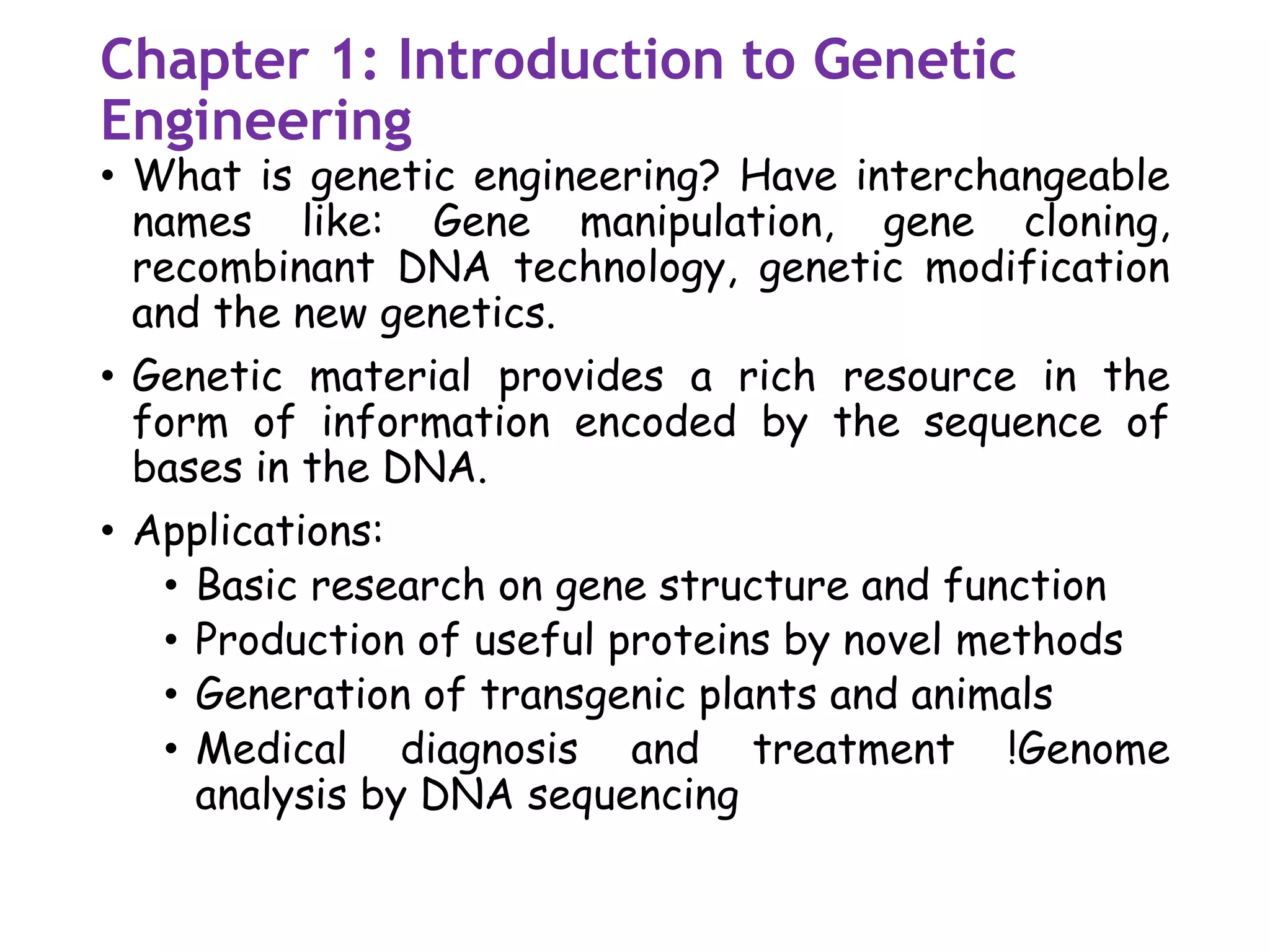
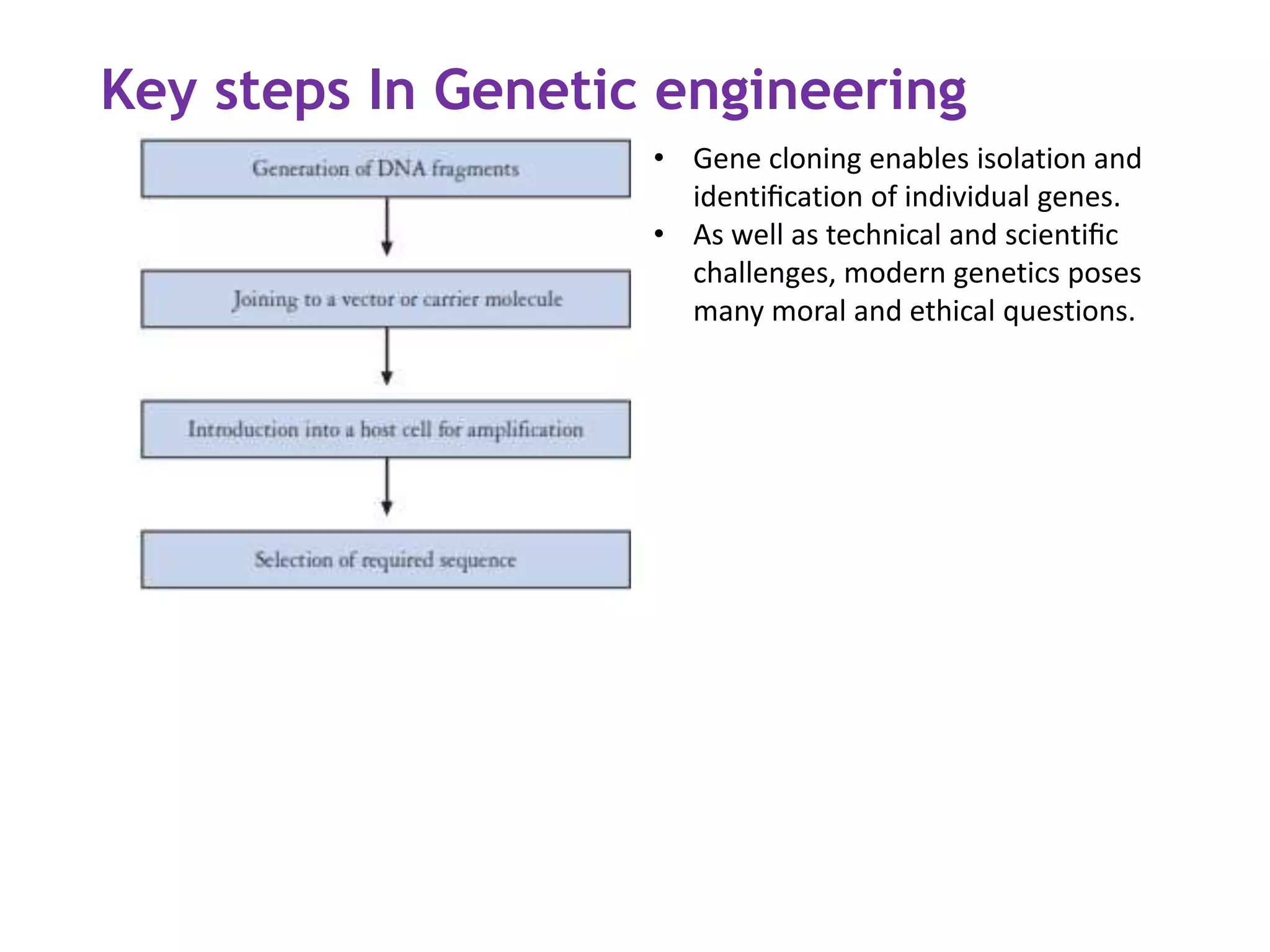
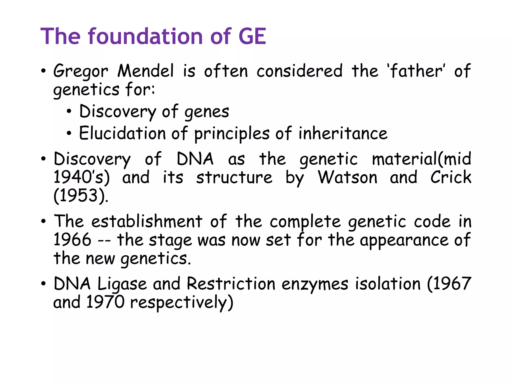
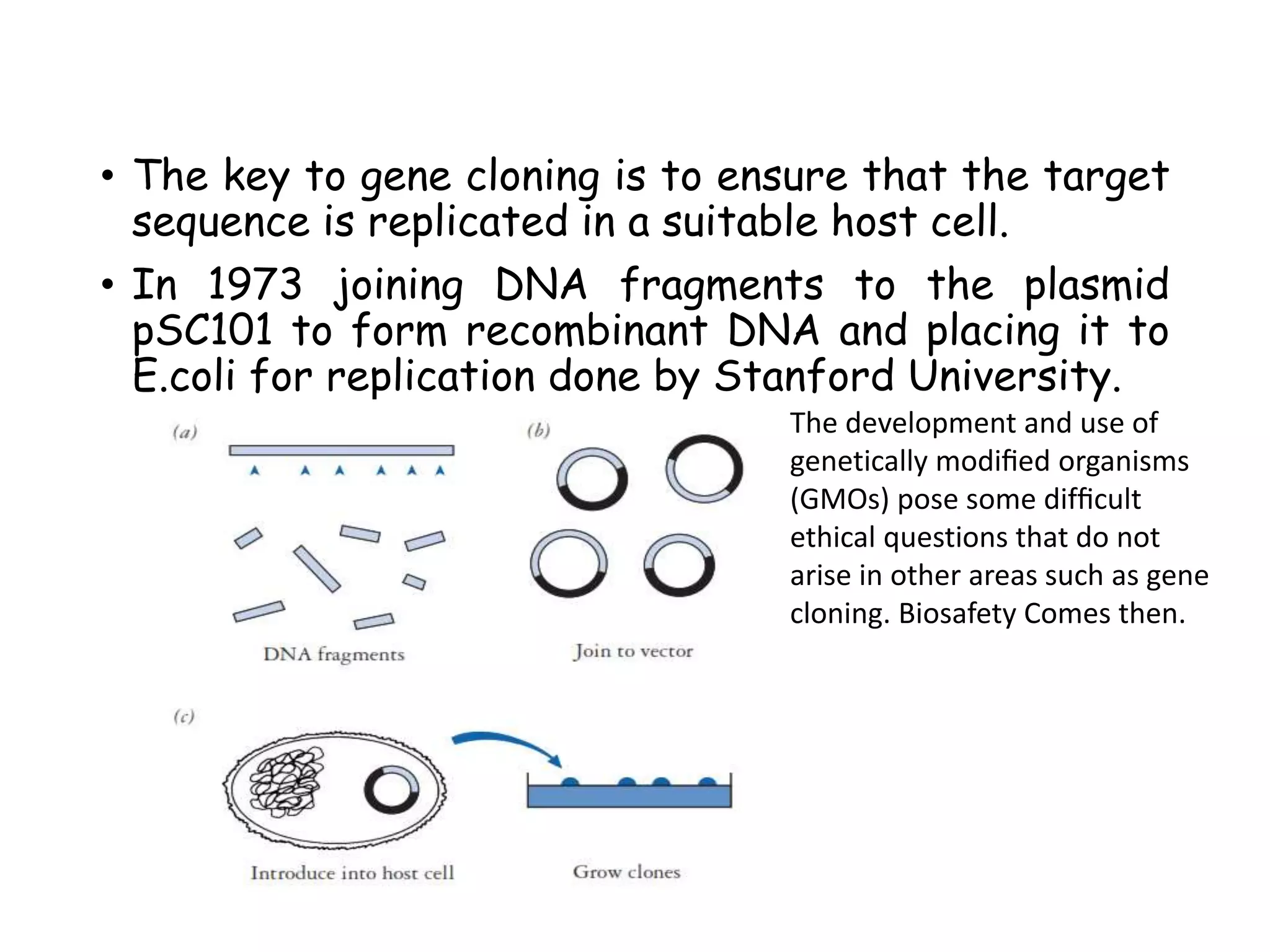
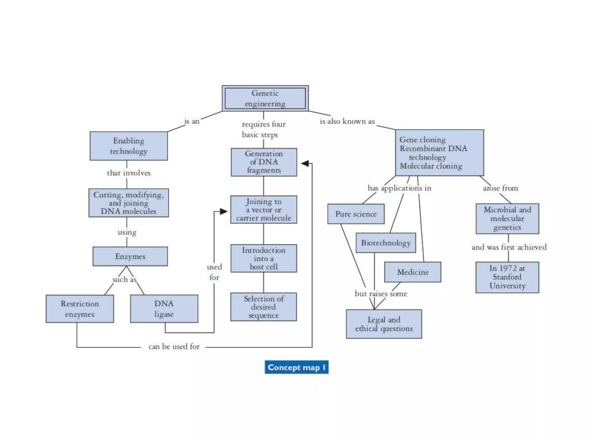
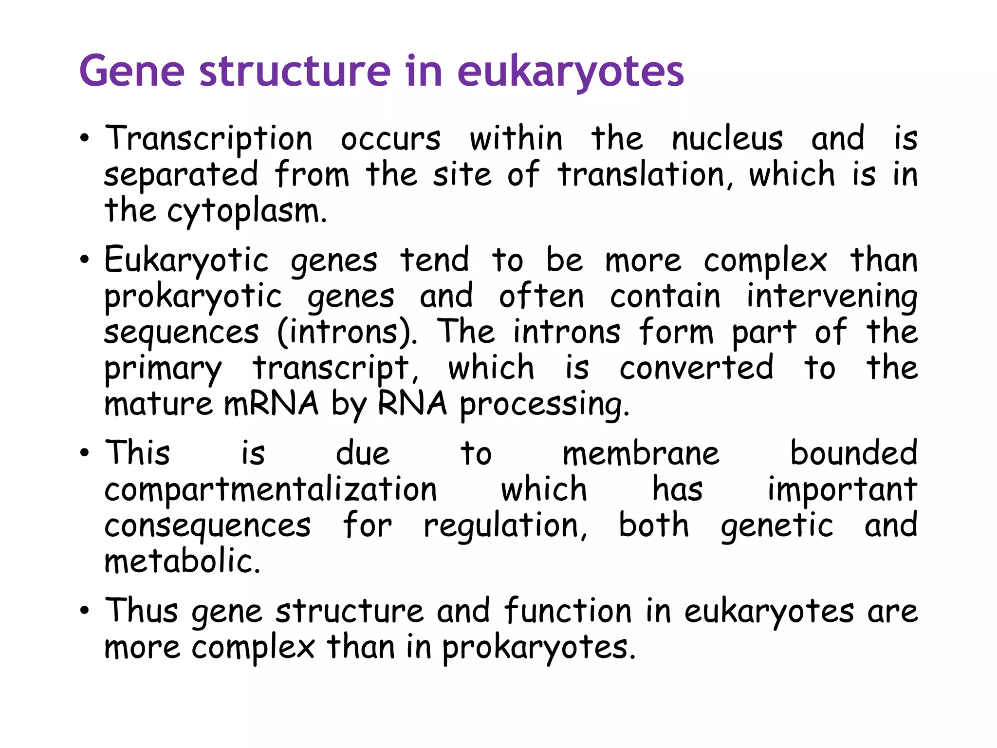
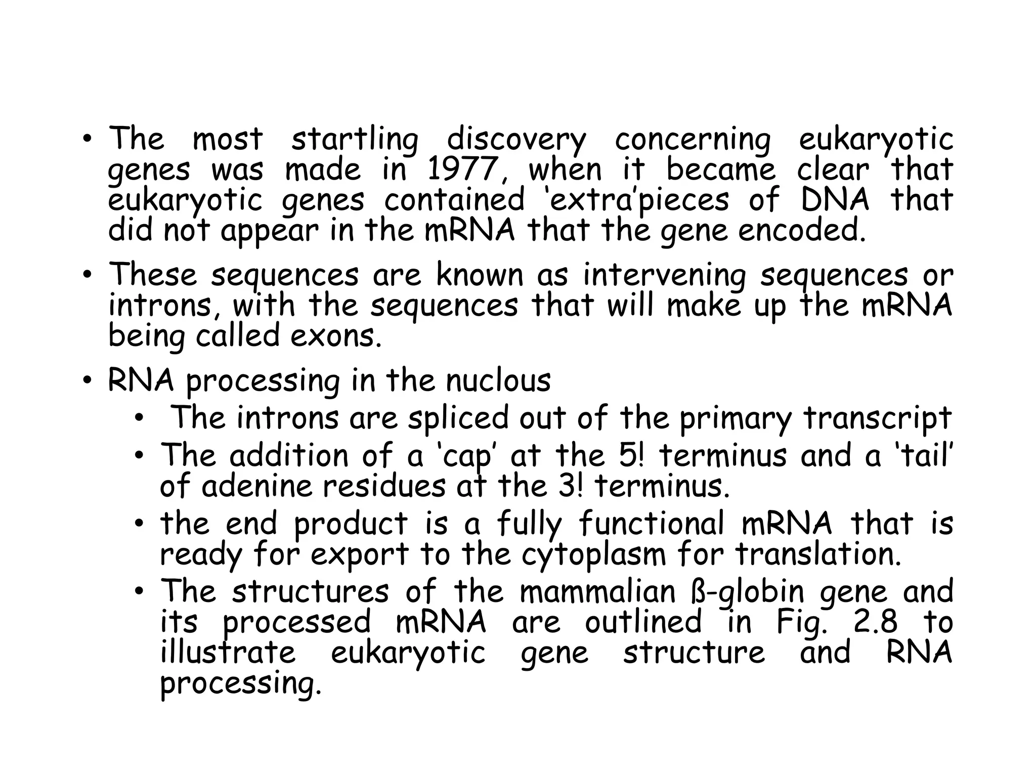
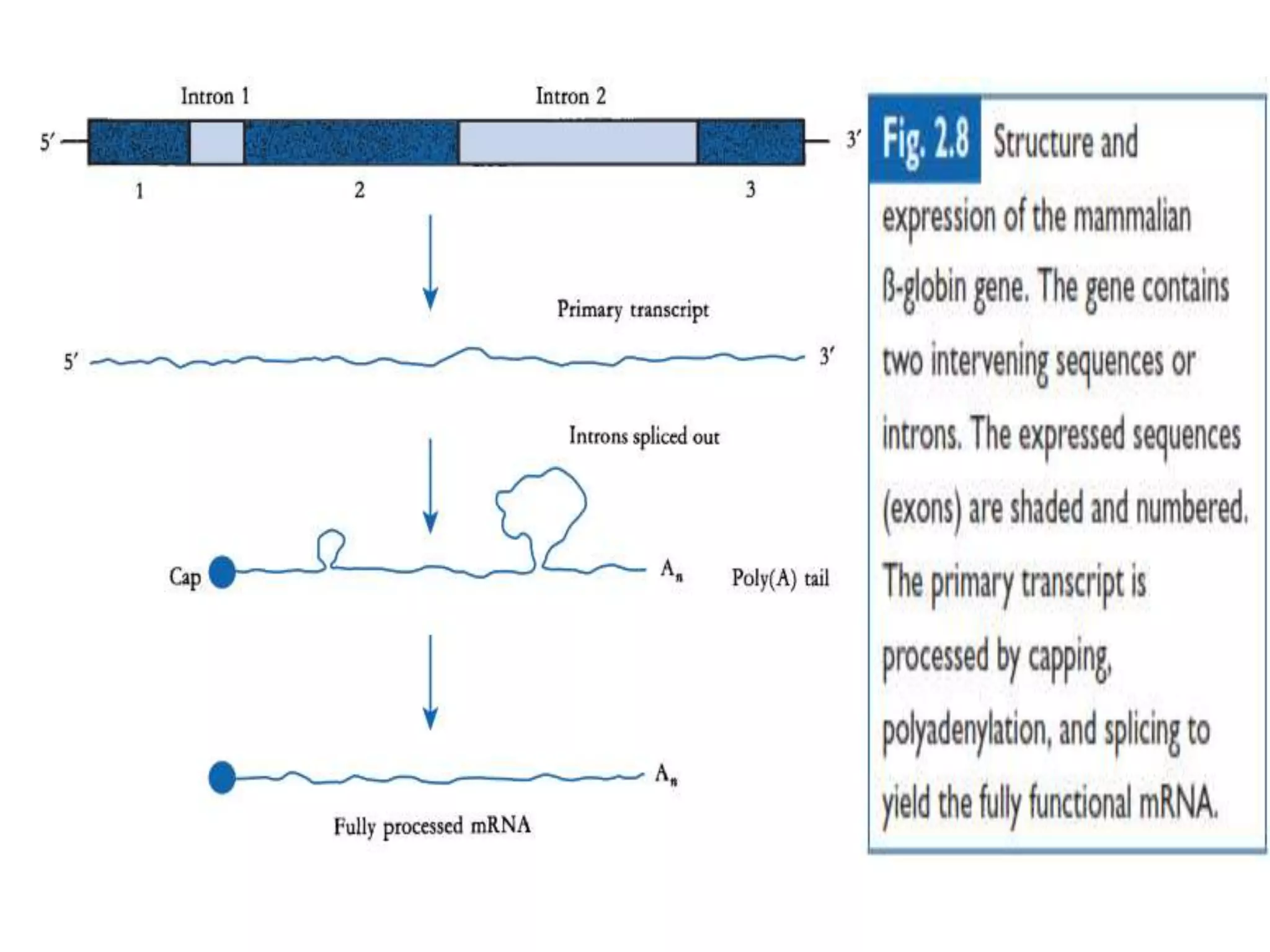
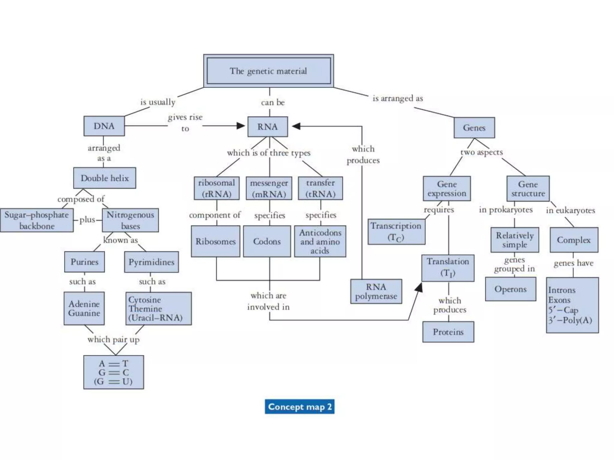
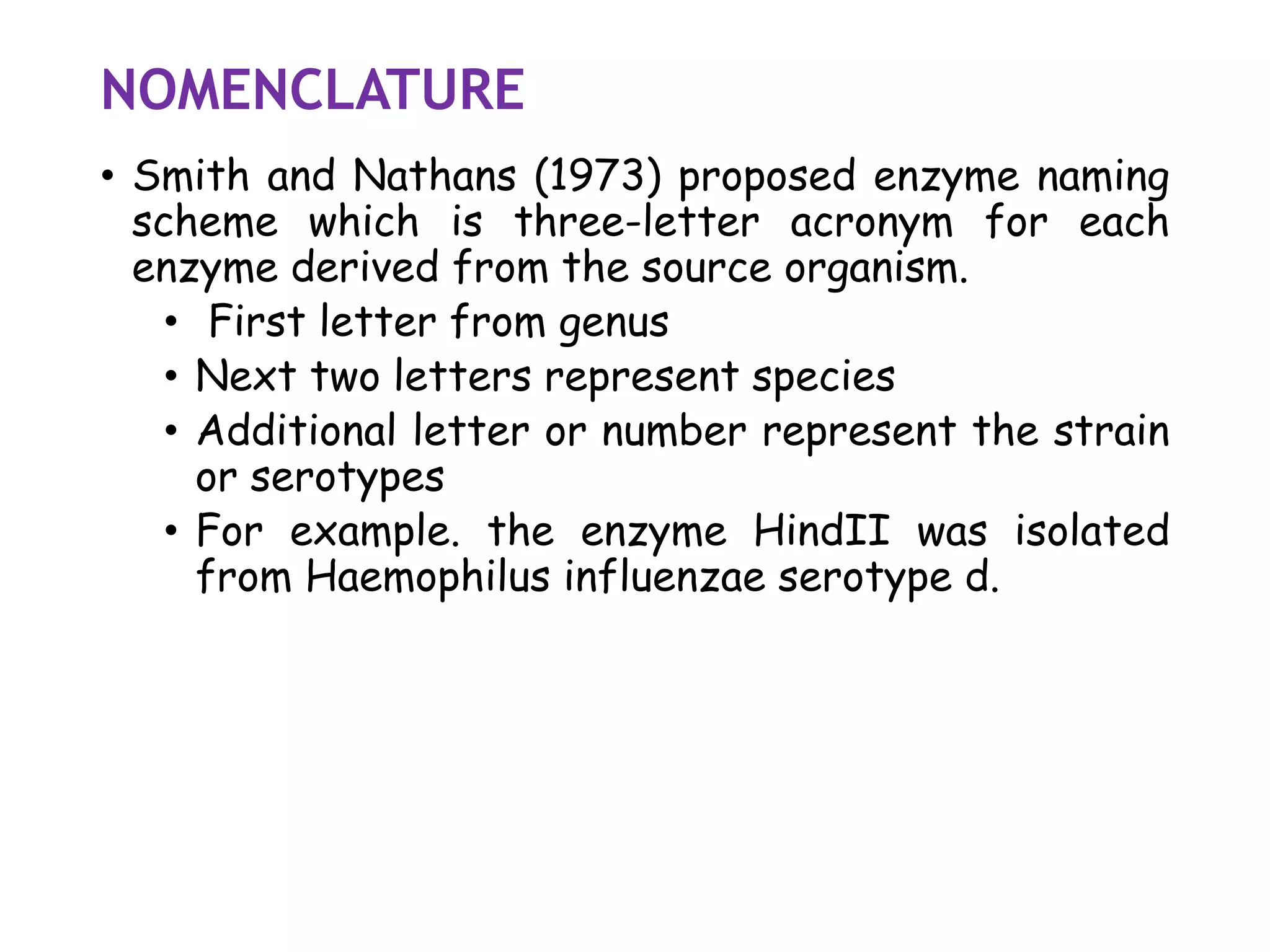
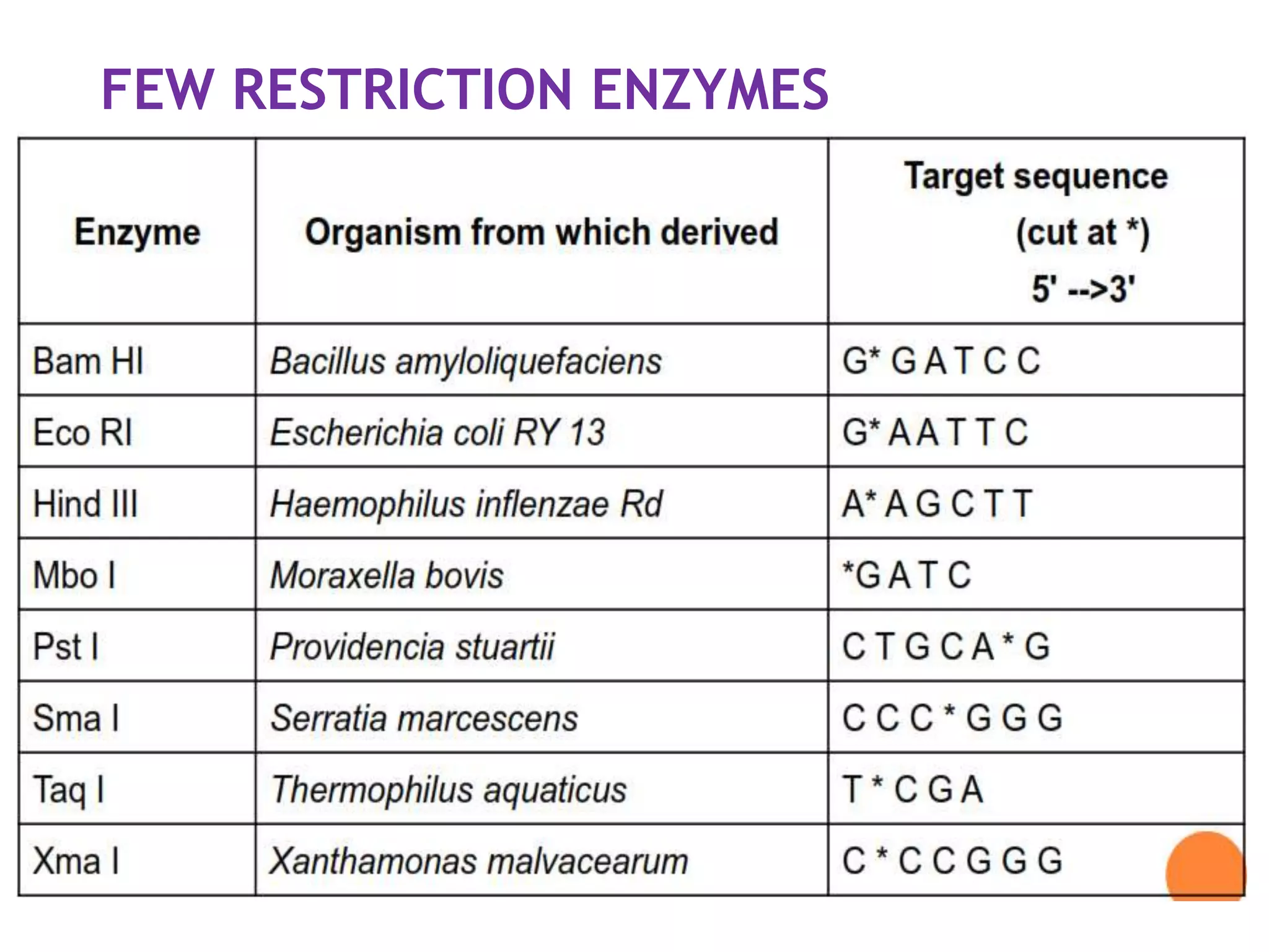
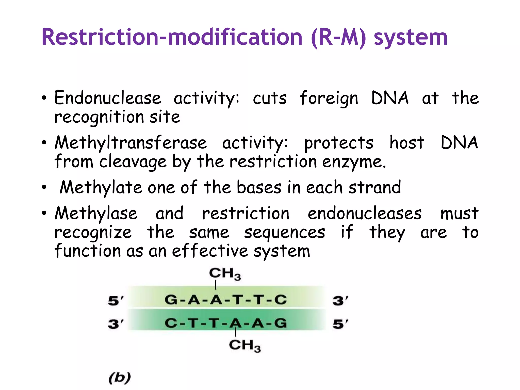
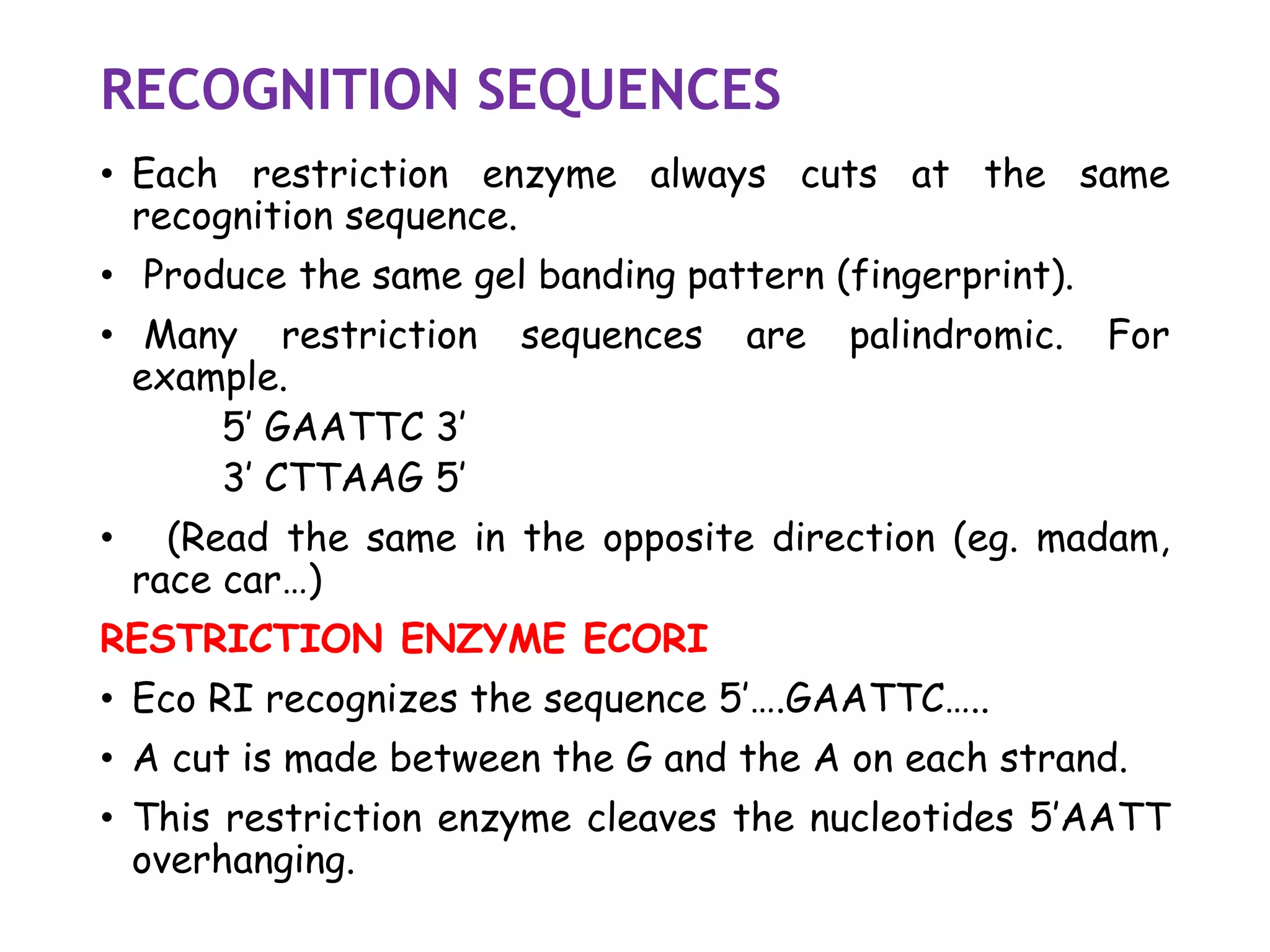
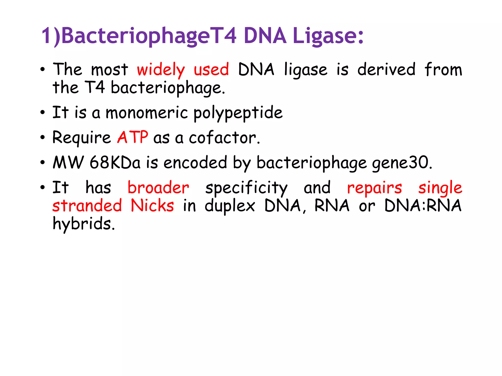
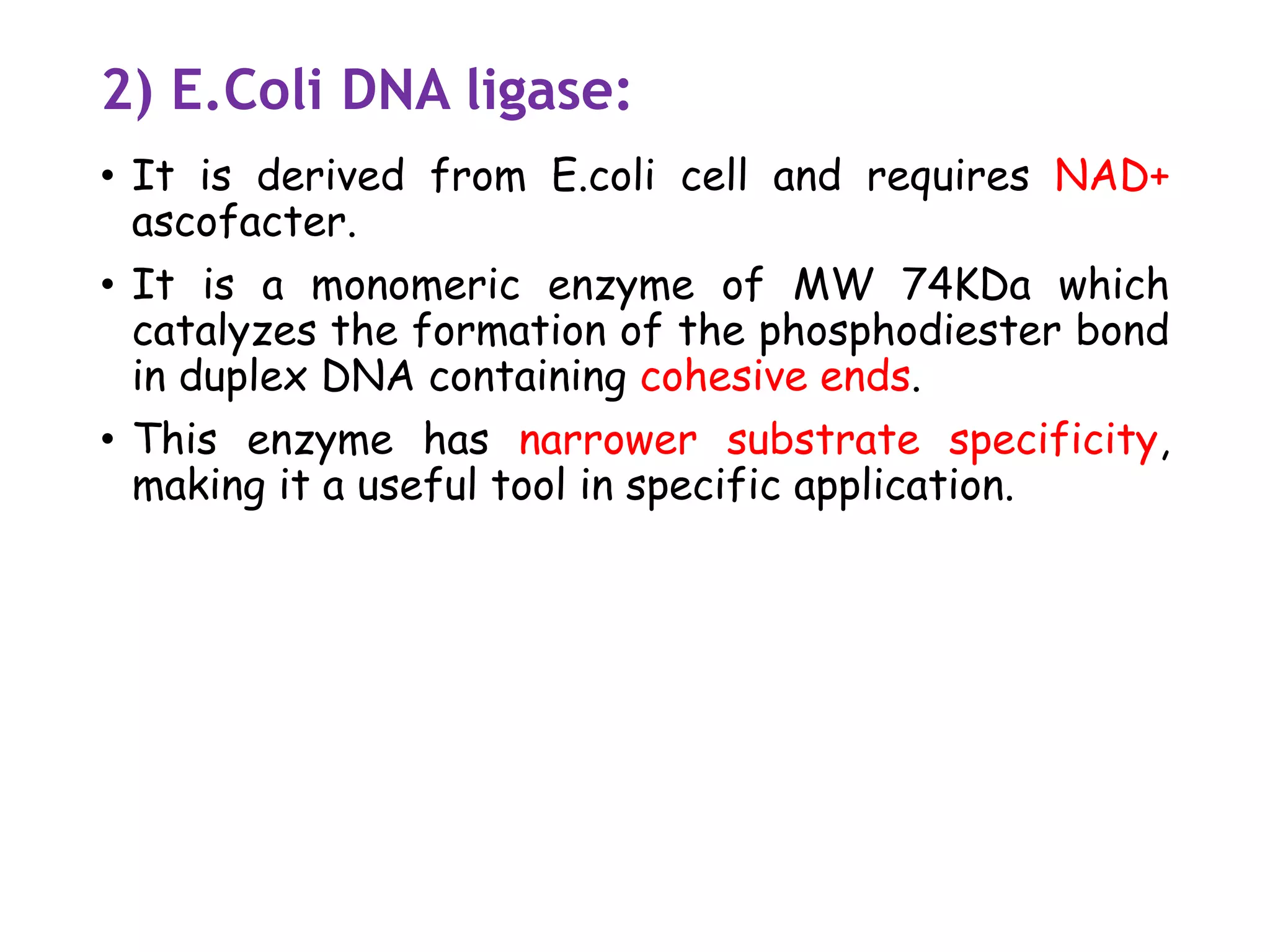
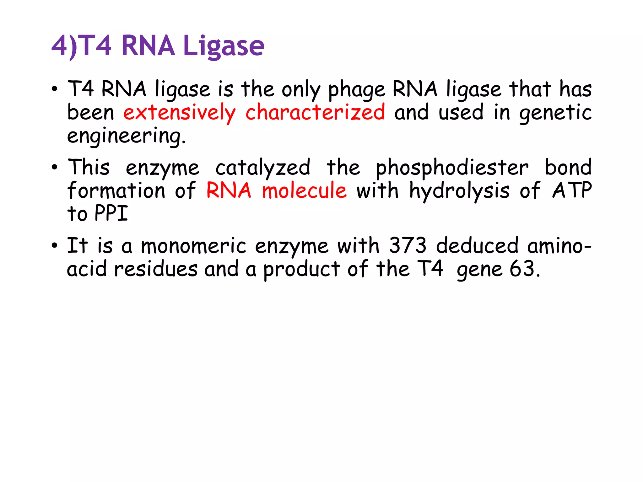
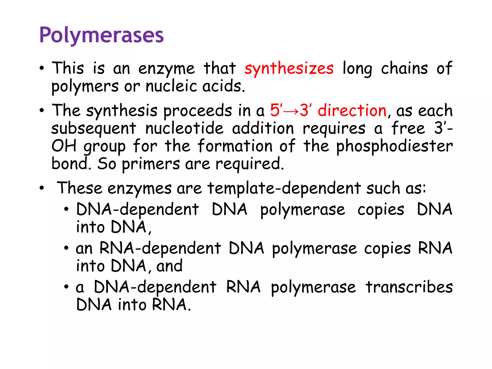
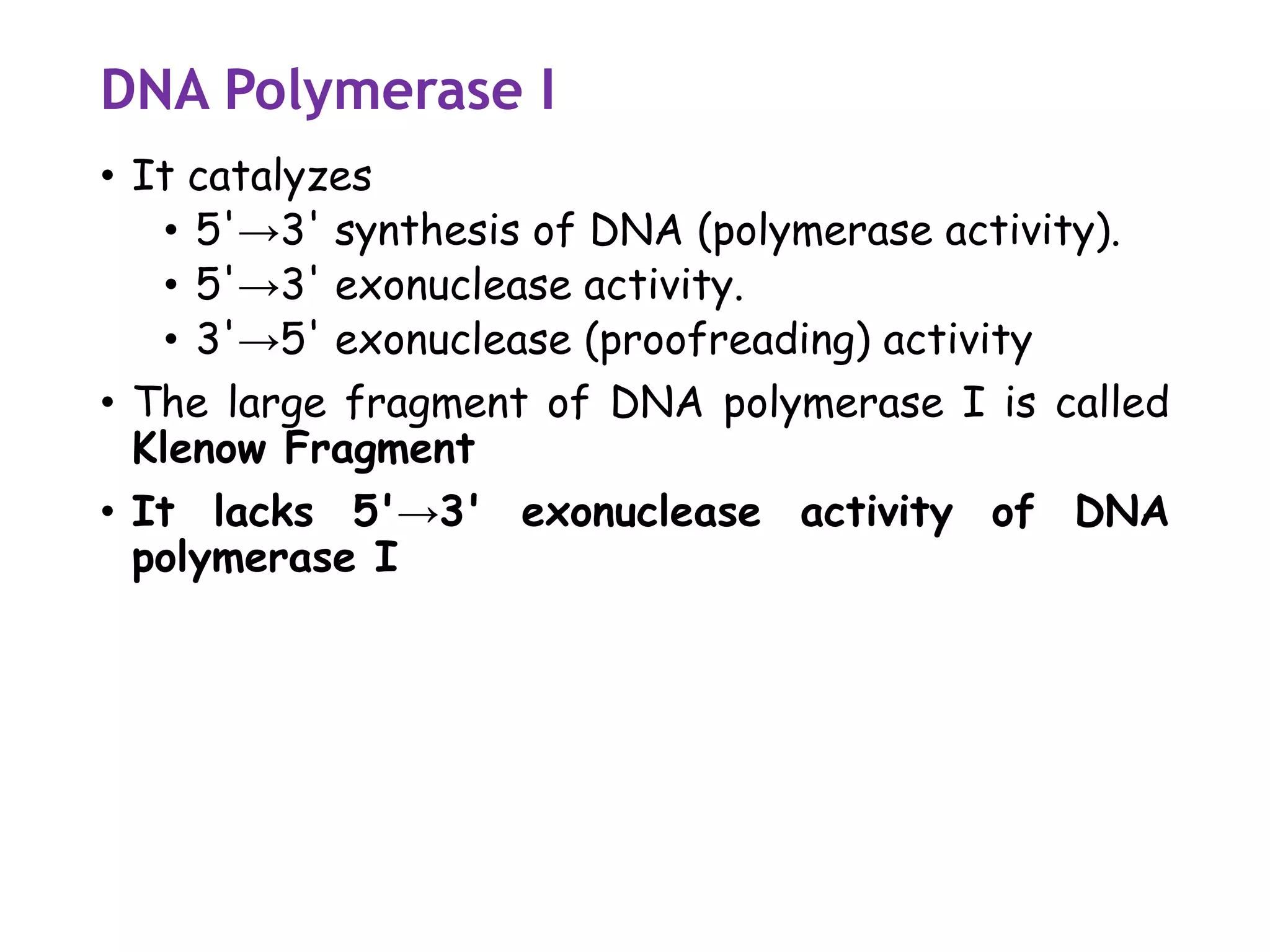
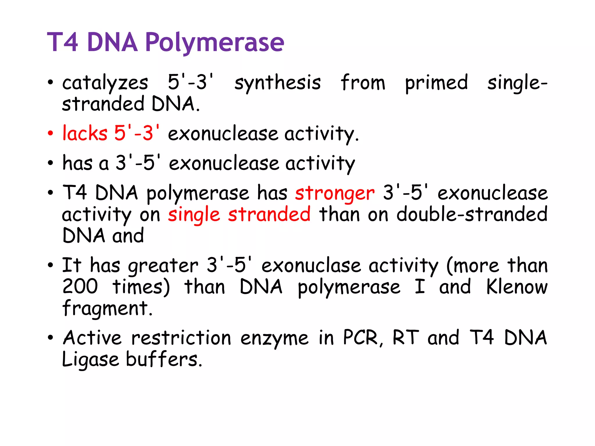
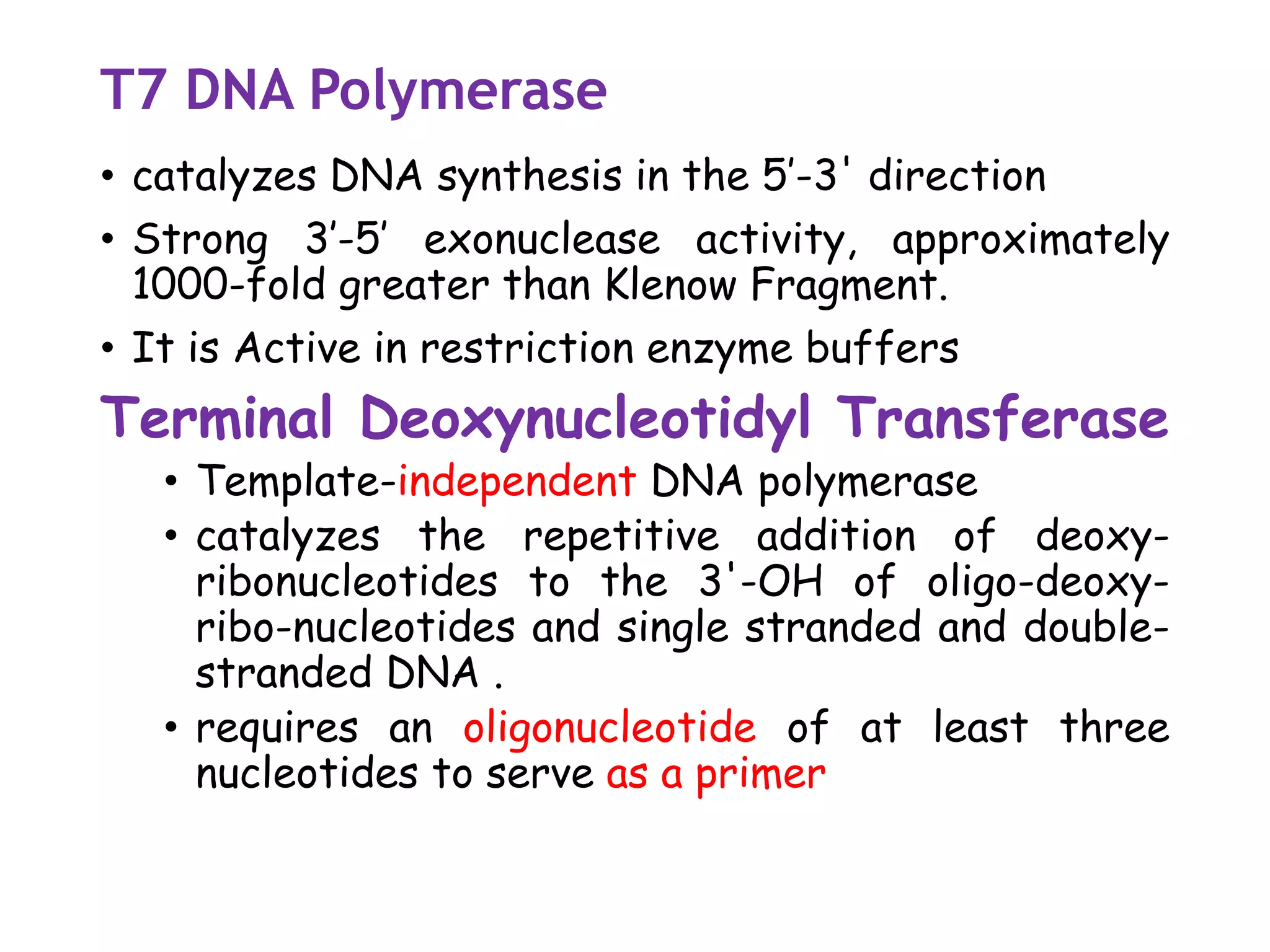
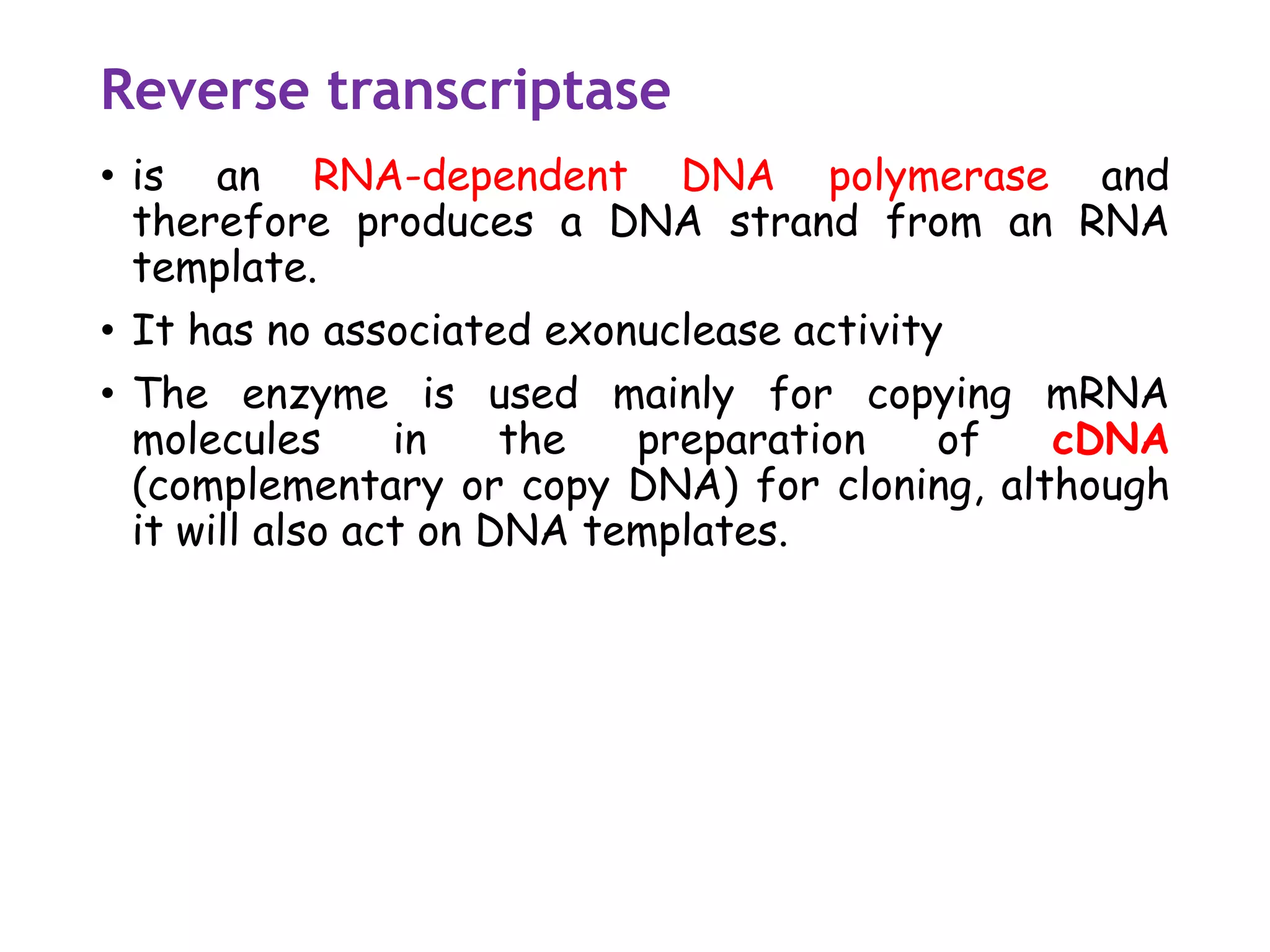
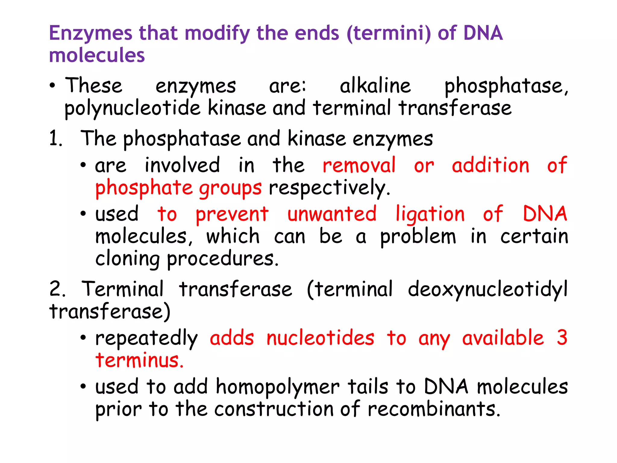
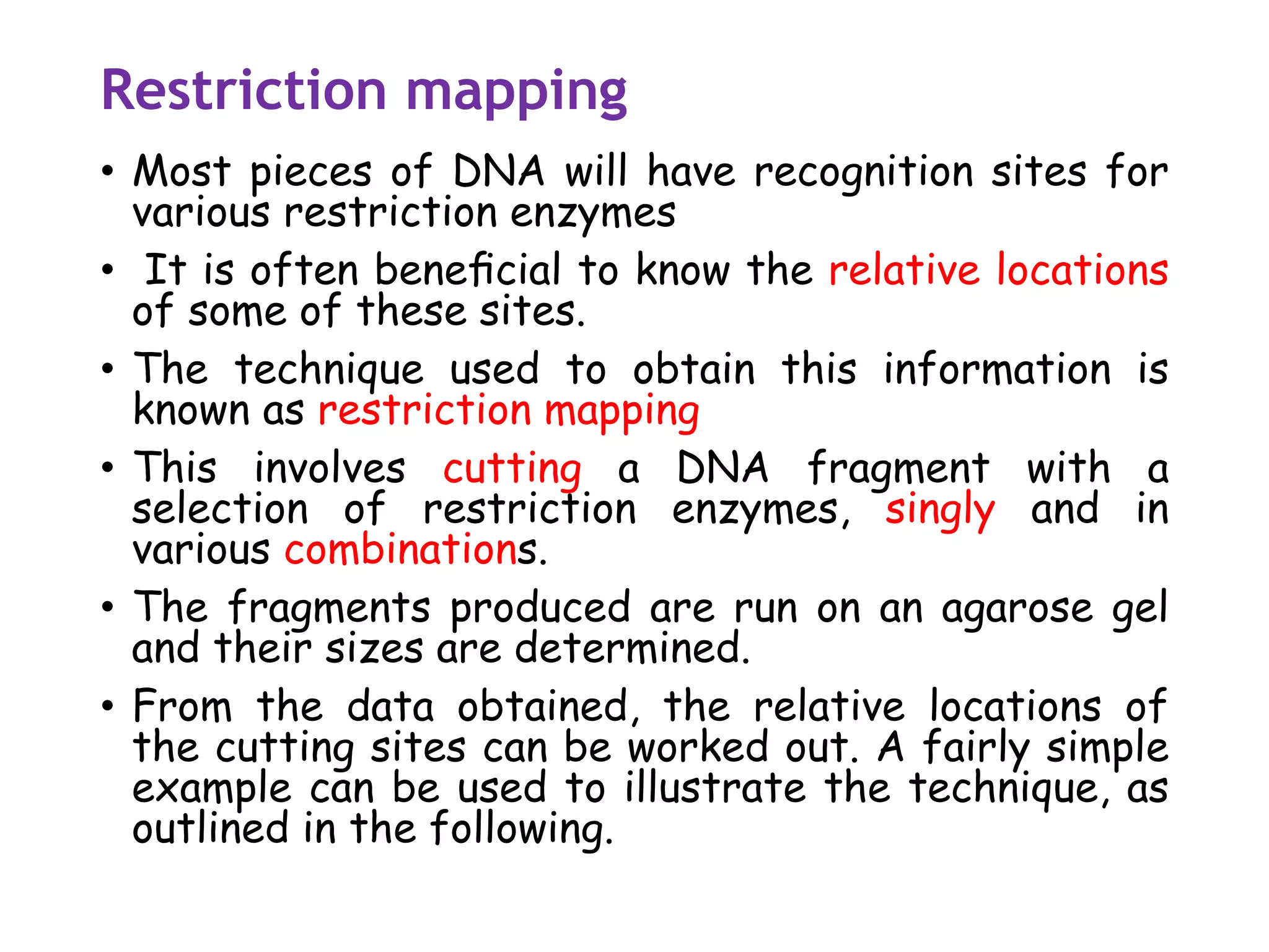
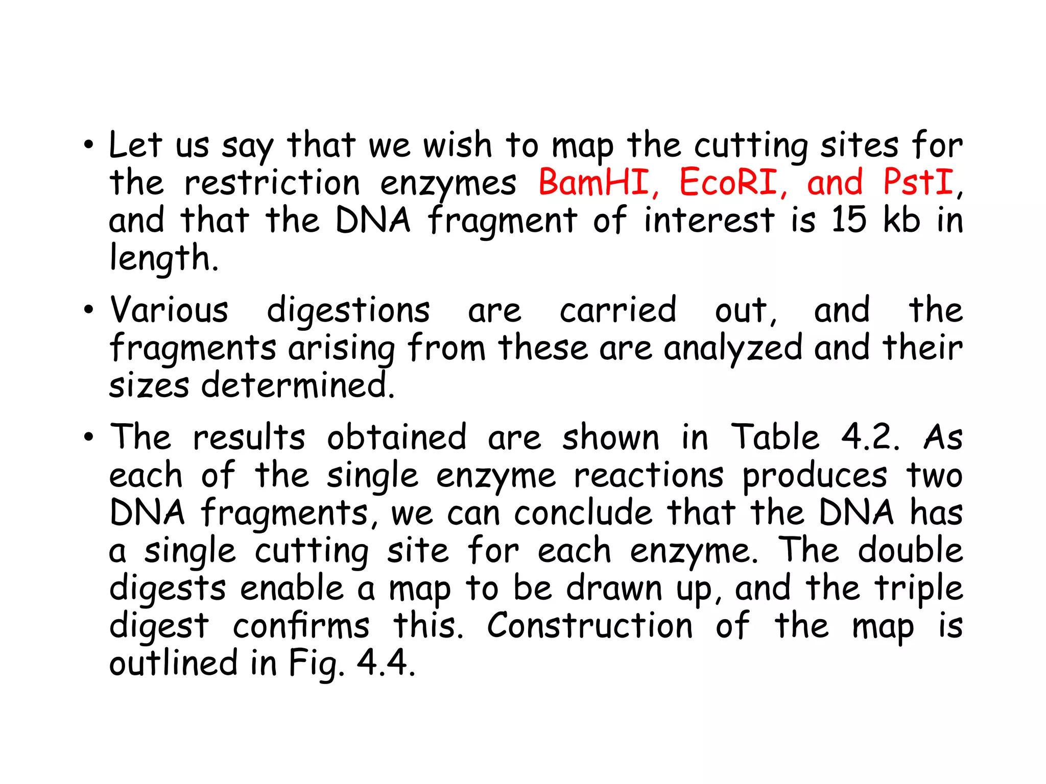
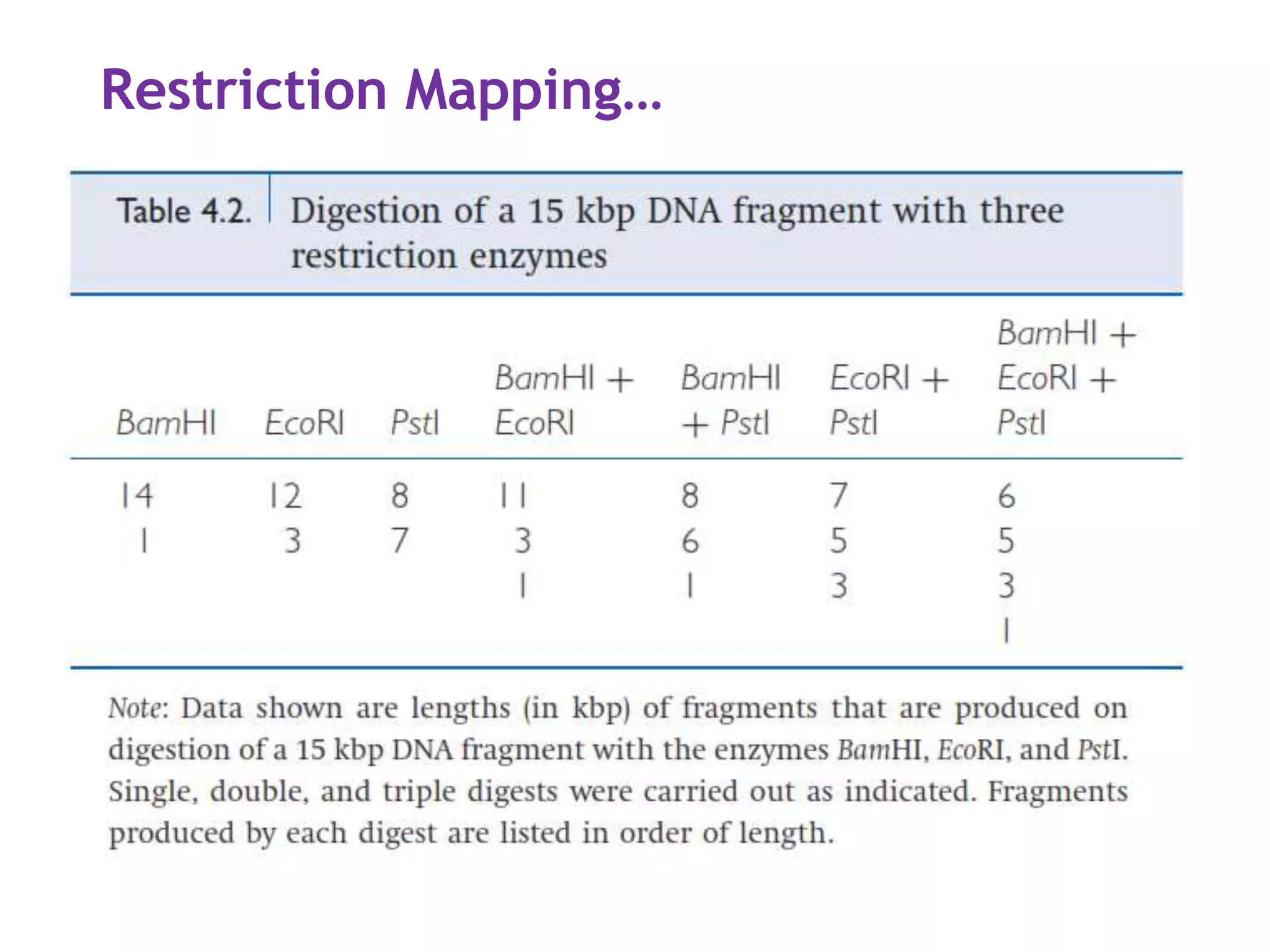
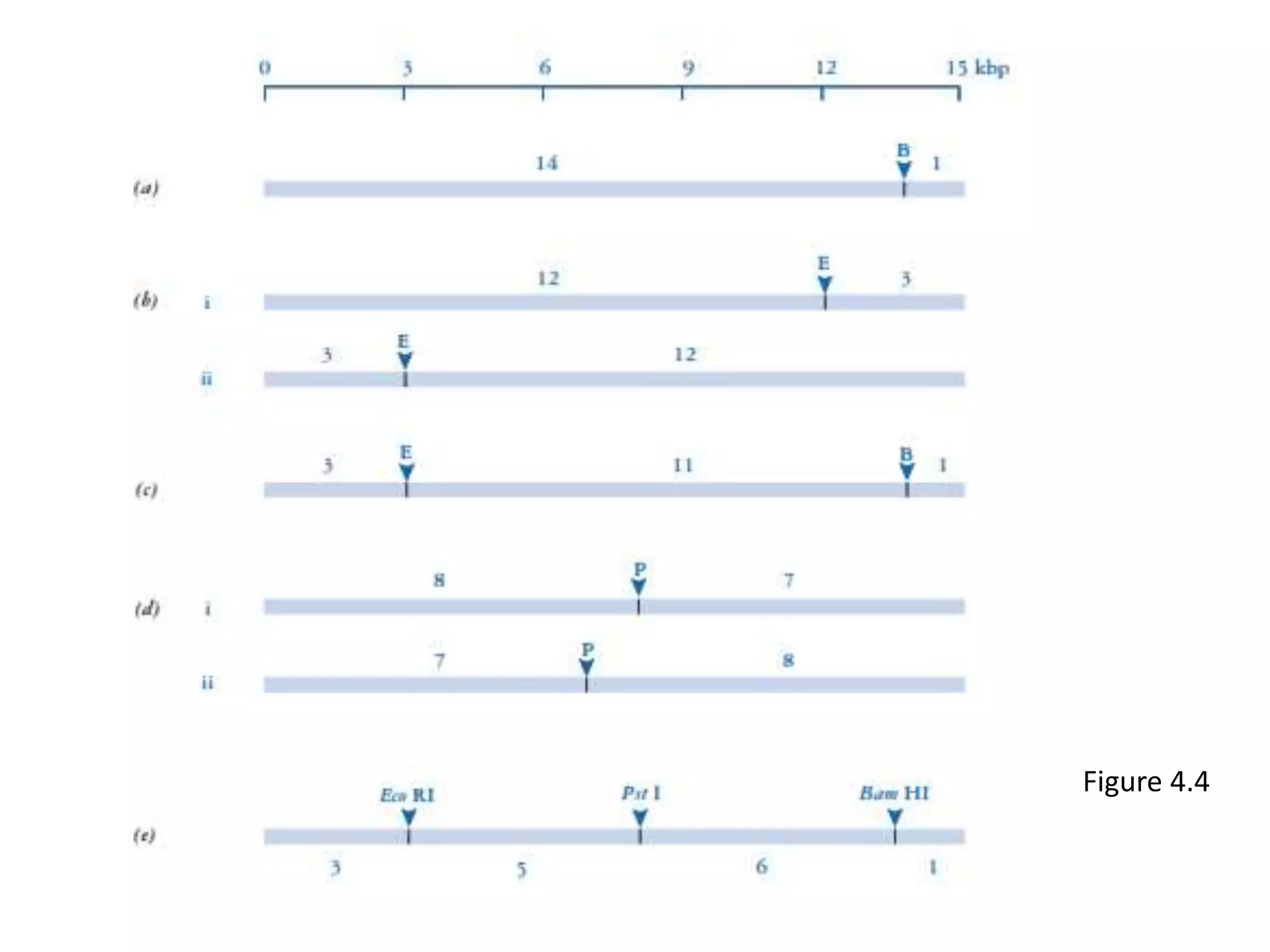
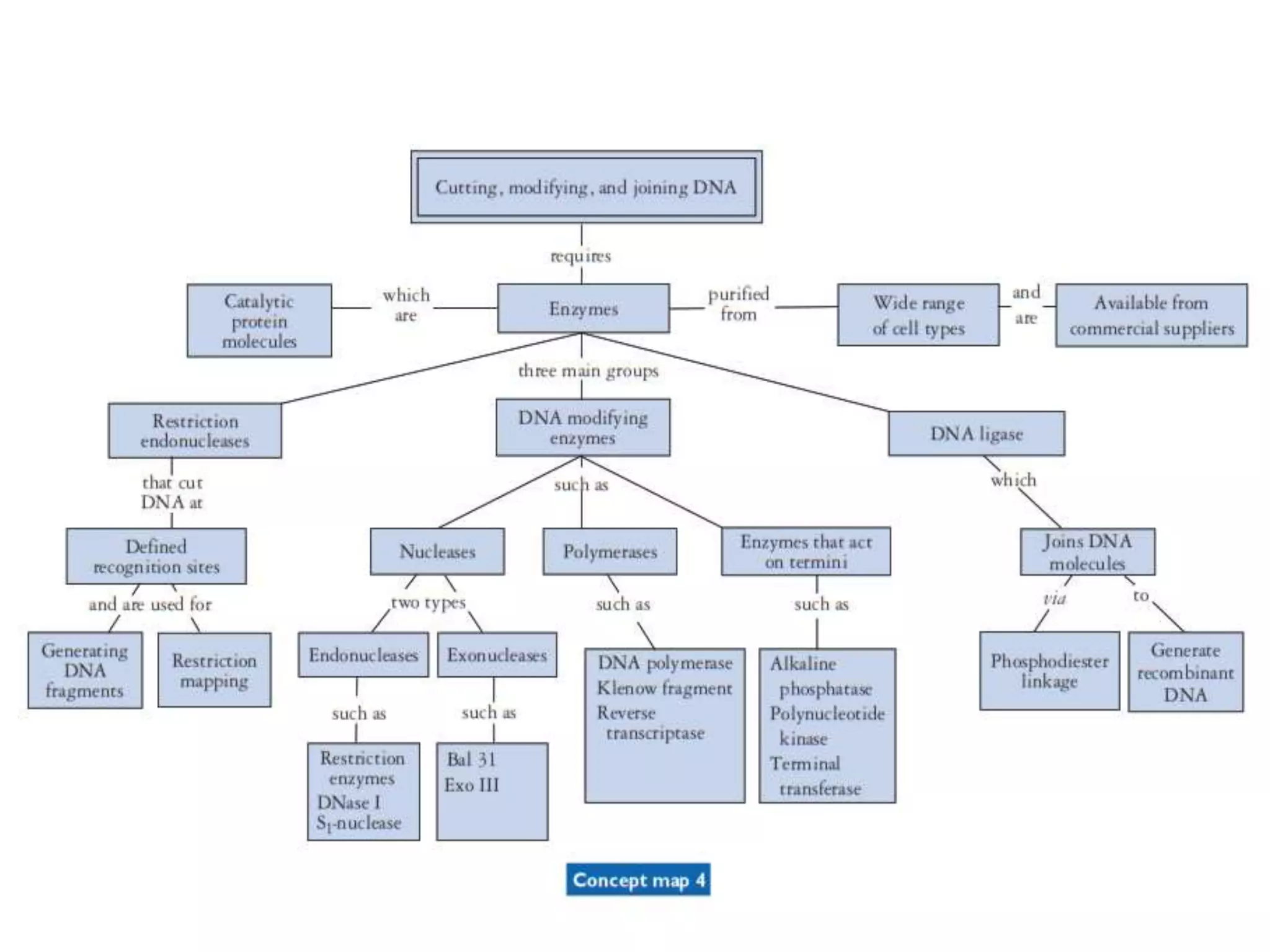
Introduction to Genetic Engineering
By: Getachew Birhanu
2020
• References:
• An introduction to Genetic Engineering 3rd edition.
Chapter 1: Introduction to Genetic
Engineering
• What is genetic engineering? Have interchangeable
names like: Gene manipulation, gene cloning,
recombinant DNA technology, genetic modification
and the new genetics.
• Genetic material provides a rich resource in the
form of information encoded by the sequence of
bases in the DNA.
• Applications:
• Basic research on gene structure and function
• Production of useful proteins by novel methods
• Generation of transgenic plants and animals
• Medical diagnosis and treatment !Genome
analysis by DNA sequencing
Key steps In Genetic engineering
• Gene cloning enables isolation and
identification of individual genes.
• As well as technical and scientific
challenges, modern genetics poses
many moral and ethical questions.
History and background
The history of genetics since 1900 (the three eras). Shaded areas
represent the periods of major development in each branch of the
subject.
The foundation of GE
• Gregor Mendel is often considered the ‘father’ of
genetics for:
• Discovery of genes
• Elucidation of principles of inheritance
• Discovery of DNA as the genetic material(mid
1940’s) and its structure by Watson and Crick
(1953).
• The establishment of the complete genetic code in
1966 -- the stage was now set for the appearance of
the new genetics.
• DNA Ligase and Restriction enzymes isolation (1967
and 1970 respectively)
• The key to gene cloning is to ensure that the target
sequence is replicated in a suitable host cell.
• In 1973 joining DNA fragments to the plasmid
pSC101 to form recombinant DNA and placing it to
E.coli for replication done by Stanford University.
The development and use of
genetically modified organisms
(GMOs) pose some difficult
ethical questions that do not
arise in other areas such as gene
cloning. Biosafety Comes then.
The flow of genetic information
• Life is directed by four nitrogenous bases: adenine (A),
guanine (G), cytosine (C), and thymine (T).
• The flow of genetic information is from DNA to RNA to
protein, via the processes of transcription (TC) and
translation (TL). This concept is known as the Central
Dogma of molecular biology.
• Two further aspects of information;
• Duplication of the genetic material prior to cell
division represents a DNA--DNA transfer, known as
DNA replication.
• some viruses have RNA instead of DNA as their
genetic material. These viruses (chiefly members of
the retrovirus group) have an enzyme called reverse
transcriptase (an RNA-dependent DNA polymerase)
that produces a double-stranded DNA molecule from
the single-stranded RNA genome.
• Thus, in these cases the flow of genetic information
is reversed with respect to the normal convention.
Gene organization
• The gene is the basic unit of genetic information.
Genes are located on chromosomes at a particular
genetic locus.
• In diploid organisms, which have their chromosomes
arranged as homologous pairs, different forms of
the same gene are known as alleles.
• The term ‘gene’is usually taken to represent the
genetic information transcribed into a single RNA
molecule, which is in turn translated into a single
protein.
• Exceptions are genes for RNA molecules (such as
rRNA and tRNA) are not translated.
• The nomenclature used for prokaryotic cells is
slightly different because of the way that their
genes are organized.
The anatomy of a gene
• There is the potential for confusion with the
nomenclature of the two DNA strands, which may
be called coding/non-coding, sense/antisense,
plus/minus, transcribed/non-transcribed, or
template/non-template.
• Recommendations from the International Union of
Biochemistry and the International Union of Pure
and Applied Chemistry favour the terms coding/non-
coding, with the coding strand of DNA taken to be
the mRNA-like strand.
• The terms template and non-template will be used
to describe DNA strands when there is not
necessarily any coding function involved, as in the
copying of DNA strands during cloning procedures.
• Thus, genetic information is expressed by
transcription of the non-coding strand of DNA,
which produces an mRNA molecule that has the
same sequence as the coding strand of DNA
(although the RNA has uracil substituted for
thymine)
• The sequence of the coding strand is usually
reported when dealing with DNA sequence data, as
this permits easy reference to the sequence of the
RNA.
• In addition to the sequence of bases that specifies
the codons in a protein-coding gene, there are other
important regulatory sequences associated with
genes. A site for starting transcription is required,
and this encompasses a region that binds RNA
polymerase, known as the promoter (P), and a
specific start point for transcription (TC).
• A stop site for transcription (tC) is also required.
• From TC start to tC stop is sometimes called the
transcriptional unit, that is, the DNA region that is
copied into RNA.
• Within this transcriptional unit, there may be
regulatory sites for translation, namely a start site
(TL) and a stop signal (tL).
• Other sequences involved in the control of gene
expression may be present either upstream or
downstream from the gene itself.
Gene structure in prokaryotes
• Genes in prokaryotes tend to be grouped together
in operons, with several genes under the control of a
single regulatory region.
• Perhaps the best known example of this
arrangement is the lac operon (Fig. 2.7), which codes
for the enzymes responsible for lactose catabolism.
Gene struct..
• The fact that structural genes in prokaryotes are
often grouped together means that the transcribed
mRNA may contain information for more than one
protein.
• Such a molecule is known as a polycistronic mRNA,
with the term cistron equating to the ‘gene’ as we
have defined it (i.e. encoding one protein).
• Thus, much of the genetic information in bacteria is
expressed via polycistronic mRNAs whose synthesis
is regulated in accordance with the needs of the cell
at any given time.
• This system is flexible and efficient, and it enables
the cell to adapt quickly to changing environmental
conditions.
Gene structure in eukaryotes
• Transcription occurs within the nucleus and is
separated from the site of translation, which is in
the cytoplasm.
• Eukaryotic genes tend to be more complex than
prokaryotic genes and often contain intervening
sequences (introns). The introns form part of the
primary transcript, which is converted to the
mature mRNA by RNA processing.
• This is due to membrane bounded
compartmentalization which has important
consequences for regulation, both genetic and
metabolic.
• Thus gene structure and function in eukaryotes are
more complex than in prokaryotes.
• The most startling discovery concerning eukaryotic
genes was made in 1977, when it became clear that
eukaryotic genes contained ‘extra’pieces of DNA that
did not appear in the mRNA that the gene encoded.
• These sequences are known as intervening sequences or
introns, with the sequences that will make up the mRNA
being called exons.
• RNA processing in the nuclous
• The introns are spliced out of the primary transcript
• The addition of a ‘cap’ at the 5! terminus and a ‘tail’
of adenine residues at the 3! terminus.
• the end product is a fully functional mRNA that is
ready for export to the cytoplasm for translation.
• The structures of the mammalian ß-globin gene and
its processed mRNA are outlined in Fig. 2.8 to
illustrate eukaryotic gene structure and RNA
processing.
DNA Manipulating Enzymes
1. Restriction enzymes -- cutting DNA
• Type II restriction endonucleases
• Use of restriction endonucleases
• Restriction mapping
2. DNA modifying enzymes
• Nucleases
• Polymerases
• Enzymes that modify the ends of DNA molecules
3. DNA ligase -- joining DNA molecules
Restriction enzymes
• In 1962, Werner Arber, a Swiss biochemist,
provided the first evidence for the existence of
"molecular scissors" that could cut DNA. He showed
that E. coli bacteria have an enzymatic “immune
system” that recognizes and destroys foreign DNA,
and modifies native DNA to prevent self
destruction
• By the early 1970s these enzymes started to be
identified and purified. It was shown that each
species of bacteria had its own population of a
SPECIFIC restriction enzyme.
• Each enzyme recognized its own specific sequence
of DNA bases to cut external DNA.
• Smith,Nathans and Arber were awarded the Nobel
prize for Physiology and Medicine in 1978 for the
discovery of endonucleases.
Restriction enzymes…
• Restriction enzymes act as a ‘protection’ system for
bacteria in that they hydrolyse exogenous DNA
that is not methylated by the host modification
enzyme.
• This protect bacteria from bacteriophages (Viruses
• The modification enzyme of the system (a
methylase) modifies the host DNA by methylation
of particular bases in the restriction enzyme’s
recognition sequence, which prevents the enzyme
from cutting the DNA.
• Restriction Enzymes are also known as molecular
scessor.
Basis of RE categorization
• Their composition.
• Enzyme co-factor requirement.
• The nature of their target sequence.
• position of their DNA cleavage site relative to the
target sequence
NOMENCLATURE
• Smith and Nathans (1973) proposed enzyme naming
scheme which is three-letter acronym for each
enzyme derived from the source organism.
• First letter from genus
• Next two letters represent species
• Additional letter or number represent the strain
or serotypes
• For example. the enzyme HindII was isolated
from Haemophilus influenzae serotype d.
FEW RESTRICTION ENZYMES
Restriction-modification (R-M) system
• Endonuclease activity: cuts foreign DNA at the
recognition site
• Methyltransferase activity: protects host DNA
from cleavage by the restriction enzyme.
• Methylate one of the bases in each strand
• Methylase and restriction endonucleases must
recognize the same sequences if they are to
function as an effective system
RECOGNITION SEQUENCES
• Each restriction enzyme always cuts at the same
recognition sequence.
• Produce the same gel banding pattern (fingerprint).
• Many restriction sequences are palindromic. For
example.
5’ GAATTC 3’
3’ CTTAAG 5’
• (Read the same in the opposite direction (eg. madam,
race car…)
RESTRICTION ENZYME ECORI
• Eco RI recognizes the sequence 5’….GAATTC…..
• A cut is made between the G and the A on each strand.
• This restriction enzyme cleaves the nucleotides 5’AATT
overhanging.
• These are known as “sticky ends” because hydrogen
bonds are available to “stick” to a complimentary
3’TTAA.
• Note: Restriction enzymes don’t stop with one cut!
They continue to cut at every recognition sequence
on a DNA strand
NUCLEASES
• Nucleases are the group of enzymes which cuts
DNA at specific sites. They are of two types:-
• 1) Exo-nucleases
• 2)Endo-nucleases
EXONUCLEASES:
• These enzymes act upon genome and digest the base
pairs on 5' or 3' ends of single stranded DNA, these
are of following:
• a) λ Exonuclease
• b) Exonuclease III
• a) λ Exonuclease:-This enzyme removes nucleotide
from 5’ end of double stranded DNA.
• b)Exonuclease III:- The Enzyme removes nucleotide
from 3’ end of the double stranded DNA fragment.
ENDONUCLEASES
• They act upon genetic material and cleave the
double stranded DNA at any point except the ends.
• They produce internal cuts, called cleavage, In the
DNA molecule
• examples:
• a) S1-Nuclease- It degrades the single stranded
DNA OR Single stranded protrusion of double
stranded DNA with cohesive ends.
• b)Bal 31 nuclease-it degrade simultaneously both
3’ and 5’ strands of DNA and they possess blunt
end at both termini.
Restriction Endonucleases
• These are the group of ezymes which cuts the DNA
within its inner site & are also called as Molecular
Scissors.
• The are first isolated from the Ecoli cell.
• They are of three types :-Type 1,2,3.
Type I:
• These are the complex nucleases which functions
simultaneously as endonucleases and methylase and
they require Mg++ and ATP as cofactor.
• The recognition site will be of 15bp long and
cleavage site is about 1000bp away from the
recognition site.
• ex:ECOR1,SAL1,ECOK,ECOB etc.
Type II:
• They have separate activity for endonucleases,
methylation of DNA and requires mg++ as
cofactor.
• This enzyme cleaves the DNA molecule 100bp away
from the recognition site and these sites have
palindromic sequence.
• ex: HIND III,HSU-I, XmaI, SmaI, BamHI etc.
Type III:
• This enzyme has both nuclease as well as
methylation activity and requires both ATP and
Mg++ and S-Adenosyl Methionine(AdoMet) as
cofactor.
• It cleaves the DNA 25-27bp away from the
recognition site at non-palindromic sequence.
• ex :HpaII, MboII, Fok I etc.
ARTIFICIAL RESTRICTION ENZYMES
• Generated by fusing a natural or engineered DNA
binding domain to a nuclease domain
• can target large DNA sites (up to 36 bp)
• can be engineered to bind to desired DNA
sequences
STICKY END CUTTERS
Most restriction enzymes make staggered cuts.
Staggered cuts produce single stranded
"sticky-ends".
o
o
DNA from different sources
easily because of sticky-end
can be spliced
overhangs.
o
Hindlll
•
A - A - T - T - C T 7
I
IEcoRI I I I I
[ I ] ! + 1 ' ! p
( I- + C - T- T- A - A - G •
t
BLUNT END CUTTERS
o Some restriction enzymes cut DNA at opposite
o They leave blunt ended DNA fragments
base
o These are called blunt end cutters
c-T
I i
I 1
c-T
I I
A-G A-G
I
I
I
I
I
I
I
I
q1
l
I
f
f
I
I
I
1
t
Alul If
fI I It t
T-C G-A T- € G-A
c~c c~c
I I
c-c,
I
I
G - c l
q
I 1I
1Haelll I 1
- l h h h] { '- ±f ( - '
[el h h h
DNA LIGASE
• DNA ligase catalyzes the formation of
phosphodiester bond between the 5’ phosphate of
one strand of DNA or RNA and the 3’-hydroxyl of
another.
• This enzyme is used to covalently link or ligate
fragments of DNA together
• In genetic engineering it is used to seal
discontinuities in the sugar—phosphate chains that
arise when recombinant DNA is made by joining
DNA molecules from different sources.
• It can therefore be thought of as molecular glue,
which is used to stick pieces of DNA together.
• The DNA ligase used in molecular cloning differ in
their abilities to ligate non canonical substrate, such
as blunt ended duplex DNA:RNA hybrid or ssDNAs.
• Types of DNA Ligase:
• 1)BacteriophageT4 DNA ligase
• 2)E.coli DNA Ligase
• 3)Taq DNA Ligase
• 4)T4 DNA Ligase
1)BacteriophageT4 DNA Ligase:
• The most widely used DNA ligase is derived from
the T4 bacteriophage.
• It is a monomeric polypeptide
• Require ATP as a cofactor.
• MW 68KDa is encoded by bacteriophage gene30.
• It has broader specificity and repairs single
stranded Nicks in duplex DNA, RNA or DNA:RNA
hybrids.
2) E.Coli DNA ligase:
• It is derived from E.coli cell and requires NAD+
ascofacter.
• It is a monomeric enzyme of MW 74KDa which
catalyzes the formation of the phosphodiester bond
in duplex DNA containing cohesive ends.
• This enzyme has narrower substrate specificity,
making it a useful tool in specific application.
3)Taq DNA ligase [NAD+ as cofactor]
• The gene encoding thermostable ligases have been
identified from several thermophilic bacteria.
• Several of this ligase have been cloned and
expressed to high levels in E.coli
• It is used in the detection of mutation as
thermostable DNA ligase and retain their activities
after exposure to higher temp for multiple rounds
• it detects mutation in mammalian DNA during
amplification reactions.
4)T4 RNA Ligase
• T4 RNA ligase is the only phage RNA ligase that has
been extensively characterized and used in genetic
engineering.
• This enzyme catalyzed the phosphodiester bond
formation of RNA molecule with hydrolysis of ATP
to PPI
• It is a monomeric enzyme with 373 deduced amino-
acid residues and a product of the T4 gene 63.
Polymerases
• This is an enzyme that synthesizes long chains of
polymers or nucleic acids.
• The synthesis proceeds in a 5’→3’ direction, as each
subsequent nucleotide addition requires a free 3’-
OH group for the formation of the phosphodiester
bond. So primers are required.
• These enzymes are template-dependent such as:
• DNA-dependent DNA polymerase copies DNA
into DNA,
• an RNA-dependent DNA polymerase copies RNA
into DNA, and
• a DNA-dependent RNA polymerase transcribes
DNA into RNA.
DNA Polymerase I
• It catalyzes
• 5'→3' synthesis of DNA (polymerase activity).
• 5'→3' exonuclease activity.
• 3'→5' exonuclease (proofreading) activity
• The large fragment of DNA polymerase I is called
Klenow Fragment
• It lacks 5'→3' exonuclease activity of DNA
polymerase I
T4 DNA Polymerase
• catalyzes 5'-3' synthesis from primed single-
stranded DNA.
• lacks 5'-3' exonuclease activity.
• has a 3'-5' exonuclease activity
• T4 DNA polymerase has stronger 3'-5' exonuclease
activity on single stranded than on double-stranded
DNA and
• It has greater 3'-5' exonuclase activity (more than
200 times) than DNA polymerase I and Klenow
fragment.
• Active restriction enzyme in PCR, RT and T4 DNA
Ligase buffers.
T7 DNA Polymerase
• catalyzes DNA synthesis in the 5’-3' direction
• Strong 3’-5’ exonuclease activity, approximately
1000-fold greater than Klenow Fragment.
• It is Active in restriction enzyme buffers
Terminal Deoxynucleotidyl Transferase
• Template-independent DNA polymerase
• catalyzes the repetitive addition of deoxy-
ribonucleotides to the 3'-OH of oligo-deoxy-
ribo-nucleotides and single stranded and double-
stranded DNA .
• requires an oligonucleotide of at least three
nucleotides to serve as a primer
Reverse transcriptase
• is an RNA-dependent DNA polymerase and
therefore produces a DNA strand from an RNA
template.
• It has no associated exonuclease activity
• The enzyme is used mainly for copying mRNA
molecules in the preparation of cDNA
(complementary or copy DNA) for cloning, although
it will also act on DNA templates.
Enzymes that modify the ends (termini) of DNA
molecules
• These enzymes are: alkaline phosphatase,
polynucleotide kinase and terminal transferase
1. The phosphatase and kinase enzymes
• are involved in the removal or addition of
phosphate groups respectively.
• used to prevent unwanted ligation of DNA
molecules, which can be a problem in certain
cloning procedures.
2. Terminal transferase (terminal deoxynucleotidyl
transferase)
• repeatedly adds nucleotides to any available 3
terminus.
• used to add homopolymer tails to DNA molecules
prior to the construction of recombinants.
Restriction mapping
• Most pieces of DNA will have recognition sites for
various restriction enzymes
• It is often beneficial to know the relative locations
of some of these sites.
• The technique used to obtain this information is
known as restriction mapping
• This involves cutting a DNA fragment with a
selection of restriction enzymes, singly and in
various combinations.
• The fragments produced are run on an agarose gel
and their sizes are determined.
• From the data obtained, the relative locations of
the cutting sites can be worked out. A fairly simple
example can be used to illustrate the technique, as
outlined in the following.
• Let us say that we wish to map the cutting sites for
the restriction enzymes BamHI, EcoRI, and PstI,
and that the DNA fragment of interest is 15 kb in
length.
• Various digestions are carried out, and the
fragments arising from these are analyzed and their
sizes determined.
• The results obtained are shown in Table 4.2. As
each of the single enzyme reactions produces two
DNA fragments, we can conclude that the DNA has
a single cutting site for each enzyme. The double
digests enable a map to be drawn up, and the triple
digest confirms this. Construction of the map is
outlined in Fig. 4.4.
Restriction Mapping…
Figure 4.4
• Restriction mapping.
(a) The 15 kb fragment yields two fragments of 14
and 1 kb when cut with BamHI.
(b) The EcoRI fragments of 12 and 3 kb can be
orientated in two ways with respect to the BamHI
site, as shown in (b)i and (b)ii.
(c) The BamHI/EcoRI double digest gives fragments
of 11, 3, and 1 kb, and therefore the relative
positions of the BamHI and EcoRI sites are as
shown in (c).
(d) Similar reasoning with the orientation of the 8 and
7 kb PstI fragments (d) gives the final map (e).
1.introduction to genetic engineering and restriction enzymes

1.introduction to genetic engineering and restriction enzymes

  • 1.
    Introduction to GeneticEngineering By: Getachew Birhanu 2020 • References: • An introduction to Genetic Engineering 3rd edition.
  • 2.
    Chapter 1: Introductionto Genetic Engineering • What is genetic engineering? Have interchangeable names like: Gene manipulation, gene cloning, recombinant DNA technology, genetic modification and the new genetics. • Genetic material provides a rich resource in the form of information encoded by the sequence of bases in the DNA. • Applications: • Basic research on gene structure and function • Production of useful proteins by novel methods • Generation of transgenic plants and animals • Medical diagnosis and treatment !Genome analysis by DNA sequencing
  • 3.
    Key steps InGenetic engineering • Gene cloning enables isolation and identification of individual genes. • As well as technical and scientific challenges, modern genetics poses many moral and ethical questions.
  • 4.
    History and background Thehistory of genetics since 1900 (the three eras). Shaded areas represent the periods of major development in each branch of the subject.
  • 5.
    The foundation ofGE • Gregor Mendel is often considered the ‘father’ of genetics for: • Discovery of genes • Elucidation of principles of inheritance • Discovery of DNA as the genetic material(mid 1940’s) and its structure by Watson and Crick (1953). • The establishment of the complete genetic code in 1966 -- the stage was now set for the appearance of the new genetics. • DNA Ligase and Restriction enzymes isolation (1967 and 1970 respectively)
  • 6.
    • The keyto gene cloning is to ensure that the target sequence is replicated in a suitable host cell. • In 1973 joining DNA fragments to the plasmid pSC101 to form recombinant DNA and placing it to E.coli for replication done by Stanford University. The development and use of genetically modified organisms (GMOs) pose some difficult ethical questions that do not arise in other areas such as gene cloning. Biosafety Comes then.
  • 8.
    The flow ofgenetic information • Life is directed by four nitrogenous bases: adenine (A), guanine (G), cytosine (C), and thymine (T). • The flow of genetic information is from DNA to RNA to protein, via the processes of transcription (TC) and translation (TL). This concept is known as the Central Dogma of molecular biology. • Two further aspects of information; • Duplication of the genetic material prior to cell division represents a DNA--DNA transfer, known as DNA replication. • some viruses have RNA instead of DNA as their genetic material. These viruses (chiefly members of the retrovirus group) have an enzyme called reverse transcriptase (an RNA-dependent DNA polymerase) that produces a double-stranded DNA molecule from the single-stranded RNA genome. • Thus, in these cases the flow of genetic information is reversed with respect to the normal convention.
  • 11.
    Gene organization • Thegene is the basic unit of genetic information. Genes are located on chromosomes at a particular genetic locus. • In diploid organisms, which have their chromosomes arranged as homologous pairs, different forms of the same gene are known as alleles. • The term ‘gene’is usually taken to represent the genetic information transcribed into a single RNA molecule, which is in turn translated into a single protein. • Exceptions are genes for RNA molecules (such as rRNA and tRNA) are not translated. • The nomenclature used for prokaryotic cells is slightly different because of the way that their genes are organized.
  • 12.
    The anatomy ofa gene • There is the potential for confusion with the nomenclature of the two DNA strands, which may be called coding/non-coding, sense/antisense, plus/minus, transcribed/non-transcribed, or template/non-template. • Recommendations from the International Union of Biochemistry and the International Union of Pure and Applied Chemistry favour the terms coding/non- coding, with the coding strand of DNA taken to be the mRNA-like strand. • The terms template and non-template will be used to describe DNA strands when there is not necessarily any coding function involved, as in the copying of DNA strands during cloning procedures.
  • 13.
    • Thus, geneticinformation is expressed by transcription of the non-coding strand of DNA, which produces an mRNA molecule that has the same sequence as the coding strand of DNA (although the RNA has uracil substituted for thymine) • The sequence of the coding strand is usually reported when dealing with DNA sequence data, as this permits easy reference to the sequence of the RNA. • In addition to the sequence of bases that specifies the codons in a protein-coding gene, there are other important regulatory sequences associated with genes. A site for starting transcription is required, and this encompasses a region that binds RNA polymerase, known as the promoter (P), and a specific start point for transcription (TC).
  • 14.
    • A stopsite for transcription (tC) is also required. • From TC start to tC stop is sometimes called the transcriptional unit, that is, the DNA region that is copied into RNA. • Within this transcriptional unit, there may be regulatory sites for translation, namely a start site (TL) and a stop signal (tL). • Other sequences involved in the control of gene expression may be present either upstream or downstream from the gene itself.
  • 15.
    Gene structure inprokaryotes • Genes in prokaryotes tend to be grouped together in operons, with several genes under the control of a single regulatory region. • Perhaps the best known example of this arrangement is the lac operon (Fig. 2.7), which codes for the enzymes responsible for lactose catabolism.
  • 16.
    Gene struct.. • Thefact that structural genes in prokaryotes are often grouped together means that the transcribed mRNA may contain information for more than one protein. • Such a molecule is known as a polycistronic mRNA, with the term cistron equating to the ‘gene’ as we have defined it (i.e. encoding one protein). • Thus, much of the genetic information in bacteria is expressed via polycistronic mRNAs whose synthesis is regulated in accordance with the needs of the cell at any given time. • This system is flexible and efficient, and it enables the cell to adapt quickly to changing environmental conditions.
  • 17.
    Gene structure ineukaryotes • Transcription occurs within the nucleus and is separated from the site of translation, which is in the cytoplasm. • Eukaryotic genes tend to be more complex than prokaryotic genes and often contain intervening sequences (introns). The introns form part of the primary transcript, which is converted to the mature mRNA by RNA processing. • This is due to membrane bounded compartmentalization which has important consequences for regulation, both genetic and metabolic. • Thus gene structure and function in eukaryotes are more complex than in prokaryotes.
  • 18.
    • The moststartling discovery concerning eukaryotic genes was made in 1977, when it became clear that eukaryotic genes contained ‘extra’pieces of DNA that did not appear in the mRNA that the gene encoded. • These sequences are known as intervening sequences or introns, with the sequences that will make up the mRNA being called exons. • RNA processing in the nuclous • The introns are spliced out of the primary transcript • The addition of a ‘cap’ at the 5! terminus and a ‘tail’ of adenine residues at the 3! terminus. • the end product is a fully functional mRNA that is ready for export to the cytoplasm for translation. • The structures of the mammalian ß-globin gene and its processed mRNA are outlined in Fig. 2.8 to illustrate eukaryotic gene structure and RNA processing.
  • 21.
    DNA Manipulating Enzymes 1.Restriction enzymes -- cutting DNA • Type II restriction endonucleases • Use of restriction endonucleases • Restriction mapping 2. DNA modifying enzymes • Nucleases • Polymerases • Enzymes that modify the ends of DNA molecules 3. DNA ligase -- joining DNA molecules
  • 22.
    Restriction enzymes • In1962, Werner Arber, a Swiss biochemist, provided the first evidence for the existence of "molecular scissors" that could cut DNA. He showed that E. coli bacteria have an enzymatic “immune system” that recognizes and destroys foreign DNA, and modifies native DNA to prevent self destruction • By the early 1970s these enzymes started to be identified and purified. It was shown that each species of bacteria had its own population of a SPECIFIC restriction enzyme. • Each enzyme recognized its own specific sequence of DNA bases to cut external DNA. • Smith,Nathans and Arber were awarded the Nobel prize for Physiology and Medicine in 1978 for the discovery of endonucleases.
  • 23.
    Restriction enzymes… • Restrictionenzymes act as a ‘protection’ system for bacteria in that they hydrolyse exogenous DNA that is not methylated by the host modification enzyme. • This protect bacteria from bacteriophages (Viruses • The modification enzyme of the system (a methylase) modifies the host DNA by methylation of particular bases in the restriction enzyme’s recognition sequence, which prevents the enzyme from cutting the DNA. • Restriction Enzymes are also known as molecular scessor.
  • 24.
    Basis of REcategorization • Their composition. • Enzyme co-factor requirement. • The nature of their target sequence. • position of their DNA cleavage site relative to the target sequence
  • 25.
    NOMENCLATURE • Smith andNathans (1973) proposed enzyme naming scheme which is three-letter acronym for each enzyme derived from the source organism. • First letter from genus • Next two letters represent species • Additional letter or number represent the strain or serotypes • For example. the enzyme HindII was isolated from Haemophilus influenzae serotype d.
  • 26.
  • 27.
    Restriction-modification (R-M) system •Endonuclease activity: cuts foreign DNA at the recognition site • Methyltransferase activity: protects host DNA from cleavage by the restriction enzyme. • Methylate one of the bases in each strand • Methylase and restriction endonucleases must recognize the same sequences if they are to function as an effective system
  • 28.
    RECOGNITION SEQUENCES • Eachrestriction enzyme always cuts at the same recognition sequence. • Produce the same gel banding pattern (fingerprint). • Many restriction sequences are palindromic. For example. 5’ GAATTC 3’ 3’ CTTAAG 5’ • (Read the same in the opposite direction (eg. madam, race car…) RESTRICTION ENZYME ECORI • Eco RI recognizes the sequence 5’….GAATTC….. • A cut is made between the G and the A on each strand. • This restriction enzyme cleaves the nucleotides 5’AATT overhanging.
  • 29.
    • These areknown as “sticky ends” because hydrogen bonds are available to “stick” to a complimentary 3’TTAA. • Note: Restriction enzymes don’t stop with one cut! They continue to cut at every recognition sequence on a DNA strand
  • 30.
    NUCLEASES • Nucleases arethe group of enzymes which cuts DNA at specific sites. They are of two types:- • 1) Exo-nucleases • 2)Endo-nucleases EXONUCLEASES: • These enzymes act upon genome and digest the base pairs on 5' or 3' ends of single stranded DNA, these are of following: • a) λ Exonuclease • b) Exonuclease III • a) λ Exonuclease:-This enzyme removes nucleotide from 5’ end of double stranded DNA. • b)Exonuclease III:- The Enzyme removes nucleotide from 3’ end of the double stranded DNA fragment.
  • 31.
    ENDONUCLEASES • They actupon genetic material and cleave the double stranded DNA at any point except the ends. • They produce internal cuts, called cleavage, In the DNA molecule • examples: • a) S1-Nuclease- It degrades the single stranded DNA OR Single stranded protrusion of double stranded DNA with cohesive ends. • b)Bal 31 nuclease-it degrade simultaneously both 3’ and 5’ strands of DNA and they possess blunt end at both termini.
  • 32.
    Restriction Endonucleases • Theseare the group of ezymes which cuts the DNA within its inner site & are also called as Molecular Scissors. • The are first isolated from the Ecoli cell. • They are of three types :-Type 1,2,3. Type I: • These are the complex nucleases which functions simultaneously as endonucleases and methylase and they require Mg++ and ATP as cofactor. • The recognition site will be of 15bp long and cleavage site is about 1000bp away from the recognition site. • ex:ECOR1,SAL1,ECOK,ECOB etc.
  • 33.
    Type II: • Theyhave separate activity for endonucleases, methylation of DNA and requires mg++ as cofactor. • This enzyme cleaves the DNA molecule 100bp away from the recognition site and these sites have palindromic sequence. • ex: HIND III,HSU-I, XmaI, SmaI, BamHI etc. Type III: • This enzyme has both nuclease as well as methylation activity and requires both ATP and Mg++ and S-Adenosyl Methionine(AdoMet) as cofactor. • It cleaves the DNA 25-27bp away from the recognition site at non-palindromic sequence. • ex :HpaII, MboII, Fok I etc.
  • 34.
    ARTIFICIAL RESTRICTION ENZYMES •Generated by fusing a natural or engineered DNA binding domain to a nuclease domain • can target large DNA sites (up to 36 bp) • can be engineered to bind to desired DNA sequences
  • 35.
    STICKY END CUTTERS Mostrestriction enzymes make staggered cuts. Staggered cuts produce single stranded "sticky-ends". o o DNA from different sources easily because of sticky-end can be spliced overhangs. o Hindlll • A - A - T - T - C T 7 I IEcoRI I I I I [ I ] ! + 1 ' ! p ( I- + C - T- T- A - A - G • t
  • 36.
    BLUNT END CUTTERS oSome restriction enzymes cut DNA at opposite o They leave blunt ended DNA fragments base o These are called blunt end cutters c-T I i I 1 c-T I I A-G A-G I I I I I I I I q1 l I f f I I I 1 t Alul If fI I It t T-C G-A T- € G-A c~c c~c I I c-c, I I G - c l q I 1I 1Haelll I 1 - l h h h] { '- ±f ( - ' [el h h h
  • 37.
    DNA LIGASE • DNAligase catalyzes the formation of phosphodiester bond between the 5’ phosphate of one strand of DNA or RNA and the 3’-hydroxyl of another. • This enzyme is used to covalently link or ligate fragments of DNA together • In genetic engineering it is used to seal discontinuities in the sugar—phosphate chains that arise when recombinant DNA is made by joining DNA molecules from different sources. • It can therefore be thought of as molecular glue, which is used to stick pieces of DNA together.
  • 38.
    • The DNAligase used in molecular cloning differ in their abilities to ligate non canonical substrate, such as blunt ended duplex DNA:RNA hybrid or ssDNAs. • Types of DNA Ligase: • 1)BacteriophageT4 DNA ligase • 2)E.coli DNA Ligase • 3)Taq DNA Ligase • 4)T4 DNA Ligase
  • 39.
    1)BacteriophageT4 DNA Ligase: •The most widely used DNA ligase is derived from the T4 bacteriophage. • It is a monomeric polypeptide • Require ATP as a cofactor. • MW 68KDa is encoded by bacteriophage gene30. • It has broader specificity and repairs single stranded Nicks in duplex DNA, RNA or DNA:RNA hybrids.
  • 40.
    2) E.Coli DNAligase: • It is derived from E.coli cell and requires NAD+ ascofacter. • It is a monomeric enzyme of MW 74KDa which catalyzes the formation of the phosphodiester bond in duplex DNA containing cohesive ends. • This enzyme has narrower substrate specificity, making it a useful tool in specific application.
  • 41.
    3)Taq DNA ligase[NAD+ as cofactor] • The gene encoding thermostable ligases have been identified from several thermophilic bacteria. • Several of this ligase have been cloned and expressed to high levels in E.coli • It is used in the detection of mutation as thermostable DNA ligase and retain their activities after exposure to higher temp for multiple rounds • it detects mutation in mammalian DNA during amplification reactions.
  • 42.
    4)T4 RNA Ligase •T4 RNA ligase is the only phage RNA ligase that has been extensively characterized and used in genetic engineering. • This enzyme catalyzed the phosphodiester bond formation of RNA molecule with hydrolysis of ATP to PPI • It is a monomeric enzyme with 373 deduced amino- acid residues and a product of the T4 gene 63.
  • 43.
    Polymerases • This isan enzyme that synthesizes long chains of polymers or nucleic acids. • The synthesis proceeds in a 5’→3’ direction, as each subsequent nucleotide addition requires a free 3’- OH group for the formation of the phosphodiester bond. So primers are required. • These enzymes are template-dependent such as: • DNA-dependent DNA polymerase copies DNA into DNA, • an RNA-dependent DNA polymerase copies RNA into DNA, and • a DNA-dependent RNA polymerase transcribes DNA into RNA.
  • 44.
    DNA Polymerase I •It catalyzes • 5'→3' synthesis of DNA (polymerase activity). • 5'→3' exonuclease activity. • 3'→5' exonuclease (proofreading) activity • The large fragment of DNA polymerase I is called Klenow Fragment • It lacks 5'→3' exonuclease activity of DNA polymerase I
  • 45.
    T4 DNA Polymerase •catalyzes 5'-3' synthesis from primed single- stranded DNA. • lacks 5'-3' exonuclease activity. • has a 3'-5' exonuclease activity • T4 DNA polymerase has stronger 3'-5' exonuclease activity on single stranded than on double-stranded DNA and • It has greater 3'-5' exonuclase activity (more than 200 times) than DNA polymerase I and Klenow fragment. • Active restriction enzyme in PCR, RT and T4 DNA Ligase buffers.
  • 46.
    T7 DNA Polymerase •catalyzes DNA synthesis in the 5’-3' direction • Strong 3’-5’ exonuclease activity, approximately 1000-fold greater than Klenow Fragment. • It is Active in restriction enzyme buffers Terminal Deoxynucleotidyl Transferase • Template-independent DNA polymerase • catalyzes the repetitive addition of deoxy- ribonucleotides to the 3'-OH of oligo-deoxy- ribo-nucleotides and single stranded and double- stranded DNA . • requires an oligonucleotide of at least three nucleotides to serve as a primer
  • 47.
    Reverse transcriptase • isan RNA-dependent DNA polymerase and therefore produces a DNA strand from an RNA template. • It has no associated exonuclease activity • The enzyme is used mainly for copying mRNA molecules in the preparation of cDNA (complementary or copy DNA) for cloning, although it will also act on DNA templates.
  • 48.
    Enzymes that modifythe ends (termini) of DNA molecules • These enzymes are: alkaline phosphatase, polynucleotide kinase and terminal transferase 1. The phosphatase and kinase enzymes • are involved in the removal or addition of phosphate groups respectively. • used to prevent unwanted ligation of DNA molecules, which can be a problem in certain cloning procedures. 2. Terminal transferase (terminal deoxynucleotidyl transferase) • repeatedly adds nucleotides to any available 3 terminus. • used to add homopolymer tails to DNA molecules prior to the construction of recombinants.
  • 49.
    Restriction mapping • Mostpieces of DNA will have recognition sites for various restriction enzymes • It is often beneficial to know the relative locations of some of these sites. • The technique used to obtain this information is known as restriction mapping • This involves cutting a DNA fragment with a selection of restriction enzymes, singly and in various combinations. • The fragments produced are run on an agarose gel and their sizes are determined. • From the data obtained, the relative locations of the cutting sites can be worked out. A fairly simple example can be used to illustrate the technique, as outlined in the following.
  • 50.
    • Let ussay that we wish to map the cutting sites for the restriction enzymes BamHI, EcoRI, and PstI, and that the DNA fragment of interest is 15 kb in length. • Various digestions are carried out, and the fragments arising from these are analyzed and their sizes determined. • The results obtained are shown in Table 4.2. As each of the single enzyme reactions produces two DNA fragments, we can conclude that the DNA has a single cutting site for each enzyme. The double digests enable a map to be drawn up, and the triple digest confirms this. Construction of the map is outlined in Fig. 4.4.
  • 51.
  • 52.
  • 53.
    • Restriction mapping. (a)The 15 kb fragment yields two fragments of 14 and 1 kb when cut with BamHI. (b) The EcoRI fragments of 12 and 3 kb can be orientated in two ways with respect to the BamHI site, as shown in (b)i and (b)ii. (c) The BamHI/EcoRI double digest gives fragments of 11, 3, and 1 kb, and therefore the relative positions of the BamHI and EcoRI sites are as shown in (c). (d) Similar reasoning with the orientation of the 8 and 7 kb PstI fragments (d) gives the final map (e).