The larynx is broad above, where it presents the form of a triangular box flattened behind and at the sides, and bounded in front by a prominent vertical ridge. Below, it is narrow and cylindrical. It is composed of cartilages, which are connected together by ligaments and moved by numerous muscles. It is lined by mucous membrane continuous above with that of the pharynx and below with that of the trachea.
introduction of neck and boundaries of neck , superficial fascia and structures present with in it, deep cervical fascia types and most importantly spaces with in it mainly about Retro-pharyngeal spaces and applied anatomy along with incision markings.
introduction of neck and boundaries of neck , superficial fascia and structures present with in it, deep cervical fascia types and most importantly spaces with in it mainly about Retro-pharyngeal spaces and applied anatomy along with incision markings.
Anatomy of larynx is a complicated topic for many students. This is our attempt at making the topic a little easier for them to understand with the practical aspects of learning the anatomy.
Larynx is the voice box present in the neck above trachea and also forms an important pathway for air passage for breathing. The most important structure in the neck so as to support our survival nad the disease which are quite common causes of change in voice as a complaint, it becomes even more important to understand it's exact anatomy for students in medical field so as to diagnose and treat the patient correctly. As they say you can only diagnose a disease when you know what a normal structure looks like. Anatomy of neck is very sophisticated in ways it accomodates many evident blood vessels and nerves along with the thyroid gland which all reside in close proximity with larynx. And all the structures pertaining to larynx as in cartilages, ligaments, vocal folds and epiglottis are equally delicate and can have injury if person operating does not have the correct knowledge of anatomy of larynx along with its physiology. The most common of pathologies of larynx relate to the vocal cord dysfunction due to physiological or anatomical disturbance in their structure and can be very distressing to the patient, hence the need to understand it's anatomy and physiology.
The Larynx: Anatomy, Function, and Disorders
Introduction
The larynx, commonly known as the voice box, is a vital structure in the human body responsible for a multitude of functions, the most prominent of which is voice production. This complex organ plays a crucial role in breathing, swallowing, and protecting the airway. Understanding the anatomy, function, and common disorders of the larynx is essential for grasping its significance in our daily lives. In this comprehensive 2000-word essay, we will explore the larynx in detail, delving into its anatomy, function, the mechanics of voice production, common laryngeal disorders, and their treatment.
I. Anatomy of the Larynx
The larynx is a complex structure located in the neck, connecting the lower part of the pharynx to the trachea. It comprises several cartilages, muscles, ligaments, and other anatomical components that work together to facilitate various functions. To understand the larynx better, it is crucial to break down its anatomy into its constituent parts.
Cartilages
A. Thyroid Cartilage: The thyroid cartilage, often referred to as the Adam's apple, is the most prominent and easily palpable cartilage of the larynx. It is made up of two fused plates and provides structural support to the front of the larynx.
B. Cricoid Cartilage: The cricoid cartilage is a ring-like structure that sits just below the thyroid cartilage. It plays a crucial role in connecting the larynx to the trachea and provides structural support.
C. Epiglottis: The epiglottis is a leaf-shaped cartilage located behind the tongue root. It acts as a lid to cover the entrance of the trachea during swallowing, preventing food and liquids from entering the airway.
D. Arytenoid Cartilages: These paired cartilages are located on top of the cricoid cartilage. They play a pivotal role in controlling vocal cord tension and movement.
E. Corniculate and Cuneiform Cartilages: These smaller cartilages are positioned within the aryepiglottic folds and aid in maintaining the laryngeal structure.
Muscles
A. Intrinsic Laryngeal Muscles: These muscles are responsible for controlling the position and tension of the vocal cords. Key intrinsic muscles include the cricothyroid, thyroarytenoid, lateral cricoarytenoid, posterior cricoarytenoid, and interarytenoid muscles.
B. Extrinsic Laryngeal Muscles: Extrinsic muscles are responsible for moving the larynx as a whole, helping with functions such as swallowing and speech. The sternothyroid, thyrohyoid, and omohyoid muscles are examples of extrinsic laryngeal muscles.
Vocal Cords
The vocal cords, or vocal folds, are a pair of muscular structures located within the larynx. They are composed of layers of mucous membrane, muscle, and connective tissue. The true vocal cords, also known as the vocal ligaments, are the structures primarily responsible for sound production. They are capable of opening and closing rapidly to produce sound when air flows through them.
Palestine last event orientationfvgnh .pptxRaedMohamed3
An EFL lesson about the current events in Palestine. It is intended to be for intermediate students who wish to increase their listening skills through a short lesson in power point.
Instructions for Submissions thorugh G- Classroom.pptxJheel Barad
This presentation provides a briefing on how to upload submissions and documents in Google Classroom. It was prepared as part of an orientation for new Sainik School in-service teacher trainees. As a training officer, my goal is to ensure that you are comfortable and proficient with this essential tool for managing assignments and fostering student engagement.
2024.06.01 Introducing a competency framework for languag learning materials ...Sandy Millin
http://sandymillin.wordpress.com/iateflwebinar2024
Published classroom materials form the basis of syllabuses, drive teacher professional development, and have a potentially huge influence on learners, teachers and education systems. All teachers also create their own materials, whether a few sentences on a blackboard, a highly-structured fully-realised online course, or anything in between. Despite this, the knowledge and skills needed to create effective language learning materials are rarely part of teacher training, and are mostly learnt by trial and error.
Knowledge and skills frameworks, generally called competency frameworks, for ELT teachers, trainers and managers have existed for a few years now. However, until I created one for my MA dissertation, there wasn’t one drawing together what we need to know and do to be able to effectively produce language learning materials.
This webinar will introduce you to my framework, highlighting the key competencies I identified from my research. It will also show how anybody involved in language teaching (any language, not just English!), teacher training, managing schools or developing language learning materials can benefit from using the framework.
The Roman Empire A Historical Colossus.pdfkaushalkr1407
The Roman Empire, a vast and enduring power, stands as one of history's most remarkable civilizations, leaving an indelible imprint on the world. It emerged from the Roman Republic, transitioning into an imperial powerhouse under the leadership of Augustus Caesar in 27 BCE. This transformation marked the beginning of an era defined by unprecedented territorial expansion, architectural marvels, and profound cultural influence.
The empire's roots lie in the city of Rome, founded, according to legend, by Romulus in 753 BCE. Over centuries, Rome evolved from a small settlement to a formidable republic, characterized by a complex political system with elected officials and checks on power. However, internal strife, class conflicts, and military ambitions paved the way for the end of the Republic. Julius Caesar’s dictatorship and subsequent assassination in 44 BCE created a power vacuum, leading to a civil war. Octavian, later Augustus, emerged victorious, heralding the Roman Empire’s birth.
Under Augustus, the empire experienced the Pax Romana, a 200-year period of relative peace and stability. Augustus reformed the military, established efficient administrative systems, and initiated grand construction projects. The empire's borders expanded, encompassing territories from Britain to Egypt and from Spain to the Euphrates. Roman legions, renowned for their discipline and engineering prowess, secured and maintained these vast territories, building roads, fortifications, and cities that facilitated control and integration.
The Roman Empire’s society was hierarchical, with a rigid class system. At the top were the patricians, wealthy elites who held significant political power. Below them were the plebeians, free citizens with limited political influence, and the vast numbers of slaves who formed the backbone of the economy. The family unit was central, governed by the paterfamilias, the male head who held absolute authority.
Culturally, the Romans were eclectic, absorbing and adapting elements from the civilizations they encountered, particularly the Greeks. Roman art, literature, and philosophy reflected this synthesis, creating a rich cultural tapestry. Latin, the Roman language, became the lingua franca of the Western world, influencing numerous modern languages.
Roman architecture and engineering achievements were monumental. They perfected the arch, vault, and dome, constructing enduring structures like the Colosseum, Pantheon, and aqueducts. These engineering marvels not only showcased Roman ingenuity but also served practical purposes, from public entertainment to water supply.
Biological screening of herbal drugs: Introduction and Need for
Phyto-Pharmacological Screening, New Strategies for evaluating
Natural Products, In vitro evaluation techniques for Antioxidants, Antimicrobial and Anticancer drugs. In vivo evaluation techniques
for Anti-inflammatory, Antiulcer, Anticancer, Wound healing, Antidiabetic, Hepatoprotective, Cardio protective, Diuretics and
Antifertility, Toxicity studies as per OECD guidelines
Introduction to AI for Nonprofits with Tapp NetworkTechSoup
Dive into the world of AI! Experts Jon Hill and Tareq Monaur will guide you through AI's role in enhancing nonprofit websites and basic marketing strategies, making it easy to understand and apply.
Read| The latest issue of The Challenger is here! We are thrilled to announce that our school paper has qualified for the NATIONAL SCHOOLS PRESS CONFERENCE (NSPC) 2024. Thank you for your unwavering support and trust. Dive into the stories that made us stand out!
Operation “Blue Star” is the only event in the history of Independent India where the state went into war with its own people. Even after about 40 years it is not clear if it was culmination of states anger over people of the region, a political game of power or start of dictatorial chapter in the democratic setup.
The people of Punjab felt alienated from main stream due to denial of their just demands during a long democratic struggle since independence. As it happen all over the word, it led to militant struggle with great loss of lives of military, police and civilian personnel. Killing of Indira Gandhi and massacre of innocent Sikhs in Delhi and other India cities was also associated with this movement.
Model Attribute Check Company Auto PropertyCeline George
In Odoo, the multi-company feature allows you to manage multiple companies within a single Odoo database instance. Each company can have its own configurations while still sharing common resources such as products, customers, and suppliers.
Macroeconomics- Movie Location
This will be used as part of your Personal Professional Portfolio once graded.
Objective:
Prepare a presentation or a paper using research, basic comparative analysis, data organization and application of economic information. You will make an informed assessment of an economic climate outside of the United States to accomplish an entertainment industry objective.
Overview on Edible Vaccine: Pros & Cons with Mechanism
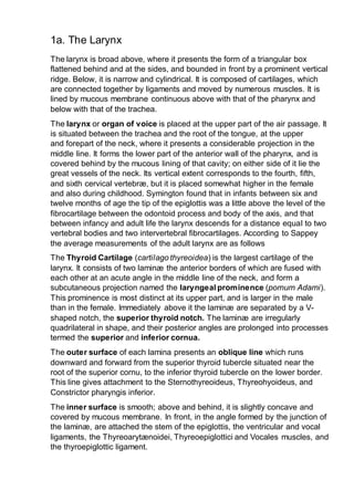
1a. the larynx
1. 1a. The Larynx
The larynx is broad above, where it presents the form of a triangular box
flattened behind and at the sides, and bounded in front by a prominent vertical
ridge. Below, it is narrow and cylindrical. It is composed of cartilages, which
are connected together by ligaments and moved by numerous muscles. It is
lined by mucous membrane continuous above with that of the pharynx and
below with that of the trachea.
The larynx or organ of voice is placed at the upper part of the air passage. It
is situated between the trachea and the root of the tongue, at the upper
and forepart of the neck, where it presents a considerable projection in the
middle line. It forms the lower part of the anterior wall of the pharynx, and is
covered behind by the mucous lining of that cavity; on either side of it lie the
great vessels of the neck. Its vertical extent corresponds to the fourth, fifth,
and sixth cervical vertebræ, but it is placed somewhat higher in the female
and also during childhood. Symington found that in infants between six and
twelve months of age the tip of the epiglottis was a little above the level of the
fibrocartilage between the odontoid process and body of the axis, and that
between infancy and adult life the larynx descends for a distance equal to two
vertebral bodies and two intervertebral fibrocartilages. According to Sappey
the average measurements of the adult larynx are as follows
The Thyroid Cartilage (cartilago thyreoidea) is the largest cartilage of the
larynx. It consists of two laminæ the anterior borders of which are fused with
each other at an acute angle in the middle line of the neck, and form a
subcutaneous projection named the laryngeal prominence (pomum Adami).
This prominence is most distinct at its upper part, and is larger in the male
than in the female. Immediately above it the laminæ are separated by a V-
shaped notch, the superior thyroid notch. The laminæ are irregularly
quadrilateral in shape, and their posterior angles are prolonged into processes
termed the superior and inferior cornua.
The outer surface of each lamina presents an oblique line which runs
downward and forward from the superior thyroid tubercle situated near the
root of the superior cornu, to the inferior thyroid tubercle on the lower border.
This line gives attachment to the Sternothyreoideus, Thyreohyoideus, and
Constrictor pharyngis inferior.
The inner surface is smooth; above and behind, it is slightly concave and
covered by mucous membrane. In front, in the angle formed by the junction of
the laminæ, are attached the stem of the epiglottis, the ventricular and vocal
ligaments, the Thyreoarytænoidei, Thyreoepiglottici and Vocales muscles, and
the thyroepiglottic ligament.
2. The upper border is concave behind and convex in front; it gives attachment
to the corresponding half of the hyothyroid membrane.
The lower border is concave behind, and nearly straight in front, the two
parts being separated by the inferior thyroid tubercle. A small part of it in and
near the middle line is connected to the cricoid cartilage by the middle
cricothyroid ligament.
The posterior border, thick and rounded, receives the insertions of the
Stylopharyngeus and Pharyngopalatinus. It ends above, in the superior cornu,
and below, in the inferior cornu. The superior cornu is long and narrow,
directed upward, backward, and medialward, and ends in a conical extremity,
which gives attachment to the lateral hyothyroid ligament. The inferior
cornu is short and thick; it is directed downward, with a slight inclination
forward and medialward, and presents, on the medial side of its tip, a small
oval articular facet for articulation with the side of the cricoid cartilage.
During infancy the laminæ of the thyroid cartilage are joined to each other by
a narrow, lozenge-shaped strip, named the intrathyroid cartilage. This strip
extends from the upper to the lower border of the cartilage in the middle line,
and is distinguished from the laminæ by being more transparent and more
flexible.
The Cricoid Cartilage (cartilago cricoidea) is smaller, but thicker and stronger
than the thyroid, and forms the lower and posterior parts of the wall of the
larynx. It consists of two parts: a posterior quadrate lamina, and a
narrow anterior arch, one-fourth or one-fifth of the depth of the lamina.
The lamina (lamina cartilaginis cricoideæ; posterior portion) is deep and
broad, and measures from above downward about 2 or 3 cm.; on its posterior
surface, in the middle line, is a vertical ridge to the lower part of which are
attached the longitudinal fibers of the esophagus; and on either side of this a
broad depression for the Cricoarytænoideus posterior.
The arch (arcus cartilaginis cricoideæ; anterior portion) is narrow and convex,
and measures vertically from 5 to 7 mm.; it affords attachment externally in
front and at the sides to the Cricothyreiodei, and behind, to part of the
Constrictor pharyngis inferior.
On either side, at the junction of the lamina with the arch, is a small round
articular surface, for articulation with the inferior cornu of the thyroid cartilage.
The lower border of the cricoid cartilage is horizontal, and connected to the
highest ring of the trachea by the cricotracheal ligament.
The upper border runs obliquely upward and backward, owing to the
great depth of the lamina. It gives attachment, in front, to the middle
cricothyroid ligament; at the side, to the conus elasticus and the
3. Cricoarytænoidei laterales; behind, it presents, in the middle, a shallow notch,
and on either side of this is a smooth, oval, convex surface, directed upward
and lateralward, for articulation with the base of an arytenoid cartilage.
The inner surface of the cricoid cartilage is smooth, and lined by mucous
membrane.
The Arytenoid Cartilages (cartilagines arytænoideæ) are two in number,
and situated at the upper border of the lamina of the cricoid cartilage, at the
back of the larynx. Each is pyramidal in form, and has three surfaces, a base,
and an apex.
The posterior surface is a triangular, smooth, concave, and gives attachment
to the Arytænoidei obliquus and transversus.
The antero-lateral surface is somewhat convex and rough. On it, near the
apex of the cartilage, is a rounded elevation (colliculus) from which a ridge
(crista arcuata) curves at first backward and then downward and forward to
the vocal process. The lower part of this crest intervenes between two
depressions or foveæ, an upper, triangular, and a lower oblong in shape; the
latter gives attachment to the Vocalis muscle.
The medial surface is narrow, smooth, and flattened, covered by mucous
membrane, and forms the lateral boundary of the intercartilaginous part of the
rima glottidis.
The base of each cartilage is broad, and on it is a concave smooth surface,
for articulation with the cricoid cartilage. Its lateral angle is short, rounded, and
prominent; it projects backward and lateralward, and is termed the muscular
process; it gives insertion to the Cricoarytænoideus posterior behind, and to
the Cricoarytænoideus lateralis in front. Its anterior angle, also prominent, but
more pointed, projects horizontally forward; it gives attachment to the vocal
ligament, and is called the vocal process.
The apex of each cartilage is pointed, curved backward and medialward, and
surmounted by a small conical, cartilaginous nodule, the corniculate
cartilage.
The Corniculate Cartilages (cartilagines corniculatæ; cartilages of Santorini)
are two small conical nodules consisting of yellow elastic cartilage, which
articulate with the summits of the arytenoid cartilages and serve to prolong
them backward and medialward. They are situated in the posterior parts of the
aryepiglottic folds of mucous membrane, and are sometimes fused with the
arytenoid cartilages.
The Cuneiform Cartilages (cartilagines cuneiformes; cartilages of Wrisberg)
are two small, elongated pieces of yellow elastic cartilage, placed one on
either side, in the aryepiglottic fold, where they give rise to small whitish
4. elevations on the surface of the mucous membrane, just in front of the
arytenoid cartilages.
The Epiglottis (cartilago epiglottica) is a thin lamella of fibrocartilage of a
yellowish color, shaped like a leaf, and projecting obliquely upward behind the
root of the tongue, in front of the entrance to the larynx. The free extremity is
broad and rounded; the attached part or stem is long, narrow, and connected
by the thyroepiglottic ligament to the angle formed by the two laminæ of the
thyroid cartilage, a short distance below the superior thyroid notch. The lower
part of its anterior surface is connected to the upper border of the body of the
hyoid bone by an elastic ligamentous band, the hyoepiglottic ligament.
The anterior or lingual surface is curved forward, and covered on its upper,
free part by mucous membrane which is reflected on to the sides and root of
the tongue, forming a median and two lateral glossoepiglottic folds; the
lateral folds are partly attached to the wall of the pharynx. The depressions
between the epiglottis and the root of the tongue, on either side of the median
fold, are named the valleculæ. The lower part of the anterior surface lies
behind the hyoid bone, the hyothyroid membrane, and upper part of the
thyroid cartilage, but is separated from these structures by a mass of fatty
tissue.
The posterior or laryngeal surface is smooth, concave from side to side,
concavo-convex from above downward; its lower part projects backward as an
elevation, the tubercle or cushion. When the mucous membrane is removed,
the surface of the cartilage is seen to be indented by a number of small pits, in
which mucous glands are lodged. To its sides the aryepiglottic folds are
attached.
Structure.—The corniculate and cuneiform cartilages, the epiglottis, and the
apices of the arytenoids at first consist of hyaline cartilage, but later elastic
fibers are deposited in the matrix, converting them into yellow fibrocartilage,
which shows little tendency to calcification. The thyroid, cricoid, and the
greater part of the arytenoids consist of hyaline cartilage, and become more or
less ossified as age advances. Ossification commences about the twenty-fifth
year in the thyroid cartilage, and somewhat later in the cricoid and arytenoids;
by the sixty-fifth year these cartilages may be completely converted into bone.
5. Ligaments.—The ligaments of the larynx (Figs. 951, 952) are extrinsic, i.
e., those connecting the thyroid cartilage and epiglottis with the hyoid bone,
and the cricoid cartilage with the trachea; and intrinsic, those which connect
the several cartilages of the larynx to each other.
Extrinsic Ligaments.—The ligaments connecting the thyroid cartilage with
the hyoid bone are the hyothyroid membrane, and a middle and two lateral
hyothyroid ligaments.
The Hyothyroid Membrane (membrana hyothyreoidea; thyrohyoid
membrane) is a broad, fibro-elastic layer, attached below to the upper border
of the thyroid cartilage and to the front of its superior cornu, and above to the
upper margin of the posterior surface of the body and greater cornua of the
hyoid bone, thus passing behind the posterior surface of the body of the
hyoid, and being separated from it by a mucous bursa, which facilitates the
upward movement of the larynx during deglutition. Its middle thicker part is
termed the middle hyothyroid ligament (ligamentum hyothyreoideum
medium; middle thyrohyoid ligament), its lateral thinner portions are pierced
by the superior laryngeal vessels and the internal branch of the superior
laryngeal nerve. Its anterior surface is in relation with the Thyreohyoideus,
Sternohyoideus, and Omohyoideus, and with the body of the hyoid bone.
6. The Lateral Hyothyroid Ligament (ligamentum hyothyreoideum laterale;
lateral thyrohyoid ligament) is a round elastic cord, which forms the posterior
border of the hyothyroid membrane and passes between the tip of the
superior cornu of the thyroid cartilage and the extremity of the greater cornu of
the hyoid bone. A small cartilaginous nodule (cartilago triticea), sometimes
bony, is frequently found in it.
The Epiglottis is connected with the hyoid bone by an elastic band,
the hyoepiglottic ligament (ligamentum hyoepiglotticum), which extends
from the anterior surface of the epiglottis to the upper border of the body of
the hyoid bone. The glossoepiglottic folds of mucous membrane (page 1075)
may also be considered as extrinsic ligaments of the epiglottis.
The Cricotracheal Ligament (ligamentum cricotracheale) connects the
cricoid cartilage with the first ring of the trachea. It resembles the fibrous
membrane which connects the cartilaginous rings of the trachea to each
other.
Intrinsic Ligaments.—Beneath the mucous membrane of the larynx is a
broad sheet of fibrous tissue containing many elastic fibers, and termed
the elastic membrane of the larynx. It is subdivided on either side by the
interval between the ventricular and vocal ligaments, the upper portion
extends between the arytenoid cartilage and the epiglottis and is often poorly
defined; the lower part is a well-marked membrane forming, with its fellow of
the opposite side, the conus elasticus which connects the thyroid, cricoid, and
arytenoid cartilages to one another. In addition the joints between the
individual cartilages are provided with ligaments.
The Conus Elasticus (cricothyroid membrane) is composed mainly of yellow
elastic tissue. It consists of an anterior and two lateral portions. The anterior
part or middle cricothyroid ligament (ligamentum cricothyreoideum
medium; central part of cricothyroid membrane) is thick and strong, narrow
above and broad below. It connects together the front parts of the contiguous
margins of the thyroid and cricoid cartilages. It is overlapped on either side by
the Cricothyreoideus, but between these is subcutaneous; it is crossed
horizontally by a small anastomotic arterial arch, formed by the junction of the
two cricothyroid arteries, branches of which pierce it. The lateral portions are
thinner and lie close under the mucous membrane of the larynx; they extend
from the superior border of the cricoid cartilage to the inferior margin of the
vocal ligaments, with which they are continuous. These ligaments may
therefore be regarded as the free borders of the lateral portions of the conus
elasticus, and extend from the vocal processes of the arytenoid cartilages to
the angle of the thyroid cartilage about midway between its upper and lower
borders.
7. Each arytenoid cartilage is connected to the cricoid by a capsule and a
posterior cricoarytenoid ligament. The capsule (capsula articularis
cricoarytenoidea) is thin and loose, and is attached to the margins of the
articular surfaces. The posterior cricoarytenoid ligament (ligamentum
cricoarytenoideum posterius) extends from the cricoid to the medial and back
part of the base of the arytenoid.
The thyroepiglottic ligament (ligamentum thyreoepiglotticum) is a long,
slender, elastic cord which connects the stem of the epiglottis with the angle
of the thyroid cartilage, immediately beneath the superior thyroid notch, above
the attachment of the ventricular ligaments.
Movements.—The articulation between the inferior cornu of the thyroid
cartilage and the cricoid cartilage on either side is a diarthrodial one, and
permits of rotatory and gliding movements. The rotatory movement is one in
which the cricoid cartilage rotates upon the inferior cornua of the thyroid
cartilage around an axis passing transversely through both joints. The gliding
movement consists in a limited shifting of the cricoid on the thyroid in different
directions
The articulation between the arytenoid cartilages and the cricoid is also a
diarthrodial one, and permits of two varieties of movement: one is a rotation of
the arytenoid on a vertical axis, whereby the vocal process is moved
lateralward or medialward, and the rima glottidis increased or diminished; the
other is a gliding movement, and allows the arytenoid cartilages to approach
or recede from each other; from the direction and slope of the articular
surfaces lateral gliding is accompanied by a forward and downward
movement. The two movements of gliding and rotation are associated, the
medial gliding being connected with medialward rotation, and the lateral
gliding with lateralward rotation. The posterior cricoarytenoid ligaments limit
the forward movement of the arytenoid cartilages on the cricoid.
Interior of the Larynx (Figs. 953, 954).—The cavity of the larynx (cavum
laryngis) extends from the laryngeal entrance to the lower border of the cricoid
cartilage where it is continuous with that of the trachea. It is divided into two
parts by the projection of the vocal folds, between which is a narrow triangular
fissure or chink, the rima glottidis. The portion of the cavity of the larynx
above the vocal folds is called the vestibule; it is wide and triangular in
shape, its base or anterior wall presenting, however, about its center the
backward projection of the tubercle of the epiglottis. It contains the ventricular
folds, and between these and the vocal folds are the ventricles of the
larynx. The portion below the vocal folds is at first of an elliptical form, but
lower down it widens out, assumes a circular form, and is continuous with the
tube of the trachea.
8. The entrance of the larynx (Fig. 955) is a triangular opening, wide in front,
narrow behind, and sloping obliquely downward and backward. It is bounded,
in front, by the epiglottis; behind, by the apices of the arytenoid cartilages, the
corniculate cartilages, and the interarytenoid notch; and on either side, by a
fold of mucous membrane, enclosing ligamentous and muscular fibers,
stretched between the side of the epiglottis and the apex of the arytenoid
cartilage; this is the aryepiglottic fold, on the posterior part of the margin of
which the cuneiform cartilage forms a more or less distinct whitish
prominence, the cuneiform tubercle.
9.
10. The Ventricular Folds (plicœ ventriculares; superior or false vocal cords) are
two thick folds of mucous membrane, each enclosing a narrow band of fibrous
tissue, the ventricular ligament which is attached in front to the angle of the
thyroid cartilage immediately below the attachment of the epiglottis, and
behind to the antero-lateral surface of the arytenoid cartilage, a short distance
above the vocal process. The lower border of this ligament, enclosed in
mucous membrane, forms a free crescentic margin, which constitutes the
upper boundary of the ventricle of the larynx.
The Vocal Folds (plicœ vocales; inferior or true vocal cords) are concerned in
the production of sound, and enclose two strong bands, named the vocal
ligaments (ligamenta vocales; inferior thyroarytenoid). Each ligament consists
of a band of yellow elastic tissue, attached in front to the angle of the thyroid
cartilage, and behind to the vocal process of the arytenoid. Its lower border is
continuous with the thin lateral part of the conus elasticus. Its upper border
forms the lower boundary of the ventricle of the larynx. Laterally, the Vocalis
muscle lies parallel with it. It is covered medially by mucous membrane, which
is extremely thin and closely adherent to its surface.
11.
12. The Ventricle of the Larynx (ventriculus laryngis [Morgagnii]; laryngeal
sinus) is a fusiform fossa, situated between the ventricular and vocal folds on
either side, and extending nearly their entire length. The fossa is
bounded, above, by the free crescentic edge of the ventricular fold; below, by
the straight margin of the vocal fold; laterally, by the mucous membrane
covering the corresponding Thyreoarytænoideus. The anterior part of the
ventricle leads up by a narrow opening into a cecal pouch of mucous
membrane of variable size called the appendix.
The appendix of the laryngeal ventricle (appendix ventriculi laryngis;
laryngeal saccule) is a membranous sac, placed between the ventricular fold
and the inner surface of the thyroid cartilage, occasionally extending as far as
its upper border or even higher; it is conical in form, and curved slightly
backward. On the surface of its mucous membrane are the openings of sixty
or seventy mucous glands, which are lodged in the submucous areolar tissue.
This sac is enclosed in a fibrous capsule, continuous below with the
ventricular ligament. Its medial surface is covered by a few delicate muscular
fasciculi, which arise from the apex of the arytenoid cartilage and become lost
in the aryepiglottic fold of mucous membrane; laterally it is separated from the
13. thyroid cartilage by the Thyreoepiglotticus. These muscles compress the sac,
and express the secretion it contains upon the vocal folds to lubricate their
surfaces.
The Rima Glottidis (Fig. 956) is the elongated fissure or chink between the
vocal folds in front, and the bases and vocal processes of the arytenoid
cartilages behind. It is therefore subdivided into a larger anterior
intramembranous part (glottis vocalis), which measures about three-fifths of
the length of the entire aperture, and a posterior intercartilaginous part (glottis
respiratoria). Posteriorly it is limited by the mucous membrane passing
between the arytenoid cartilages. The rima glottidis is the narrowest part of
the cavity of the larynx, and its level corresponds with the bases of the
arytenoid cartilages. Its length, in the male, is about 23 mm.; in the female
from 17 to 18 mm. The width and shape of the rima glottidis vary with the
movements of the vocal folds and arytenoid cartilages during respiration and
phonation. In the condition of rest, i. e., when these structures are
uninfluenced by muscular action, as in quiet respiration, the intramembranous
part is triangular, with its apex in front and its base behind—the latter being
represented by a line, about 8 mm. long, connecting the anterior ends of the
vocal processes, while the medial surfaces of the arytenoids are parallel to
each other, and hence the intercartilaginous part is rectangular. During
14. extreme adduction of the vocal folds, as in the emission of a high note, the
intramembranous part is reduced to a linear slit by the apposition of the vocal
folds, while the intercartilaginous part is triangular, its apex corresponding to
the anterior ends of the vocal processes of the arytenoids, which are
approximated by the medial rotation of the cartilages. Conversely in extreme
abduction of the vocal folds, as in forced inspiration, the arytenoids and their
vocal processes are rotated lateralward, and the intercartilaginous part is
triangular in shape but with its apex directed backward. In this condition the
entire glottis is somewhat lozenge-shaped, the sides of the intramembranous
part diverging from before backward, those of the intercartilaginous part
diverging from behind forward—the widest part of the aperture corresponding
with the attachments of the vocal folds to the vocal processes.
Muscles.—The muscles of the larynx are extrinsic, passing between the
larynx and parts around—these have been described in the section on
Myology; and intrinsic, confined entirely to the larynx.
The intrinsic muscles are:
The Cricothyreoideus (Cricothyroid) (Fig. 957), triangular in form, arises from
the front and lateral part of the cricoid cartilage; its fibers diverge, and are
arranged in two groups. The lower fibers constitute a pars obliqua and slant
backward and lateralward to the anterior border of the inferior cornu; the
anterior fibers, forming a pars recta, run upward, backward, and lateralward
to the posterior part of the lower border of the lamina of the thyroid cartilage.
The medial borders of the two muscles are separated by a triangular interval,
occupied by the middle cricothyroid ligament.
15. The Cricoarytænoideus posterior (posterior cricoarytenoid) (Fig.
958) arises from the broad depression on the corresponding half of the
posterior surface of the lamina of the cricoid cartilage; its fibers run upward
and lateralward, and converge to be inserted into the back of the muscular
process of the arytenoid cartilage. The uppermost fibers are nearly horizontal,
the middle oblique, and the lowest almost vertical.
The Cricoarytænoideus lateralis (lateral cricoarytenoid) (Fig. 959) is
smaller than the preceding, and of an oblong form. It arises from the upper
border of the arch of the cricoid cartilage, and, passing obliquely upward and
backward, is inserted into the front of the muscular process of the arytenoid
cartilage.
FIG. 957– Side view of the larynx, showing muscular attachments.
17. FIG. 959– Muscles of larynx. Side view. Right lamina of thyroid cartilage
removed.
The Arytænoideus (Fig. 958) is a single muscle, filling up the posterior
concave surfaces of the arytenoid cartilages. It arises from the posterior
surface and lateral border of one arytenoid cartilage, and is inserted into the
corresponding parts of the opposite cartilage. It consists of oblique and
transverse parts. The Arytænoideus obliquus, the more superficial, forms
two fasciculi, which pass from the base of one cartilage to the apex of the
opposite one, and therefore cross each other like the limbs of the letter X; a
few fibers are continued around the lateral margin of the cartilage, and are
prolonged into the aryepiglottic fold; they are sometimes described as a
separate muscle, the Aryepiglotticus. The Arytænoideus
transversus crosses transversely between the two cartilages.
The Thyreoarytænoideus (Thyroarytenoid) (Figs. 959, 960) is a broad, thin,
muscle which lies parallel with and lateral to the vocal fold, and supports the
wall of the ventricle and its appendix. It arises in front from the lower half of
the angle of the thyroid cartilage, and from the middle cricothyroid ligament.
18. Its fibers pass backward and lateralward, to be inserted into the base and
anterior surface of the arytenoid cartilage. The lower and deeper fibers of the
muscle can be differentiated as a triangular band which is inserted into the
vocal process of the arytenoid cartilage, and into the adjacent portion of its
anterior surface; it is termed the Vocalis, and lies parallel with the vocal
ligament, to which it is adherent.
FIG. 960– Muscles of the larynx, seen from above. (Enlarged.)
A considerable number of the fibers of the Thyreoarytænoideus are
prolonged into the aryepiglottic fold, where some of them become lost, while
others are continued to the margin of the epiglottis. They have received a
distinctive name, Thyreoepiglotticus, and are sometimes described as a
separate muscle. A few fibers extend along the wall of the ventricle from the
lateral wall of the arytenoid cartilage to the side of the epiglottis and constitute
the Ventricularis muscle.
Actions.—In considering the actions of the muscles of the larynx, they may
be conveniently divided into two groups, vix.: 1. Those which open and close
the glottis. 2. Those which regulate the degree of tension of the vocal folds.
19. The Cricoarytœnoidei posteriores separate the vocal folds, and, consequently,
open the glottis, by rotating the arytenoid cartilages outward around a vertical
axis passing through the cricoarytenoid joints; so that their vocal processes
and the vocal folds attached to them become widely separated.
The Cricoarytœnoidei laterales close the glottis by rotating the arytenoid
cartilages inward, so as to approximate their vocal processes.
The Arytœnoideus approximates the arytenoid cartilages, and thus closes the
opening of the glottis, especially at its back part.
The Cricothyreoidei produce tension and elongation of the vocal folds by
drawing up the arch of the cricoid cartilage and tilting back the upper border of
its lamina; the distance between the vocal processes and the angle of the
thyroid is thus increased, and the folds are consequently elongated
The Thyreoarytœnoidei, consisting of two parts having different attachments
and different directions, are rather complicated as regards their action. Their
main use is to draw the arytenoid cartilages forward toward the thyroid, and
thus shorten and relax the vocal folds. But, owing to the connection of the
deeper portion with the vocal fold, this part, if acting separately, is supposed
to modify its elasticity and tension, while the lateral portion rotates the
arytenoid cartilage inward, and thus narrows the rima glottidis by bringing the
two vocal folds together.
Mucous Membrane.—The mucous membrane of the larynx is continuous
above with that lining the mouth and pharynx, and is prolonged through the
trachea and bronchi into the lungs. It lines the posterior surface and the upper
part of the anterior surface of the epiglottis, to which it is closely adherent, and
forms the aryepiglottic folds which bound the entrance of the larynx. It lines
the whole of the cavity of the larynx; forms, by its reduplication, the chief part
of the ventricular fold, and, from the ventricle, is continued into the ventricular
appendix. It is then reflected over the vocal ligament, where it is thin, and very
intimately adherent; covers the inner surface of the conus elasticus and
cricoid cartilage; and is ultimately continuous with the lining membrane of the
trachea. The anterior surface and the upper half of the posterior surface of the
epiglottis, the upper part of the aryepiglottic folds and the vocal folds are
covered by stratified squamous epithelium; all the rest of the laryngeal
mucous membrane is covered by columnar ciliated cells, but patches of
stratified squamous epithelium are found in the mucous membrane above the
glottis.
Glands.—The mucous membrane of the larynx is furnished with numerous
mucous secreting glands, the orifices of which are found in nearly every part;
they are very plentiful upon the epiglottis, being lodged in little pits in its
substance; they are also found in large numbers along the margin of the
20. aryepiglottic fold, in front of the arytenoid cartilages, where they are termed
the arytenoid glands. They exist also in large numbers in the ventricular
appendages. None are found on the free edges of the vocal folds.
Vessels and Nerves.—The chief arteries of the larynx are the laryngeal
branches derived from the superior and inferior thyroid. The veins accompany
the arteries; those accompanying the superior laryngeal artery join the
superior thyroid vein which opens into the internal jugular vein; while those
accompanying the inferior laryngeal artery join the inferior thyroid vein which
opens into the innominate vein. The lymphatic vessels consist of two sets,
superior and inferior. The former accompany the superior laryngeal artery and
pierce the hyothyroid membrane, to end in the glands situated near the
bifurcation of the common carotid artery. Of the latter, some pass through the
middle cricothyroid ligament and open into a gland lying in front of that
ligament or in front of the upper part of the trachea, while others pass to the
deep cervical glands and to the glands accompanying the inferior thyroid
artery. The nerves are derived from the internal and external branches of the
superior laryngeal nerve, from the recurrent nerve, and from the sympathetic.
The internal laryngeal branch is almost entirely sensory, but some motor
filaments are said to be carried by it to the Arytænoideus. It enters the larynx
by piercing the posterior part of the hyothyroid membrane above the superior
laryngeal vessels, and divides into a branch which is distributed to both
surfaces of the epiglottis, a second to the aryepiglottic fold, and a third, the
largest, which supplies the mucous membrane over the back of the larynx and
communicates with the recurrent nerve. The external laryngeal branch
supplies the Cricothyreoideus. The recurrent nerve passes upward beneath
the lower border of the Constrictor pharyngis inferior immediately behind the
cricothyroid joint. It supplies all the muscles of the larynx except the
Cricothyreoideus, and perhaps a part of the Arytænoideus. The sensory
branches of the laryngeal nerves form subepithelial plexuses, from which
fibers pass to end between the cells covering the mucous membrane.
Over the posterior surface of the epiglottis, in the aryepiglottic folds, and less
regularly in some other parts, taste-buds, similar to those in the tongue, are
found